Explanation:
Epidural hematoma is most likely given the history of a head injury, loss of consciousness, lucidity, and subsequent decompensation. The middle meningeal artery is the vessel that is most frequently implicated in epidural hematomas, and hematoma evacuation is recommended. Because the blood lies between the dura and the skull, a biconvex-shaped hemorrhage is frequently seen on imaging. A deadly epidural hemorrhage can occur in up to 20% of cases. On imaging, subdural hematomas have a crescent-shaped appearance and hardly ever feature the typical epidural “lucid period.” Instead, they experience agony and bewilderment that worsen with time. Bleeds from subdural hematomas are caused by the bridging veins.
Explanation:
The MAC is the lowest amount of anesthetic vapor required to inhibit a physical response in 50% of patients to a common surgical stimulus. Less fat soluble, less powerful, yet faster onset with higher MAC. (xanax) More lipid soluble, more powerful, but longer onset, when MAC is lower. This little story, which goes, “When MAC is LOW he is FAT and SLOW,” is a helpful method to memorize it. Typical MAC values include: 104 Nitrous oxide * Halothane – 0.75 * Chloroform – 0.5 * Methoxyflurane – 0.16 * Xenon – 72 * Desflurane – 6 * Ethyl Ether – 3.2 * Sevoflurane – 2 * Enflurane – 1.7 * Isoflurane – 1.2 *
Explanation:
Keep the tube suctioned while gauging the output. Hemothorax typically results in diminished ipsilateral breath sounds and muted percussion on that side. The pleural cavity of a typical adult male can retain 4 or more liters of blood, therefore exsanguination can happen even in the absence of any other outward signs of bleeding. The instantaneous return of 1500 ml would have been a sign of OR. Indications for exploratory surgery include bleeding with instability after all other causes of bleeding have been ruled out, output >250 mL/hour x 4 hours, or 2500 mL/24 hours.

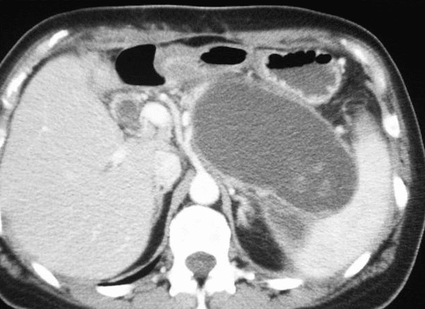
Explanation:
The head of the pancreas hosts around one-third of the pancreatic pseudocysts, while the tail hosts two-thirds of them. The most frequent causes in adults are alcohol and gallstone pancreatitis, whereas the most frequent cause in children is trauma .
Explanation:
This is a Zenker’s diverticulum. All of the options listed are possible life-threatening complications, but the most common is aspiration.
Explanation:
RQ=CO2/O2 By producing an overabundance of calories and carbs to be turned into fat, overfeeding might result in patients needing artificial breathing and intubation for a longer period of time. A RQ>1.0 would arise from this. This process results in an increase in CO2 generation, which must be offset by an increase in the patient’s respiratory effort. As a result, the respiratory system might become so worn out that weaning becomes challenging or even impossible. Since oxygen consumption rises while the body is turning stored energy into sugar, starvation would result in an RQ 0.7. (breaking down glycogen, adipose tissues).
Explanation:
The patient has burned 27% of her body (9% for arms, 9% for upper back , and 9% for lower back). Use the Parkland Formula (MEMORIZE THIS!!): 4mL x Weight in kg x % burned Replace fluid 4ml/kg x kg of pt x %burned, replace half over first 8 hours and the rest over the next 16. This should be calculated from time burned, not time of admission.