ECMO Life Support: Complete Guide to Extracorporeal Membrane Oxygenation
🎯 Complete guide to ECMO life support: how the circuit works, neonatal & adult indications, COVID applications, costs, and survival data.

Extracorporeal membrane oxygenation in neonates remains one of the most impactful applications of ecmo life support, offering a temporary cardiopulmonary bypass outside the body that allows critically diseased lungs and hearts to rest and recover. Since the first successful neonatal ECMO run in 1975, the technology has grown from an experimental last resort into a standard-of-care intervention across pediatric intensive care units and adult cardiac surgery centers throughout the United States. Understanding how this support system works—and when it is appropriate—is essential for any clinician preparing for ECMO certification or bedside practice.
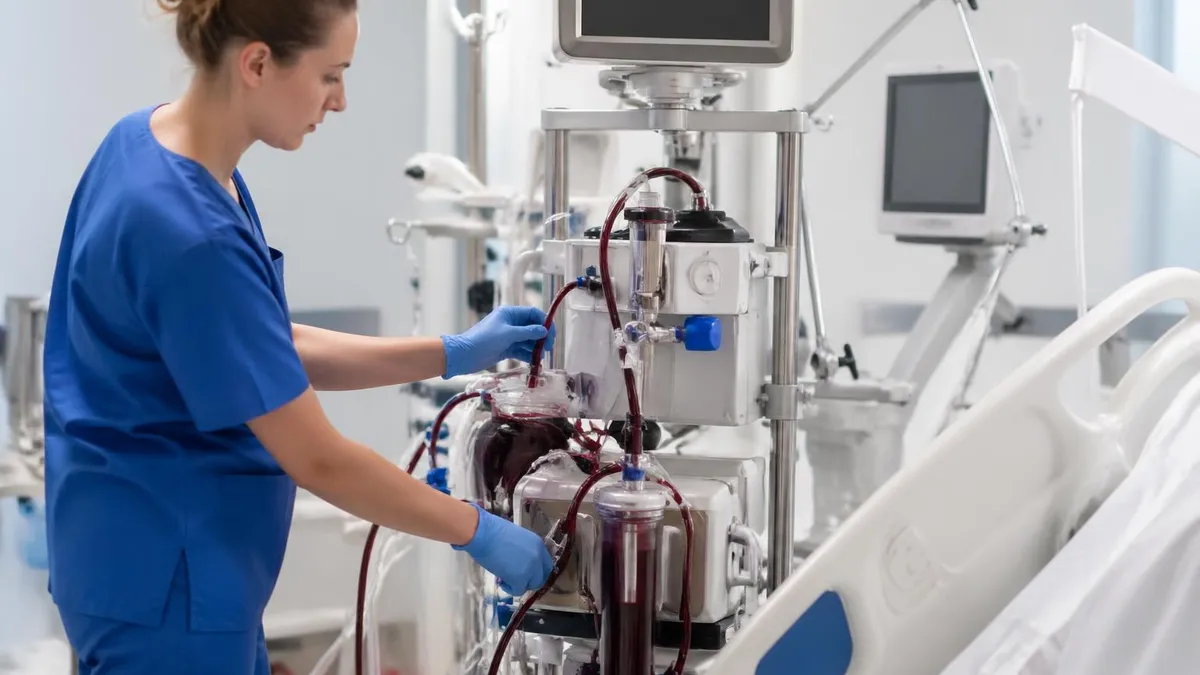
At its core, the extracorporeal membrane oxygenation procedure involves removing venous blood from the patient, passing it through an artificial membrane lung that adds oxygen and removes carbon dioxide, then returning it to the patient either venously or arterially depending on the clinical goal. This continuous circuit, driven by a centrifugal or roller pump, can sustain gas exchange even when the native lungs are severely damaged by meconium aspiration, persistent pulmonary hypertension, diaphragmatic hernia, or acute respiratory distress syndrome. The machine does the work of both the lungs and, in veno-arterial configurations, the heart.
The extracorporeal membrane oxygenation circuit consists of several precisely engineered components: drainage and return cannulas, tubing, a centrifugal pump head, an oxygenator membrane, a heat exchanger, and an array of pressure and flow sensors. Each element must function in harmony to deliver adequate oxygen delivery and prevent complications such as hemolysis, thrombosis, air embolism, or circuit failure. Clinicians managing the circuit must continuously balance sweep gas flow, blood flow rate, anticoagulation targets, and patient hemodynamics to optimize outcomes.
Venovenous extracorporeal membrane oxygenation is used primarily for isolated respiratory failure, while veno-arterial ECMO provides combined cardiac and pulmonary support. The distinction matters enormously because cannulation sites, hemodynamic goals, weaning strategies, and complication profiles differ significantly between the two modes. Venovenous configurations drain from and return blood to the venous system, leaving native cardiac output intact and requiring patients to maintain some level of cardiac function. Veno-arterial configurations can fully replace cardiac output but carry additional risks related to retrograde aortic flow.
Extracorporeal membrane oxygenation for adults has grown substantially over the past two decades, driven largely by the 2009 H1N1 influenza pandemic and the later surge of extracorporeal membrane oxygenation COVID applications during 2020 and 2021. High-volume ECMO centers demonstrated that carefully selected adult patients with refractory respiratory failure could achieve survival rates of 50 to 60 percent with appropriate ECMO support—outcomes that would have been impossible with mechanical ventilation alone. These results catalyzed expansion of ECMO programs at academic medical centers and created strong demand for credentialed ECMO specialists.
The extracorporeal membrane oxygenation machine price is a common concern for hospitals considering program development. Capital equipment costs for a modern ECMO system range from roughly $100,000 to $300,000 per unit, while disposable circuit costs per run average $15,000 to $30,000. Total per-patient hospital costs including ICU stay frequently exceed $200,000, making program sustainability dependent on adequate case volume, skilled personnel, and robust reimbursement pathways. Despite the cost, ECMO remains highly cost-effective relative to its alternatives when used in appropriately selected patients.
This guide walks through every dimension of ECMO life support: circuit anatomy and function, neonatal and pediatric indications, adult applications including COVID-19, the VV versus VA decision framework, pharmacologic management, and the knowledge domains tested on ECMO certification examinations. Whether you are a respiratory therapist, perfusionist, intensivist, or nurse preparing for the Certified ECMO Specialist (CES) exam or simply building clinical competency, this resource provides the depth and breadth you need to succeed.
ECMO Life Support by the Numbers

How the ECMO Circuit Works: Step-by-Step Flow
Venous Drainage
Pump & Flow Generation
Membrane Oxygenation
Heat Exchange
Return to Patient
Continuous Monitoring
Extracorporeal membrane oxygenation in neonates represents the historical foundation of the entire field and remains the indication with the highest survival rates reported in the Extracorporeal Life Support Organization (ELSO) registry. Neonates are considered candidates when they suffer from reversible cardiopulmonary failure and have failed maximal conventional therapy, typically defined as an oxygenation index above 40 or a pH below 7.15 despite optimal ventilator management, inhaled nitric oxide, and surfactant therapy. The underlying diagnoses most commonly treated with neonatal ECMO include meconium aspiration syndrome, persistent pulmonary hypertension of the newborn, congenital diaphragmatic hernia, respiratory distress syndrome, and sepsis-related cardiac dysfunction.
Meconium aspiration syndrome accounts for approximately 30 percent of neonatal ECMO cases in the United States and carries overall survival rates near 95 percent with ECMO support. The pathophysiology involves chemical pneumonitis, mechanical airway obstruction, and severe hypoxic respiratory failure that can rapidly become refractory to all conventional measures. When oxygenation index thresholds are crossed, the window for initiating ECMO is narrow—delays increase the risk of end-organ injury to the brain, kidneys, and heart that will compound recovery even after adequate oxygenation is restored.
Congenital diaphragmatic hernia represents a more complex challenge because the underlying lung hypoplasia is not fully reversible. ECMO does not repair the hernia but provides a bridge to surgical correction while allowing pulmonary hypertension to be managed pharmacologically. Survival rates for CDH on ECMO are lower than for other neonatal diagnoses, averaging 50 to 60 percent, but ECMO remains a critical tool in high-volume centers managing this population. Timing of surgical repair relative to ECMO cannulation remains an active area of research, with some centers favoring repair on ECMO and others preferring decannulation first.
Persistent pulmonary hypertension of the newborn is the final common pathway for many neonatal respiratory emergencies. When pulmonary vascular resistance remains high after birth, the right-to-left shunting at the ductus arteriosus and foramen ovale maintains severe hypoxemia that quickly becomes life-threatening. Inhaled nitric oxide, a selective pulmonary vasodilator, reduces the need for ECMO in many PPHN cases but is insufficient in the most severe presentations. ECMO essentially bypasses the obstructed pulmonary circuit, allowing time for vasoconstriction to resolve and the normal postnatal decline in pulmonary vascular resistance to occur.
Neonatal cannulation is typically performed via the right common carotid artery and right internal jugular vein for veno-arterial support. The carotid artery sacrifice required for venous-arterial cannulation has historically raised concerns about long-term neurodevelopmental outcomes, though most outcome data demonstrate that ECMO-treated neonates achieve reasonable developmental trajectories compared to their diagnosis-matched peers who survive without ECMO. Some centers now use cervical or femoral approaches with smaller cannulas to preserve the carotid, though technical challenges limit widespread adoption in the smallest neonates.
Pediatric ECMO—defined as patients from 30 days to 18 years—presents a distinct set of indications and technical challenges compared to neonatal ECMO. Cardiac diagnoses, particularly post-operative congenital heart disease and myocarditis, dominate the pediatric ECMO population. Bridge-to-recovery strategies are attempted first; if recovery is not expected, bridge-to-transplant or bridge-to-ventricular assist device may be considered depending on center resources and patient suitability. Pediatric respiratory ECMO is also performed for ARDS, pneumonia, and bronchiolitis in infants who fail conventional ventilation and high-frequency oscillation.
The ELSO registry, which captures outcomes from over 500 centers worldwide, reports that pediatric cardiac ECMO survival to discharge averages 40 to 50 percent, while pediatric respiratory ECMO survival approaches 60 percent. These figures underline both the severity of illness in this population and the meaningful life-saving impact of ECMO when conventional therapy has been exhausted. Clinicians managing these patients must possess deep knowledge of both the technical circuit management and the underlying disease pathophysiology to optimize each patient's chance of recovery.
Venovenous Extracorporeal Membrane Oxygenation vs VA ECMO: Key Differences
Venovenous extracorporeal membrane oxygenation is the preferred mode for isolated respiratory failure because it avoids arterial cannulation and preserves native cardiac output. Blood is drained from the venous system, oxygenated through the membrane lung, and returned to the right atrium, where it mixes with native venous blood before traversing the pulmonary circulation. Adequate cardiac function is a prerequisite; patients with significant cardiac dysfunction are not suitable candidates for VV support alone.
Common cannulation strategies for VV-ECMO include the bicaval dual-lumen cannula (a single-cannula approach through the right internal jugular vein), which allows drainage from both the superior and inferior vena cava while returning oxygenated blood directly toward the tricuspid valve. This configuration is especially popular in adults because it eliminates femoral cannulation, improves patient mobility, and reduces recirculation. Flow rates of 4 to 6 L/min in adults typically achieve arterial oxygen saturations of 85 to 95 percent, depending on native cardiac output and residual pulmonary function.

Benefits and Risks of ECMO Life Support
- +Provides complete cardiac and pulmonary support when all conventional therapies have failed
- +Allows injured lungs to rest at low ventilator settings, reducing ventilator-induced lung injury
- +Bridges patients to recovery, transplant, or durable mechanical circulatory support devices
- +Proven survival benefit in neonatal respiratory failure with registry-reported rates above 75 percent
- +Enables prone positioning and rehabilitation in awake, ambulatory VV-ECMO patients
- +Rapidly reversible and removable once native organ function recovers sufficiently
- −Systemic anticoagulation requirement creates bleeding risk including intracranial hemorrhage
- −Circuit thrombosis and oxygenator failure require emergency circuit changes under pressure
- −Hemolysis from pump and cannula shear forces can cause renal injury and hemoglobinuria
- −VA-ECMO increases LV afterload, risking ventricular distension and pulmonary edema
- −Carotid artery ligation in neonatal VA-ECMO raises potential long-term neurodevelopmental concerns
- −High cost and resource intensity limits availability outside of specialized tertiary centers
ECMO Management Checklist: Daily Circuit and Patient Assessment
- ✓Verify circuit blood flow rate, RPM, and sweep gas settings match ordered parameters at start of each shift
- ✓Assess pre- and post-membrane pressure differential to detect early oxygenator clotting or failure
- ✓Check all circuit connections, tubing, and clamps for integrity, air bubbles, or visible thrombus
- ✓Confirm anticoagulation status: check ACT, anti-Xa, or heparin level per institutional protocol
- ✓Perform head-to-toe patient assessment including neurological status, urine output, and skin perfusion
- ✓Review arterial blood gas results and adjust FiO2, sweep flow, and ventilator settings accordingly
- ✓Obtain daily point-of-care echocardiography to assess ventricular function and guide weaning
- ✓Document circuit hours and cumulative blood product transfusions in the ECMO flowsheet
- ✓Assess cannula insertion sites for bleeding, hematoma, limb ischemia, or signs of infection
- ✓Conduct a structured team huddle documenting ECMO goals, potential weaning timeline, and escalation plan
The Oxygenation Index Threshold for ECMO Referral
An oxygenation index (OI = MAP × FiO2 × 100 / PaO2) consistently above 40 in neonates or above 25 in pediatric and adult patients despite optimal conventional therapy is a widely accepted threshold for ECMO referral. Early referral to an ECMO center—before irreversible end-organ damage occurs—is associated with significantly better survival and neurological outcomes. Do not wait for cardiac arrest to call the ECMO team.
Extracorporeal membrane oxygenation for adults underwent its most dramatic expansion during the COVID-19 pandemic, when extracorporeal membrane oxygenation COVID applications became a defining feature of ICU care at academic medical centers around the world. The SARS-CoV-2 virus caused a distinct form of hypoxemic respiratory failure characterized by severe hypoxemia with relatively preserved lung compliance early in the disease course—a phenotype sometimes labeled silent hypoxemia. As the pandemic progressed and patient volumes overwhelmed conventional ICU capacity, ECMO centers faced the unprecedented challenge of managing large queues of potential candidates with limited circuits and staffing.
International collaborative data from the ELSO COVID-19 registry, published in The Lancet, demonstrated that adult patients with COVID-19-related ARDS placed on VV-ECMO at experienced centers achieved 60-day in-hospital survival rates of approximately 50 percent. These results, while sobering, compared favorably to the nearly 100 percent mortality expected in patients with refractory hypoxemia failing maximal ventilatory support. The data reinforced ECMO's role as a genuine rescue therapy when used with disciplined patient selection criteria rather than as a reflexive escalation for any ventilated COVID-19 patient.
Patient selection for adult ECMO follows several validated scoring frameworks. The RESP (Respiratory ECMO Survival Prediction) score stratifies adult respiratory ECMO candidates based on age, diagnosis, duration of mechanical ventilation, immune function, and several laboratory values to estimate predicted survival and guide triage decisions. The SAVE score performs analogous risk stratification for VA-ECMO candidates with cardiogenic shock. Both tools are incorporated into ELSO guidelines and are regularly tested on the Certified ECMO Specialist examination, making them essential knowledge for candidates.
Post-cardiac arrest ECMO, also called ECPR (extracorporeal cardiopulmonary resuscitation), represents an increasingly adopted application in adults with witnessed refractory ventricular fibrillation or pulseless ventricular tachycardia. Several randomized controlled trials including ARREST and INCEPTION have demonstrated survival benefits for carefully selected ECPR candidates compared to conventional ACLS, particularly when cannulation can be achieved within 60 minutes of arrest onset and when a specific hospital protocol is in place. ECPR requires a highly coordinated team capable of initiating ECMO in the emergency department or cardiac catheterization laboratory while CPR is ongoing.
Awake ECMO—or non-intubated VV-ECMO—has emerged as a strategy to avoid the harms of invasive mechanical ventilation in spontaneously breathing adult patients with respiratory failure. By providing supplemental gas exchange through the ECMO circuit while the patient breathes independently, clinicians can reduce sedation, enable early mobilization, and preserve respiratory muscle function. Successful awake ECMO requires careful patient selection (cooperative, hemodynamically stable, not excessively tachypneic), a bicaval dual-lumen cannula configuration, and an experienced team comfortable managing an ambulatory patient on ECMO.
The extracorporeal membrane oxygenation diagram most commonly encountered in training materials illustrates the full circuit pathway from venous drainage through the pump, oxygenator, and heat exchanger to the return cannula. Familiarity with the anatomical landmarks for each cannulation site—including the right internal jugular, femoral veins, femoral artery, and subclavian vein—is tested on board examinations and is critical for bedside troubleshooting. Clinicians should be able to trace circuit blood flow direction in both VV and VA configurations and identify where specific complications, such as oxygenator clotting or inlet obstruction, would manifest on the pressure-flow waveforms.
Long-term outcomes for adult ECMO survivors are an active area of research. Studies published in major critical care journals report that a significant proportion of survivors experience post-intensive care syndrome, characterized by cognitive impairment, muscle weakness, depression, and anxiety that persist for months to years after discharge. Structured rehabilitation programs, patient and family support groups, and close follow-up with pulmonology and cardiology are strongly recommended components of post-ECMO care. Understanding these long-term trajectories helps clinicians set realistic expectations during the initial goals-of-care conversations with patients and families at the bedside.

ECMO is contraindicated in patients with irreversible underlying conditions incompatible with meaningful survival, severe neurological injury precluding recovery, uncontrolled hemorrhagic coagulopathy that precludes anticoagulation, or when the patient or surrogate has clearly declined aggressive life support. Initiating ECMO without clearly defined goals and an exit strategy risks prolonging suffering without benefit. Always establish time-limited goals and reassess candidacy daily.
Pharmacologic management is among the most technically demanding aspects of ECMO life support and represents a major knowledge domain on ECMO certification examinations. Anticoagulation with unfractionated heparin (UFH) is the standard of care because of its reversibility, short half-life, and ability to be monitored with activated clotting time (ACT), anti-Xa levels, or viscoelastic assays like thromboelastography. Target ACT values typically range from 160 to 200 seconds during the early stabilization phase, with adjustments made based on bleeding risk, circuit appearance, and assay availability at each institution.
Heparin resistance is a well-recognized challenge in ECMO patients and occurs when standard heparin doses fail to achieve target ACT levels, often due to antithrombin III depletion, high factor VIII levels, or elevated acute-phase reactants. Management strategies include fresh frozen plasma infusion to replenish antithrombin, direct antithrombin III concentrate supplementation, or transitioning to alternative anticoagulants such as bivalirudin, a direct thrombin inhibitor monitored by activated partial thromboplastin time. Each of these strategies has distinct monitoring requirements, dosing protocols, and reversal options that ECMO specialists must master.
Sedation and analgesia management during ECMO is complicated by significant drug sequestration within the circuit components. The lipophilic, large-volume-of-distribution properties of many sedative agents—including fentanyl, midazolam, propofol, and dexmedetomidine—lead to drug adsorption onto the PVC tubing, silicone, and polysulfone oxygenator membrane. This reduces effective plasma concentrations and often requires substantially higher infusion rates than would be used outside of ECMO. Clinicians must also account for the circuit's impact on drug clearance when patients have concurrent acute kidney injury or hepatic dysfunction.
Vasoactive medication management on ECMO requires careful titration because circuit blood flow interacts with native hemodynamics in complex ways. In VA-ECMO patients, increasing ECMO flow reduces ventricular preload but raises afterload, potentially increasing myocardial oxygen demand in a recovering heart. Vasopressors like norepinephrine raise systemic vascular resistance and may worsen LV distension if native cardiac output is severely impaired. The goal is to wean vasopressors as ECMO flow provides hemodynamic stability, while monitoring for signs of differential hypoxia and ensuring adequate coronary and cerebral perfusion.
Nutrition support during ECMO presents unique challenges because gastrointestinal hypoperfusion, ileus, and high sedation requirements often delay enteral feeding initiation. Current ELSO guidelines recommend early enteral nutrition when feasible, targeting 20 to 25 kcal/kg/day with adequate protein of 1.5 to 2.0 g/kg/day to prevent muscle catabolism during prolonged runs. Parenteral nutrition should be reserved for cases where enteral feeding is not tolerated. Glycemic control targeting glucose values between 140 and 180 mg/dL reduces infection risk without increasing hypoglycemic events in this critically ill population.
Antimicrobial pharmacokinetics are profoundly altered by ECMO. Studies of commonly used antibiotics in ECMO patients demonstrate that hydrophilic antibiotics such as vancomycin and aminoglycosides exhibit increased volumes of distribution with relatively modest circuit sequestration, while lipophilic agents like meropenem and caspofungin can have substantially reduced free drug concentrations due to circuit binding. Therapeutic drug monitoring is strongly recommended for aminoglycosides and vancomycin in ECMO patients; for beta-lactam antibiotics, extended-infusion or continuous-infusion strategies improve target attainment at standard doses without requiring dose escalation.
Understanding the pharmacokinetic alterations introduced by ECMO is not merely academic—it directly affects patient outcomes. Underdosing of antifungal agents in an immunosuppressed ECMO patient can result in treatment failure for invasive candidiasis; underdosing of sedatives can produce awareness and agitation that leads to circuit dislodgement or self-extubation. Specialists who master the pharmacology domain earn a decisive advantage on ECMO board examinations and, more importantly, translate that knowledge into safer, more effective bedside practice for some of the most vulnerable patients in any hospital.
Preparing for ECMO certification requires a structured study approach that covers all domains tested by the Certified ECMO Specialist (CES) examination administered by the American Board of Cardiovascular Perfusion and the competency frameworks used by individual ECMO programs for credentialing their specialists. The CES examination tests circuit management, physiology, pharmacology, neonatal and pediatric populations, adult applications, troubleshooting, and emergency response. Candidates typically have clinical ECMO experience as respiratory therapists, perfusionists, registered nurses, or advanced practice providers before sitting for the exam.
The most effective preparation strategy combines systematic content review with high-yield practice testing under timed exam conditions. Beginning with a thorough review of the ELSO Guidelines—freely available on the ELSO website—provides the evidence-based framework that underpins most exam content. These guidelines cover patient selection, anticoagulation protocols, circuit management, weaning criteria, transport, and outcomes reporting. Reading them in full and annotating key recommendations with clinical context from your own bedside experience creates a powerful reference baseline.
Practice examinations are essential because the CES exam format rewards not just content recall but clinical reasoning under time pressure. Working through case-based questions that require you to interpret ABG results, circuit pressures, and hemodynamic data simultaneously mimics the cognitive demands of the actual exam and real bedside emergencies. Reviewing each incorrect answer with a focus on understanding the underlying physiologic principle—rather than simply memorizing the correct option—builds the durable knowledge that transfers to novel clinical scenarios.
Circuit troubleshooting is a high-yield domain that deserves dedicated preparation. Common emergency scenarios tested on the exam and encountered at the bedside include oxygenator failure (rising pre/post membrane pressure differential, declining post-membrane pO2), pump failure (loss of flow, loss of RPM, thrombus in pump head), air embolism (visible bubbles in tubing, sudden loss of flow), and cannula displacement (sudden change in drainage or return pressures, patient deterioration). For each scenario, candidates must know the immediate response steps: clamp order, circuit change procedure, hand cranking, and patient stabilization.
Emergency circuit change drills are a practical training component that cannot be replicated through reading alone. Most ECMO programs require specialists to complete a specified number of simulated emergency circuit changes before being credentialed for independent practice. These drills build the procedural memory needed to perform a circuit change in under five minutes—the standard expected at most high-volume centers—while simultaneously communicating with the bedside team and maintaining patient hemodynamics. Candidates who have performed these drills regularly will find the related exam questions significantly easier to reason through.
Documentation and record-keeping are often underemphasized in ECMO preparation but represent a meaningful portion of competency requirements. Accurate flowsheet documentation of circuit parameters, anticoagulation values, patient vital signs, blood gas results, and clinical decisions provides the audit trail necessary for quality improvement, regulatory compliance, and medicolegal protection. ECMO programs that participate in the ELSO registry contribute anonymized data that has generated the survival statistics and practice guidelines that now govern the field—every flowsheet entry is a contribution to that collective knowledge base.
Finally, maintaining situational awareness across a twelve-hour ECMO shift requires deliberate cognitive strategies. Fatigue management, structured handoff communication using standardized SBAR or I-PASS formats, and proactive escalation of concern to the attending physician are professional competencies that complement technical expertise. The best ECMO specialists combine deep technical knowledge with excellent communication skills, sound clinical judgment, and the discipline to follow protocols even under pressure. Approaching certification preparation with the same rigor brings you closer to that standard and dramatically improves your chances of success on exam day.
ECMO Questions and Answers

VA ECMO: Veno-Arterial Configuration, Indications, and Cannulation Guide
ECMO Extracorporeal Membrane Oxygenation Practice Test PDF (Free Printable 2026)
ECMO: How Extracorporeal Membrane Oxygenation Works and Who Needs It

ECMO Circuit: How Extracorporeal Membrane Oxygenation Works, Components, and Clinical Use

ECMO Machine: Components, How It Works, VA vs VV Types, and Clinical Management
About the Author

Educational Psychologist & Academic Test Preparation Expert
Columbia University Teachers CollegeDr. Lisa Patel holds a Doctorate in Education from Columbia University Teachers College and has spent 17 years researching standardized test design and academic assessment. She has developed preparation programs for SAT, ACT, GRE, LSAT, UCAT, and numerous professional licensing exams, helping students of all backgrounds achieve their target scores.
Join the Discussion
Connect with other students preparing for this exam. Share tips, ask questions, and get advice from people who have been there.
View discussion (7 replies)