
ECMO vs Mechanical Ventilation: Understanding Extracorporeal Membrane Oxygenation and Ventilator Support
ECMO vs mechanical ventilation explained: how each works, when clinicians choose ECMO, and what patients need to know. ✅ Updated 2026 October.

When a patient's lungs or heart fail to maintain adequate oxygen delivery, clinicians must quickly decide between escalating conventional support or transitioning to more advanced rescue therapy. The debate around ecmo vs mechanical ventilation sits at the center of modern critical care, shaping survival outcomes for newborns, children, and adults alike. Extracorporeal membrane oxygenation in neonates has become a landmark application of this technology, demonstrating that even the smallest patients can survive conditions once considered uniformly fatal when the right support is chosen at the right time.
Mechanical ventilation has been the backbone of respiratory support for decades. By delivering pressurized breaths through an endotracheal tube or tracheostomy, a ventilator assists or fully controls a patient's breathing, offloading the work of the respiratory muscles and maintaining gas exchange. It is widely available, relatively straightforward to initiate, and can be managed by most intensive care teams. However, ventilation does not rest the lungs — it continues to subject injured alveoli to stretch and pressure, a phenomenon called ventilator-induced lung injury (VILI) that can perpetuate the very damage clinicians are trying to treat.
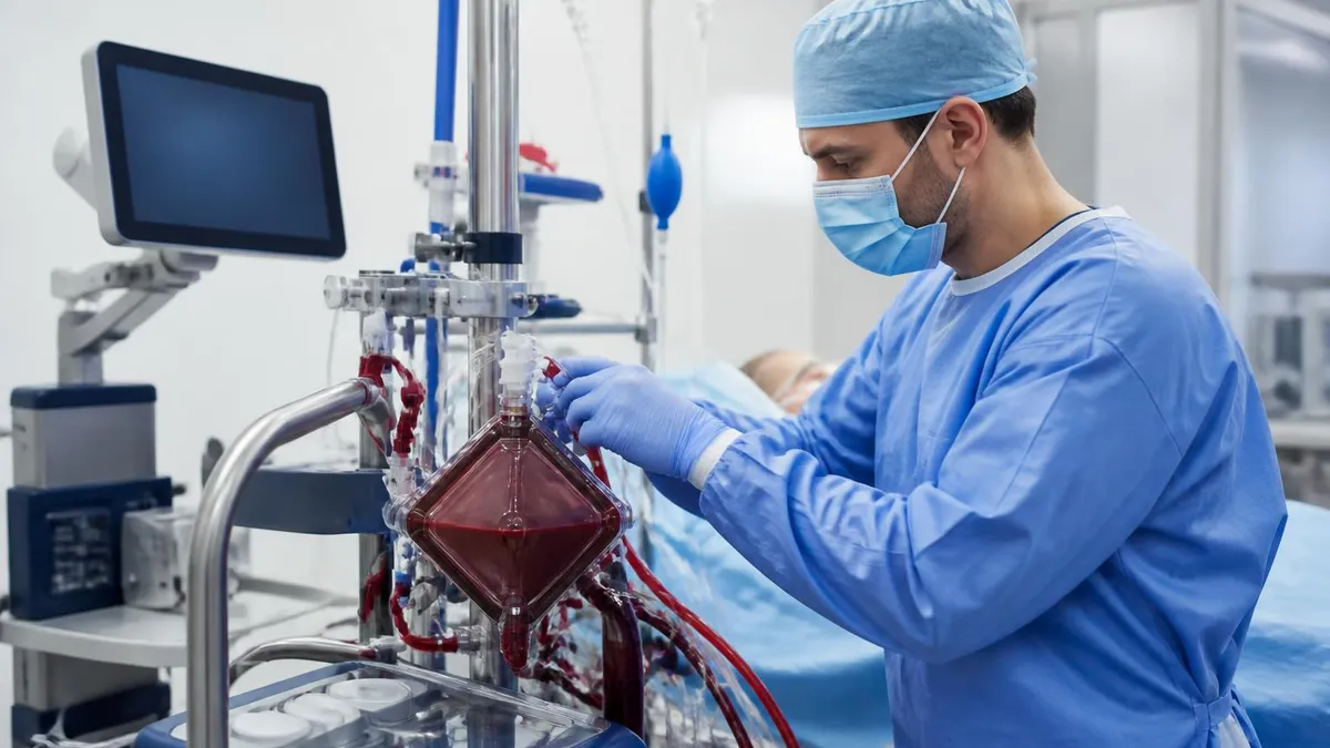
Extracorporeal membrane oxygenation, by contrast, routes blood outside the body through an artificial lung — the oxygenator — where carbon dioxide is removed and oxygen is added before blood is returned to the patient. Because gas exchange occurs external to the patient's own lungs, ECMO allows clinicians to apply ultra-protective ventilator settings or even rest the lungs almost entirely, interrupting the cycle of injury and giving tissue time to heal. This capacity to provide complete cardiopulmonary bypass in a mobile, bedside format is what distinguishes ECMO from any conventional ventilation strategy.
The extracorporeal membrane oxygenation procedure carries substantial complexity. Cannulation requires skilled surgical or interventional technique; the circuit demands continuous anticoagulation to prevent clotting; and the bedside team must monitor dozens of parameters around the clock. These demands mean ECMO is not appropriate for every critically ill patient, and understanding which clinical scenarios favor each modality is essential knowledge for ECMO specialists, respiratory therapists, intensivists, and the nursing staff who care for these patients daily.
Cost is another dimension separating the two modalities. Extracorporeal membrane oxygenation machine price can reach $100,000 to $300,000 or more for the capital equipment alone, with disposable circuit components adding thousands of dollars per run. Mechanical ventilators, while not inexpensive, are dramatically less costly and far more widely distributed — critical considerations for resource-limited settings and smaller community hospitals that may lack ECMO programs entirely.
This article unpacks the mechanisms, indications, risks, and decision frameworks that guide clinicians choosing between ECMO and mechanical ventilation. Whether you are a nursing student preparing for board examinations, a respiratory therapy candidate reviewing ECMO physiology, or a clinician seeking a structured refresher, you will find evidence-based explanations, practical comparisons, and study resources throughout this guide. Understanding both modalities in depth — their overlapping applications and their clear distinctions — is central to competent critical care practice in any modern ICU.
Throughout the following sections we will examine the extracorporeal membrane oxygenation circuit in detail, contrast venovenous and venoarterial configurations, explore the evidence base from COVID-19 to neonatal respiratory failure, and outline the clinical triggers that should prompt an ECMO consultation when mechanical ventilation has reached its limits. By the end, readers will have a nuanced, well-structured understanding of when each therapy shines and when escalation is warranted.
ECMO vs Mechanical Ventilation by the Numbers

How Mechanical Ventilation and ECMO Work: Core Mechanisms
A ventilator delivers pressurized gas through an artificial airway, controlling tidal volume, respiratory rate, PEEP, and FiO2. The patient's native lungs still perform gas exchange, meaning injured alveoli continue to be stretched with each breath, risking ventilator-induced lung injury over time.
Blood is drained from the patient, pumped through a membrane oxygenator that removes CO2 and adds O2, then returned. The lungs can rest at very low ventilator settings. The extracorporeal membrane oxygenation circuit replaces pulmonary function outside the body, giving injured tissue time to heal.
A bridge modality between conventional ventilation and ECMO, HFOV delivers tiny tidal volumes at very high frequencies to minimize lung stretch. Often used in neonates and children, it represents an intermediate escalation step before committing to the extracorporeal membrane oxygenation procedure.
Neuromuscular blockade, prone positioning, and inhaled nitric oxide are non-ECMO adjuncts that can improve oxygenation in ARDS. If these fail to maintain a PaO2/FiO2 ratio above 80 mmHg, most guidelines recommend ECMO consultation as the logical next step in the escalation ladder.
The extracorporeal membrane oxygenation circuit consists of five core components: drainage cannulas, a centrifugal or roller pump, a membrane oxygenator (artificial lung), a heat exchanger, and return cannulas. Blood exits the patient through large-bore venous drainage cannulas, typically placed in the femoral vein, internal jugular vein, or right atrium.
The pump creates continuous non-pulsatile flow — usually 3 to 6 liters per minute in adults — drawing blood through the circuit at rates calibrated to meet the patient's metabolic demands. Proper understanding of the extracorporeal membrane oxygenation circuit is essential for any clinician managing these patients, as circuit problems are a leading cause of ECMO-related morbidity.
The membrane oxygenator is the functional heart of the system. Modern oxygenators use hollow-fiber polypropylene or polymethylpentene membranes through which oxygen flows on one side while blood flows on the other, allowing oxygen to diffuse in and carbon dioxide to diffuse out across a concentration gradient. Gas exchange efficiency is characterized by the sweep gas flow rate — increasing sweep removes more CO2, while increasing FiO2 of the sweep gas raises blood oxygen content. Experienced ECMO specialists titrate both variables continuously, just as a respiratory therapist titrates ventilator settings.
Venovenous extracorporeal membrane oxygenation (VV-ECMO) is configured for isolated respiratory failure. Deoxygenated blood is drained from a large central vein, oxygenated externally, and returned to the right atrium or superior vena cava. The patient's own heart continues to pump this now-oxygenated blood through the pulmonary and systemic circulations. VV-ECMO does not provide cardiac support — if myocardial function is compromised, venoarterial (VA-ECMO) is required instead, with return blood entering the arterial system directly to supplement or replace cardiac output.
Venoarterial ECMO is more complex and carries higher procedural risk because arterial cannulation is required. In peripheral VA-ECMO, drainage occurs from the femoral vein and return blood enters the femoral artery, supplying the lower body with oxygenated blood while the heart continues to eject desaturated blood into the ascending aorta — a phenomenon called differential hypoxia or the Harlequin syndrome. Central VA-ECMO, performed by cardiac surgeons via sternotomy, avoids this phenomenon by draining the right atrium and returning to the ascending aorta, but it is generally reserved for post-cardiotomy patients who cannot be weaned from cardiopulmonary bypass.
Anticoagulation management on ECMO is a delicate balance. Heparin remains the most commonly used agent, targeting an activated clotting time (ACT) of 180 to 220 seconds or an anti-Xa level of 0.3 to 0.7 IU/mL, depending on institutional protocol. Too little anticoagulation risks circuit thrombosis, oxygenator failure, and catastrophic thromboembolism. Too much increases bleeding risk — including intracranial hemorrhage, which is the most feared ECMO complication in neonates. Bivalirudin has emerged as an alternative, particularly in patients with heparin-induced thrombocytopenia (HIT), and pediatric ECMO centers are accumulating experience with direct thrombin inhibitors in the smallest patients.
Extracorporeal membrane oxygenation for adults has grown substantially since the CESAR trial published in 2009 demonstrated improved survival when patients with severe ARDS were transferred to an ECMO-capable center. The COVID-19 pandemic further expanded adult ECMO utilization, with extracorporeal membrane oxygenation covid registries from ELSO documenting thousands of adult runs during 2020 and 2021. Outcomes varied widely by center volume — a consistent finding across ECMO literature is that high-volume centers achieve significantly better survival rates, underscoring the importance of regionalized care and timely transfer protocols when patients deteriorate on mechanical ventilation.
Understanding how the extracorporeal membrane oxygenation diagram maps blood flow through the circuit helps clinicians troubleshoot rapidly. In a typical VV-ECMO diagram, drainage from the inferior vena cava passes through the pump, then the oxygenator, then the heat exchanger, and returns via a separate cannula into the right atrium. Recirculation — when returned oxygenated blood is immediately drained back into the circuit rather than entering the pulmonary circulation — is a common problem visible on saturations: drainage saturations rise while patient saturations remain low. Repositioning cannulas or switching to a dual-lumen single cannula can resolve significant recirculation.
Venovenous Extracorporeal Membrane Oxygenation: Indications, Setup, and Monitoring
Venovenous extracorporeal membrane oxygenation is indicated when severe, reversible respiratory failure fails to respond to optimized mechanical ventilation, prone positioning, neuromuscular blockade, and inhaled vasodilators. The most widely cited threshold is a Murray Lung Injury Score above 3.0 or a PaO2/FiO2 ratio persistently below 80 mmHg on FiO2 of 1.0 and PEEP of at least 10 cm H2O. Primary diagnoses include severe ARDS from pneumonia, aspiration, trauma, and — as documented extensively in extracorporeal membrane oxygenation covid literature — viral pneumonitis causing refractory hypoxemia.
Contraindications must be weighed carefully and include unrecoverable underlying disease, prolonged high-pressure mechanical ventilation exceeding seven to ten days (associated with irreversible lung fibrosis), significant intracranial hemorrhage, and conditions precluding systemic anticoagulation. Age and comorbidities influence candidacy but are not absolute barriers. ELSO guidelines provide a framework for candidacy discussions, and most experienced ECMO centers hold multidisciplinary conferences to evaluate borderline cases before cannulation proceeds.

ECMO vs Mechanical Ventilation: Advantages and Limitations
- +Provides complete lung rest, interrupting ventilator-induced lung injury cycle
- +Supports gas exchange even when lungs are severely damaged or flooded
- +Venoarterial configuration adds hemodynamic support for combined cardiorespiratory failure
- +Enables very low FiO2 and pressure settings, reducing oxygen toxicity
- +Proven survival benefit in neonatal respiratory failure and select adult ARDS
- +Can bridge patients to lung transplantation or cardiac recovery
- −Requires specialized surgical cannulation skill and 24/7 expert staffing
- −Systemic anticoagulation increases bleeding risk including fatal intracranial hemorrhage
- −Extracorporeal membrane oxygenation machine price makes it inaccessible in resource-limited settings
- −Circuit thrombosis, oxygenator failure, and pump malfunction can cause rapid patient deterioration
- −Not appropriate for irreversible conditions or prolonged high-pressure ventilation injury
- −Limited to specialized centers — transport logistics delay initiation in rural or community settings
Pros and Cons at a Glance
| Pros | Cons |
|---|---|
| Provides complete lung rest, interrupting ventilator-induced lung injury cycle | Systemic anticoagulation increases bleeding risk including fatal intracranial hemorrhage |
| Supports gas exchange even when lungs are severely damaged or flooded | Extracorporeal membrane oxygenation machine price makes it inaccessible in resource-limited settings |
| Venoarterial configuration adds hemodynamic support for combined cardiorespiratory failure | Circuit thrombosis, oxygenator failure, and pump malfunction can cause rapid patient deterioration |
| Proven survival benefit in neonatal respiratory failure and select adult ARDS | Not appropriate for irreversible conditions or prolonged high-pressure ventilation injury |
| Can bridge patients to lung transplantation or cardiac recovery | Limited to specialized centers — transport logistics delay initiation in rural or community settings |
Clinical Decision Checklist: When to Escalate from Ventilation to ECMO
- ✓Confirm PaO2/FiO2 ratio remains below 80 mmHg on FiO2 1.0 despite optimized ventilation
- ✓Document failure of prone positioning for at least 16 consecutive hours
- ✓Verify neuromuscular blockade has been trialed and maintained for at least 48 hours
- ✓Confirm inhaled nitric oxide or inhaled prostacyclin has been administered without sustained response
- ✓Assess that the underlying cause of respiratory failure is potentially reversible
- ✓Screen for absolute contraindications: active intracranial bleeding, end-stage organ failure, advanced directives against escalation
- ✓Calculate Murray Lung Injury Score and document score above 3.0 in the chart
- ✓Evaluate duration of high-pressure mechanical ventilation — beyond 7 days significantly increases fibrosis risk
- ✓Initiate transfer or consultation with nearest ELSO-registered ECMO center without delay
- ✓Obtain informed consent from patient or surrogate after explaining ECMO risks, benefits, and alternatives
The 7-Day Ventilation Window Matters
Data from the ELSO registry and multiple single-center series consistently show that patients ventilated at high pressures for more than 7 to 10 days before ECMO initiation have significantly lower survival rates than those escalated earlier. Early ECMO consultation — ideally before lung fibrosis becomes established — is associated with better outcomes. When conventional ventilation is failing, do not wait until the patient is moribund to call the ECMO team.
Extracorporeal membrane oxygenation in neonates represents one of the most mature and evidence-rich applications of this technology. Since the first successful neonatal ECMO run in 1975 and the landmark randomized controlled trial published in the UK Collaborative ECMO Trial in 1996, neonatal ECMO has become standard of care for selected term and near-term newborns with severe respiratory failure unresponsive to maximal conventional therapy. Conditions most frequently treated include meconium aspiration syndrome, persistent pulmonary hypertension of the newborn (PPHN), congenital diaphragmatic hernia (CDH), neonatal pneumonia, and respiratory distress syndrome in near-term infants whose lungs are insufficiently mature for adequate gas exchange.
The physiologic rationale for neonatal ECMO is particularly compelling. Newborn pulmonary vascular resistance is normally high at birth and typically falls rapidly as the lungs expand and oxygen tension rises. In disease states such as PPHN, this resistance remains improved, causing right-to-left shunting through the ductus arteriosus and foramen ovale, bypassing the lungs entirely and producing profound hypoxemia that may not respond even to high-frequency ventilation and inhaled nitric oxide.
ECMO breaks this cycle by oxygenating blood externally, lowering pulmonary arterial pressure, and allowing the pulmonary vasculature time to remodel and dilate — a process that typically takes five to fourteen days in successful runs.
Neonatal ECMO candidacy criteria have evolved over decades. Most centers require gestational age of at least 34 weeks, birth weight above 2 kilograms, reversible diagnosis, and absence of lethal chromosomal anomalies or major intracranial hemorrhage prior to cannulation. An oxygenation index (OI) above 40, calculated as mean airway pressure × FiO2 × 100 divided by PaO2, is the most widely used threshold — representing a level of ventilatory support associated with 80% mortality without ECMO rescue. Some centers also apply the alveolar-arterial oxygen difference (AaDO2) above 600 mmHg for more than 4 hours as a supplementary criterion.
Congenital diaphragmatic hernia deserves special mention because it remains one of the most challenging neonatal ECMO indications with survival rates of only 50 to 60%, substantially lower than meconium aspiration syndrome where survival exceeds 90%. In CDH, the herniation of abdominal organs into the chest cavity during fetal development causes bilateral lung hypoplasia — there is simply less functional lung tissue than in other neonatal diseases, and ECMO cannot create new alveoli. ECMO in CDH serves primarily to stabilize the infant for surgical repair of the hernia, allowing pulmonary circulation to stabilize before the physiologic stress of abdominal surgery.
Extracorporeal membrane oxygenation for adults with ARDS has a more contested evidence base than neonatal applications. The CESAR trial randomized 180 adult patients to conventional management versus transfer to an ECMO center; 63% of the ECMO group survived to 6 months without disability compared with 47% in the conventional group.
However, critics noted that the conventional arm did not consistently use lung-protective ventilation. The subsequent EOLIA trial in 2018 found a non-statistically significant absolute risk reduction of 11% with ECMO, though a post-hoc Bayesian analysis suggested a 98% probability of benefit. These nuances mean adult ECMO for ARDS remains an expert-center decision requiring careful patient selection.
The extracorporeal membrane oxygenation covid experience during the pandemic provided an extraordinary natural experiment in adult ECMO. During the first COVID-19 wave, ELSO data showed survival rates of approximately 55 to 60% among carefully selected patients — comparable to pre-pandemic ARDS ECMO outcomes, despite unprecedented volumes. However, centers that exceeded their normal ECMO capacity showed worse outcomes, reinforcing that ECMO quality is closely tied to team experience and patient volume. Many centers established strict selection criteria limiting COVID ECMO to patients under 65 with limited comorbidities and fewer than 14 days of mechanical ventilation.
Weaning from ECMO and transition back to mechanical ventilation requires a systematic approach. For VV-ECMO, trials involve reducing sweep gas flow to allow CO2 to rise and assessing whether the patient's native lungs can maintain adequate gas exchange at moderate ventilator settings. In neonates, a weaning trial may last 30 to 60 minutes before decannulation is deemed safe.
For VA-ECMO cardiac weaning, flow rates are gradually reduced while echocardiography confirms maintained cardiac output and the patient is assessed for hemodynamic stability at progressively lower circuit support. Successful weaning and decannulation mark a critical milestone, after which conventional mechanical ventilation resumes and is itself weaned toward extubation.

ECMO supports gas exchange and hemodynamics while the underlying disease is treated, but it cannot reverse conditions that are inherently irreversible. Initiating ECMO in patients with end-stage pulmonary fibrosis, multi-organ failure, or unresectable malignancy without a clear bridge-to-transplant plan may prolong dying rather than extend meaningful life. Every ECMO program should define clear goals of care, timeframes for reassessment, and exit strategies before cannulation, in partnership with the patient's family and the palliative care team.
Risk management on ECMO requires anticipating complications before they escalate into life-threatening events. The most feared complication remains intracranial hemorrhage (ICH), occurring in approximately 3 to 15% of neonatal ECMO runs and carrying high mortality and long-term neurological morbidity. ICH risk is highest in the first 72 hours of a run, correlating with the most aggressive anticoagulation requirements during circuit stabilization.
Daily cranial ultrasounds in neonates and CT scanning in children and adults at the first sign of neurological change are standard practice at high-volume centers. When significant ICH is detected, the ECMO team faces the difficult decision of reducing or halting anticoagulation — accepting circuit thrombosis risk — to halt bleeding progression.
Circuit thrombosis is the mechanical mirror image of hemorrhage: insufficient anticoagulation or stagnant flow zones allow clot formation in the oxygenator, pump head, or cannulas. Fibrin and platelet aggregates in the oxygenator improve transmembrane pressure gradients and reduce gas exchange efficiency. When post-oxygenator PaO2 begins declining and the pressure gradient rises, oxygenator exchange becomes necessary — a procedure that can be accomplished without decannulating the patient, though it requires a skilled circuit team working rapidly to minimize time off ECMO support. Preventive strategies include maintaining minimum flows above 1 L/min, avoiding flow interruptions, and using heparin-bonded circuit components.
Limb ischemia is a significant complication of femoral artery cannulation in peripheral VA-ECMO. The large arterial return cannula can partially or completely obstruct distal perfusion to the ipsilateral leg, causing ischemic compartment syndrome within hours. A distal perfusion cannula — a small side-arm catheter placed in the superficial femoral artery antegrade — is standard practice in most adult VA-ECMO programs and has substantially reduced limb amputation rates. Hourly limb checks assessing color, warmth, capillary refill, and Doppler signals are mandatory nursing assessments that allow early detection before irreversible ischemia occurs.
Infection on ECMO is both common and dangerous. Cannula insertion sites, circuit connections, and indwelling lines all represent entry points for hospital-acquired pathogens. ECMO patients are inherently immunocompromised by critical illness, massive transfusion, and often prior immunosuppressive therapy. Nosocomial pneumonia, candidemia, and cannula-site infections significantly prolong ECMO runs and worsen outcomes. Strict sterile technique during cannulation, daily chlorhexidine-based site care, and early removal of unnecessary vascular lines are infection control measures that experienced ECMO centers rigorously enforce.
Hemolysis — destruction of red blood cells by shear forces within the pump — occurs to some degree in all ECMO runs and worsens as pump speeds increase and circuit components age. Plasma-free hemoglobin levels above 50 mg/dL indicate clinically significant hemolysis requiring investigation. Causes include pump thrombus, excessive RPM relative to flow, and cannula kinking. Severe hemolysis can cause acute kidney injury, jaundice, and cardiovascular instability. Centrifugal pumps cause less hemolysis than older roller pumps, which is one reason they have become the dominant pump technology in modern ECMO circuits worldwide.
Long-term outcomes after ECMO reveal that survival to hospital discharge, while the primary endpoint of most studies, does not capture the full picture. Neonatal ECMO survivors face improved rates of neurodevelopmental delay, hearing loss (particularly sensorineural, from both disease and aminoglycoside exposure), pulmonary hypertension recurrence, and behavioral difficulties requiring long-term follow-up.
ECMO survivors should be enrolled in structured developmental follow-up programs that include audiology, neurology, and developmental pediatrics at 6, 12, 24, and 36 months of age — a standard of care endorsed by ELSO and most major neonatology societies. For adult ECMO survivors, post-intensive care syndrome — encompassing cognitive impairment, anxiety, depression, and physical deconditioning — is common and responsive to structured rehabilitation programs initiated early during the ICU stay.
Educating the next generation of ECMO-competent clinicians requires more than textbook knowledge. Simulation training, circuit management practice on water loops, and supervised clinical mentorship are the cornerstones of ECMO education. Candidates preparing for credentialing examinations must master not only the pathophysiology and circuit mechanics covered here but also pharmacology, troubleshooting algorithms, and the ethical dimensions of ECMO initiation and withdrawal. Practice examinations that test these domains under timed conditions are an invaluable preparation tool — and the resources available throughout this site are specifically designed to build that exam-ready depth of understanding.
Preparing for ECMO credentialing examinations or nursing board questions related to extracorporeal support requires a structured, multi-modal study approach. Begin by mastering the foundational physiology: oxygen delivery (DO2), oxygen consumption (VO2), and the Fick principle underlie all ECMO titration decisions. Understanding that DO2 equals cardiac output multiplied by arterial oxygen content allows you to predict how changes in pump flow, hemoglobin, and oxygen saturation each contribute to tissue oxygenation — a calculation that appears regularly on ECMO specialist board examinations and critical care nursing boards alike.
Next, solidify your understanding of the differences between VV-ECMO and VA-ECMO. Know that VV-ECMO supports oxygenation and CO2 removal but provides no hemodynamic support — the heart must still generate adequate cardiac output. VA-ECMO supports both gas exchange and circulation but introduces unique complications including Harlequin syndrome, left ventricular distension (requiring LV venting in some patients), and arterial cannulation risks. Being able to identify which configuration is indicated from a clinical vignette — describing a patient with ARDS and preserved ejection fraction versus a patient with cardiogenic shock and pulmonary edema — is a high-yield exam skill.
Pharmacology on ECMO is a frequently tested and clinically critical domain. The extracorporeal membrane oxygenation circuit sequesters lipophilic and protein-bound drugs, altering their volume of distribution and effective plasma concentrations. Drugs most significantly affected include fentanyl (highly lipophilic), midazolam, and vancomycin. Standard dosing may be inadequate — leading to under-sedation or sub-therapeutic antibiotic levels — while other drugs may accumulate unexpectedly. ECMO pharmacology questions test understanding of which drug classes are most affected and how to adjust dosing strategies, often recommending therapeutic drug monitoring as the most reliable guide.
Troubleshooting circuit alarms is another high-yield exam domain. Know that a rising inlet pressure (more negative) suggests drainage obstruction — check for cannula kinking, hypovolemia, or patient positioning. A rising outlet pressure suggests return obstruction. A falling post-oxygenator PaO2 with rising transmembrane pressure difference indicates oxygenator failure. Chatter (oscillation of the drainage line) suggests inadequate venous return requiring volume resuscitation or reduced pump speed. Practice running through these troubleshooting algorithms mentally under timed conditions — the exam will present these as rapid clinical scenarios requiring immediate action identification.
Weaning strategies deserve focused review. Understand that VV-ECMO weaning is tested by reducing sweep gas flow (not pump flow) to assess the patient's native CO2 clearance capacity, then assessing oxygenation on moderate FiO2. Pump flow reduction is generally reserved for VA-ECMO cardiac weaning, where echocardiography guides the process by confirming native cardiac output recovery. Know the criteria for successful extubation after ECMO decannulation and the typical ventilatory trajectory: most neonatal ECMO patients are extubated within 24 to 72 hours of decannulation if recovery is proceeding normally.
Use practice questions actively, not passively. After answering each question, read the full rationale whether you answered correctly or not — understanding why the wrong answers are wrong is as important as knowing the right answer. Pattern recognition develops from repeated exposure: after practicing 200 to 300 ECMO questions, you will begin to recognize the specific clinical cues that distinguish VV from VA indications, the timing of complications, and the pharmacokinetic principles being tested. Spacing your practice sessions over several weeks — rather than cramming — produces significantly better long-term retention and exam performance.
Finally, integrate your ECMO knowledge with broader critical care and neonatal physiology. Exam questions rarely test ECMO in isolation — they embed circuit management decisions within complex clinical scenarios involving ventilator management, fluid balance, sepsis treatment, and family communication.
The clinician who understands how ECMO fits into the full continuum of critical care — from initial stabilization through conventional ventilation through high-frequency modes through ECMO and ultimately to recovery — is the clinician who performs best on both board examinations and real clinical calls at three in the morning when a neonate's saturations are dropping and the team is looking to the ECMO specialist for leadership.
Sample ECMO - Extracorporeal Membrane Oxygenation Practice Questions
Try these questions from our free ECMO - Extracorporeal Membrane Oxygenation practice tests. The correct answer and an explanation follow each question.
During ECMO weaning trials, which clinical parameter best confirms readiness to separate from VV-ECMO support?
- A. SpO2 > 88% at ECMO flows of 1 L/min with FiO2 0.5 and moderate vent settings
- B. SpO2 > 95% with ECMO at full flows
- C. PaO2 > 200 mmHg on ECMO
- D. Normal chest X-ray with ECMO at maximum flow
Answer: A. SpO2 > 88% at ECMO flows of 1 L/min with FiO2 0.5 and moderate vent settings
Maintaining acceptable oxygenation at minimal ECMO flows (1 L/min) with reduced FiO2 and moderate ventilation confirms sufficient native lung recovery for safe decannulation.
What term describes ECMO support initiated specifically to support a patient through a reversible cause of cardiac arrest when conventional CPR has failed?
- A. Bridge-to-recovery ECMO
- B. ECPR (Extracorporeal CPR)
- C. LVAD bridging
- D. Resuscitative ECMO
Answer: B. ECPR (Extracorporeal CPR)
ECPR is the specific term for VA-ECMO deployed as an adjunct to conventional CPR for refractory cardiac arrest.
What is the physiological rationale for targeting a lower 'lung-protective' ventilator strategy (ultra-protective ventilation) during VV-ECMO?
- A. ECMO eliminates the need for any ventilation
- B. ECMO assumes gas exchange, allowing the lungs to rest and potentially recover from VILI
- C. Lower ventilation reduces recirculation
- D. Reduced ventilation decreases sweep gas requirements
Answer: B. ECMO assumes gas exchange, allowing the lungs to rest and potentially recover from VILI
With ECMO handling gas exchange, tidal volumes and pressures can be minimized to prevent ventilator-induced lung injury (VILI) and promote lung recovery.
In VA-ECMO, how does left ventricular (LV) afterload change compared to a patient without ECMO support?
- A. LV afterload decreases due to blood volume offloading
- B. LV afterload increases because retrograde ECMO flow opposes ventricular ejection
- C. LV afterload is unchanged since ECMO bypasses the left heart
- D. LV afterload fluctuates with sweep gas adjustments
Answer: B. LV afterload increases because retrograde ECMO flow opposes ventricular ejection
Retrograde aortic flow from femoral VA-ECMO increases LV afterload, which can worsen LV distension and pulmonary edema in patients with severely impaired LV function.
Take the full ECMO - Extracorporeal Membrane Oxygenation practice test
ECMO Questions and Answers

VA ECMO Cannulation: Complete Guide to Veno-Arterial Extracorporeal Membrane Oxygenation

ECMO vs Life Support: Understanding Extracorporeal Membrane Oxygenation and Conventional Life-Sustaining Therapies

Neonatal ECMO: Extracorporeal Membrane Oxygenation in Newborns — Complete Guide
ECMO Patient Guide: What to Expect, Care, Recovery & Outcomes

ECMO Specialist vs Perfusionist: Roles, Duties, and Career Paths Compared
About the Author

Educational Psychologist & Academic Test Preparation Expert
Columbia University Teachers CollegeDr. Lisa Patel holds a Doctorate in Education from Columbia University Teachers College and has spent 17 years researching standardized test design and academic assessment. She has developed preparation programs for SAT, ACT, GRE, LSAT, UCAT, and numerous professional licensing exams, helping students of all backgrounds achieve their target scores.
Join the Discussion
Connect with other students preparing for this exam. Share tips, ask questions, and get advice from people who have been there.
View discussion (9 replies)