ECMO vs Life Support: Understanding Extracorporeal Membrane Oxygenation and Conventional Life-Sustaining Therapies
ECMO vs life support explained: how extracorporeal membrane oxygenation differs from conventional ICU care, who qualifies, and what outcomes to expect. 🎯

When conventional life support reaches its limits, physicians turn to a powerful bridge technology: extracorporeal membrane oxygenation in neonates and adults alike. Understanding ecmo vs life support is essential for clinicians, patients, and families navigating critical illness. ECMO is not simply a stronger ventilator or a more aggressive vasopressor regimen — it is a fundamentally different paradigm that temporarily replaces the function of the heart, the lungs, or both, buying time for the underlying disease to resolve or for a definitive intervention to occur.
Conventional life support encompasses the entire arsenal of standard intensive care: mechanical ventilation, vasopressors such as norepinephrine and vasopressin, inotropes like dobutamine and milrinone, intra-aortic balloon pumps, prone positioning, neuromuscular blockade, continuous renal replacement therapy, and meticulous hemodynamic monitoring. These therapies support organ function while the body heals, but they have physiologic ceilings. When a patient requires FiO₂ of 1.0 and peak pressures above 35 cmH₂O on the ventilator, or when mean arterial pressure cannot be maintained despite maximal vasopressor doses, conventional support has been exhausted.
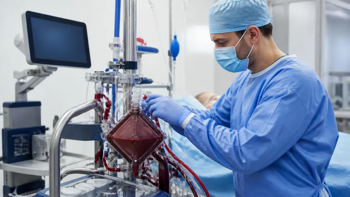
ECMO, by contrast, drains venous blood from the patient, passes it through an oxygenator membrane that adds oxygen and removes carbon dioxide, and then returns the blood either to the venous circulation (venovenous ECMO, or VV-ECMO) or to the arterial circulation (venoarterial ECMO, or VA-ECMO). The extracorporeal membrane oxygenation circuit effectively performs gas exchange and, in the VA configuration, cardiac output outside the body, allowing the native heart and lungs to rest at very low workloads. This is categorically different from any form of conventional life support.
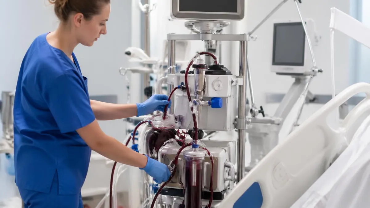
The distinction matters clinically because ECMO carries its own risk profile. Cannulation requires large-bore access, systemic anticoagulation is mandatory, and complications including bleeding, thromboembolism, cannula site infection, and limb ischemia are well-documented. Patients on ECMO require a dedicated bedside specialist around the clock, and the equipment itself — oxygenators, centrifugal pumps, heat exchangers, and continuous monitoring systems — demands specialized training to manage safely. Initiating ECMO is therefore never a casual escalation; it is a considered decision made by a multidisciplinary team.
The extracorporeal membrane oxygenation procedure has evolved dramatically since its earliest applications in the 1970s. Dr. Robert Bartlett pioneered neonatal ECMO at the University of Michigan, demonstrating survival in newborns with meconium aspiration syndrome and persistent pulmonary hypertension who had otherwise been expected to die. Today, ECMO centers across the United States manage patients ranging from premature neonates weighing under two kilograms to adults in cardiogenic shock following massive myocardial infarction. The Extracorporeal Life Support Organization (ELSO) registry now contains data on more than 200,000 ECMO runs, providing a rich evidence base for clinical decision-making.
For families, the emotional weight of the ECMO vs life support conversation is immense. Being told that a loved one has "failed conventional treatment" and that ECMO is the next step often feels like a precipice. It is helpful to understand that ECMO is not a last-ditch experimental measure — for appropriately selected patients it is a standard of care with documented survival rates. At the same time, ECMO does not guarantee recovery, and careful discussions about goals of care, expected trajectory, and quality-of-life considerations are integral to the consent process.
This article provides a comprehensive comparison of ECMO and conventional life support, covering the underlying technology, clinical indications, patient populations from neonates to adults, the extracorporeal membrane oxygenation circuit in detail, cost considerations, and the evidence base from conditions including ARDS, cardiac arrest, and COVID-19. Whether you are a clinician preparing for ECMO specialist certification or a family member trying to understand a loved one's treatment plan, this guide will give you the foundational knowledge you need.
ECMO vs Life Support by the Numbers

Core Components of the Extracorporeal Membrane Oxygenation Circuit
Large-bore venous cannula (typically 19–29 Fr in adults) inserted into the right atrium or inferior vena cava. It draws deoxygenated blood out of the patient and into the extracorporeal circuit under negative pressure from the centrifugal pump.
Magnetically levitated impeller that generates continuous flow (typically 3–7 L/min in adults). Modern pumps such as the Maquet Rotaflow and Medtronic Biopump produce less hemolysis than older roller pumps and include integrated flow sensors.
Hollow-fiber polymer membrane across which oxygen diffuses into blood and CO₂ diffuses out. Modern polymethylpentene (PMP) oxygenators maintain performance for weeks without plasma leak, unlike older silicone membranes that degraded within days.
Maintains blood temperature at 36–37°C during the extracorporeal pass, preventing dangerous hypothermia that would otherwise result from passing body temperature blood through room-temperature tubing over hours and days.
In VV-ECMO, blood returns to the right atrium via a venous cannula. In VA-ECMO, oxygenated blood is returned to the arterial circulation via the femoral or axillary artery, providing both gas exchange and hemodynamic support simultaneously.
The distinction between venovenous extracorporeal membrane oxygenation (VV-ECMO) and venoarterial extracorporeal membrane oxygenation (VA-ECMO) is the single most important conceptual divide within ECMO practice. VV-ECMO addresses respiratory failure only: blood is drained from the venous system, oxygenated by the membrane lung, and returned to the venous system just upstream of the right heart.
The patient's own cardiac function must be adequate to distribute that oxygenated blood through the pulmonary and systemic circulations. VA-ECMO, by contrast, drains venous blood and returns oxygenated blood directly into the arterial system, bypassing both the heart and lungs and providing direct mechanical circulatory support in addition to gas exchange.
VV-ECMO is the standard configuration for severe ARDS (acute respiratory distress syndrome), whether caused by pneumonia, aspiration, trauma, or as seen in large numbers of critically ill patients during the COVID-19 pandemic. The landmark CESAR trial in the United Kingdom demonstrated improved survival without severe disability at six months in adult patients with severe ARDS who were referred to an ECMO center compared with continued conventional ventilation. The eolia trial added nuance, showing that VV-ECMO did not reduce 60-day mortality as a standalone primary endpoint but did prevent treatment failure events, prompting ongoing debate about optimal patient selection and timing.
VA-ECMO is indicated when the primary problem is cardiogenic shock — the heart cannot maintain adequate output despite pharmacologic support. Etiologies include massive myocardial infarction, fulminant myocarditis, decompensated cardiomyopathy, post-cardiotomy shock following cardiac surgery, and refractory cardiac arrest (a configuration sometimes called ECPR, or extracorporeal cardiopulmonary resuscitation). In VA-ECMO the centrifugal pump provides all or most of the cardiac output, allowing the ventricles to rest and potentially recover.
However, VA-ECMO also increases afterload — the retrograde flow of oxygenated blood from the femoral artery toward the aortic valve can impede the already-failing left ventricle, a phenomenon called LV distension or the "Harlequin effect," which sometimes requires the addition of a percutaneous left ventricular assist device (Impella) alongside ECMO.
The extracorporeal membrane oxygenation procedure for VV configuration typically begins with cannulation under ultrasound and fluoroscopic guidance. In adults, a dual-lumen cannula such as the Avalon Elite may be placed in the right internal jugular vein, with the drainage lumen positioned in the inferior vena cava and the return lumen directed across the tricuspid valve toward the pulmonary artery — a single-site approach that facilitates patient mobility.
Alternatively, a bicaval configuration uses one cannula in the right femoral vein and another in the right internal jugular vein. Each configuration has distinct advantages related to recirculation rates, patient comfort, and nursing complexity.
During an ECMO run, management goals differ significantly from those of conventional mechanical ventilation. Once adequate extracorporeal gas exchange is established, the ventilator can be dialed back dramatically — typically to a rate of 10 breaths per minute, PEEP of 10 cmH₂O, and FiO₂ of 0.30–0.40.
This "lung rest" strategy is the core mechanistic rationale for ECMO in ARDS: high-pressure, high-oxygen mechanical ventilation causes ventilator-induced lung injury (VILI), and by offloading the ventilator onto the ECMO circuit, the injured lung is given time to heal without ongoing barotrauma, volutrauma, and oxygen toxicity. This is categorically impossible with any form of conventional life support alone.
Anticoagulation management during ECMO is a continuous balancing act. Systemic heparin infusion is typically titrated to maintain an anti-Xa level of 0.3–0.7 IU/mL or an activated partial thromboplastin time (aPTT) of 60–80 seconds. Too little anticoagulation risks thrombus formation in the oxygenator, which reduces gas exchange efficiency and can embolize.
Too much anticoagulation risks catastrophic bleeding complications, most feared of which is intracranial hemorrhage — a devastating complication that occurs in approximately 3–7% of neonatal ECMO runs and less frequently in adults. Teams that manage ECMO patients must check circuit pressures, sweep gas settings, and anticoagulation parameters at regular intervals, often hourly.
Patient positioning and rehabilitation during ECMO have evolved considerably. In early ECMO practice, patients were deeply sedated and immobilized to prevent cannula dislodgement. Current evidence increasingly supports early mobility programs: physical therapists work with patients who are awake and ventilated on ECMO to perform in-bed exercises, dangle at the bedside, and in some centers even ambulate with portable ECMO circuits. Awake ECMO — performing ECMO without intubation in spontaneously breathing patients — is being studied as a bridge to lung transplantation and as a means to avoid the complications of prolonged mechanical ventilation while awaiting donor organs.
Extracorporeal Membrane Oxygenation in Neonates, Pediatrics, and Adults
Extracorporeal membrane oxygenation in neonates represents the original and most extensively studied application of the technology. Newborns are placed on ECMO for conditions including meconium aspiration syndrome (MAS), persistent pulmonary hypertension of the newborn (PPHN), congenital diaphragmatic hernia (CDH), respiratory distress syndrome unresponsive to surfactant, and sepsis-related cardiorespiratory failure. The ELSO registry reports survival-to-discharge rates of approximately 74% for neonatal respiratory ECMO, compared with survival rates of less than 20% for the same conditions prior to the ECMO era. The oxygenation index (OI = FiO₂ × MAP × 100 / PaO₂) is the primary threshold used for neonatal ECMO candidacy, with an OI above 40 representing a high-risk threshold for mortality without ECMO support.
Neonatal ECMO runs carry unique challenges including a very small blood volume relative to circuit priming volume, making circuit priming with packed red blood cells and fresh frozen plasma mandatory. Cannula sizes are correspondingly small — typically 8–12 Fr arterial and 10–14 Fr venous — and right internal jugular and right common carotid artery cannulation (the "classic" neonatal approach) was historically standard, though this sacrifices the right carotid artery permanently. Venovenous neonatal ECMO using a double-lumen Avalon cannula in the right internal jugular vein has largely replaced venoarterial ECMO for respiratory indications, preserving the carotid artery and reducing stroke risk while providing equivalent gas exchange support.

ECMO vs Conventional Life Support: Advantages and Limitations
- +Provides complete cardiopulmonary bypass capability at the bedside without the operating room
- +Enables ultra-protective lung ventilation strategies impossible with conventional ventilators alone
- +Supports patients through reversible cardiac or pulmonary crises until recovery or transplant
- +ECPR can restart circulation in cardiac arrest patients who have failed standard resuscitation
- +Modern circuits allow patient mobility and in some cases awake, ambulatory ECMO
- +Documented survival benefit in neonatal ARDS, pediatric myocarditis, and select adult ARDS populations
- −Requires systemic anticoagulation, significantly increasing bleeding and intracranial hemorrhage risk
- −Large-bore cannulas cause vascular complications including limb ischemia in up to 10–15% of patients
- −Oxygenator and pump components can fail, clot, or develop plasma leak, requiring circuit changes
- −Enormous resource demand: 24/7 specialist staffing, costly disposables, and specialized nursing
- −Does not treat the underlying disease — acts only as a bridge; outcomes depend on reversibility
- −Risk of heparin-induced thrombocytopenia (HIT) and systemic inflammatory response from circuit exposure
ECMO Initiation Checklist: Key Criteria to Verify Before Cannulation
- ✓Confirm reversible etiology — document that the underlying condition has realistic potential for recovery or bridge to transplant/VAD
- ✓Verify oxygenation index or Murray score thresholds are met (OI >40 neonates; Murray score ≥3 adults for VV-ECMO)
- ✓Rule out absolute contraindications: uncontrolled systemic bleeding, non-survivable neurologic injury, multi-organ failure without recoverable cause
- ✓Obtain informed consent from patient or surrogate, including discussion of goals of care and ECMO withdrawal plan
- ✓Confirm appropriate vessel size and patency with ultrasound before selecting cannula size and site
- ✓Assemble bedside cannulation team: ECMO specialist, attending physician, perfusionist or trained nurse, and ultrasound operator
- ✓Prime the ECMO circuit with appropriate solution — crystalloid for adults, blood prime for neonates and small pediatric patients
- ✓Initiate systemic heparin bolus (typically 50–100 units/kg) and confirm ACT > 200–250 seconds before cannulation
- ✓Establish baseline neurological exam and document baseline CBC, coagulation panel, metabolic panel, and arterial blood gas
- ✓Set ventilator to lung rest settings immediately after ECMO flow is established (FiO₂ ↓, rate ↓, pressures ↓)
ECMO Is a Bridge, Not a Cure
A critical concept in ECMO management is that the technology buys time but does not treat the underlying pathology. Every day on ECMO the team must actively evaluate whether the patient is recovering ("bridge to recovery"), eligible for transplant ("bridge to transplant"), or failing to progress — in which case honest goals-of-care conversations about withdrawal of ECMO support become necessary. Centers with the best outcomes document a clear "decision timeline" at ECMO initiation and re-evaluate it at regular intervals.
The COVID-19 pandemic dramatically elevated public and clinical awareness of extracorporeal membrane oxygenation. During the first and second waves, young patients with COVID-19-related ARDS who had no comorbidities were presenting with PaO₂/FiO₂ ratios below 60 mmHg despite prone positioning, neuromuscular blockade, and optimal ventilator management — a pattern that triggered ECMO consideration at a scale never previously seen. Centers that had previously placed 20–30 patients per year on ECMO suddenly faced surges of 50–100 simultaneous ECMO candidates, revealing stark geographic inequities in access to this life-saving technology.
The extracorporeal membrane oxygenation COVID evidence base emerged rapidly. A large international study published in The Lancet in 2020 analyzed outcomes from 1,035 COVID-19 patients placed on ECMO across 213 centers. Ninety-day in-hospital mortality was 37.4% — considerably lower than the greater than 90% mortality historically seen in similar severity ARDS without ECMO, though direct comparisons are confounded by selection effects.
Notably, patients who received ECMO within the first 7 days of mechanical ventilation had significantly better outcomes than those in whom ECMO was delayed, reinforcing the importance of early referral to ECMO centers rather than exhausting all conventional options before transfer.
COVID-19 also introduced novel challenges for ECMO management. SARS-CoV-2 infection is associated with a prothrombotic state — elevated D-dimer, fibrinogen, and von Willebrand factor, along with lupus anticoagulant-like phenomena — that dramatically increased oxygenator clotting rates and necessitated more aggressive anticoagulation protocols at many centers. At the same time, the same prothrombotic milieu paradoxically increased bleeding complications, including fatal pulmonary hemorrhage in some patients. ECMO teams had to navigate an extremely narrow therapeutic window for anticoagulation management in COVID-19 that did not exist to the same degree in other ARDS etiologies.
Beyond COVID-19, VV-ECMO has an established evidence base in ARDS from other causes. Influenza A H1N1 ARDS, severe community-acquired pneumonia (particularly from Legionella and S. pneumoniae), aspiration-related ARDS, and trauma-associated ARDS are all supported by case series and registry data. The critical clinical skill is patient selection: ECMO benefits patients with a single-organ (respiratory or cardiac) failure who have a reversible etiology. Patients with multi-organ dysfunction syndrome (MODS) affecting three or more organ systems simultaneously have survival rates below 20% on ECMO and may not benefit from initiation.
The extracorporeal membrane oxygenation diagram most commonly displayed in textbooks and training materials shows a simplified schematic of VV-ECMO: a drainage cannula in the inferior vena cava or right atrium, a centrifugal pump, an oxygenator with sweep gas inlet and CO₂ outlet, a heat exchanger, and a return cannula directed toward the tricuspid valve. Understanding this circuit layout helps clinicians troubleshoot alarms.
A high inlet pressure alarm (more negative than -100 mmHg) suggests inadequate venous drainage — the cannula tip may be against a vessel wall, the patient may be hypovolemic, or the pump speed may be set too high for the circuit. A high outlet pressure alarm suggests a distal obstruction — kinked tubing, cannula malposition, or elevated systemic vascular resistance in the case of VA-ECMO.
Sweep gas management is another dimension that distinguishes ECMO from conventional ventilation. Sweep gas — typically 100% oxygen — flows countercurrent to blood through the hollow fibers of the oxygenator. Increasing sweep gas flow rates increases CO₂ removal, while increasing FiO₂ of the sweep gas (or maintaining 100% O₂) maximizes oxygen delivery across the membrane. This allows ECMO specialists to independently titrate oxygenation and CO₂ clearance — a degree of control that is impossible with a conventional ventilator, where changes to respiratory rate and tidal volume affect both gas exchange parameters simultaneously.
Weaning from ECMO is a carefully staged process. For VV-ECMO, weaning trials involve incrementally reducing sweep gas flow (the primary determinant of CO₂ removal and indirectly oxygenation) while maintaining ECMO blood flow, allowing the native lungs to take over progressively. If arterial blood gases remain acceptable on reduced or zero sweep gas for 30–60 minutes, extubation to high-flow nasal cannula or continued conventional ventilation at lung-protective settings is considered feasible.
For VA-ECMO, weaning involves reducing pump flow in steps while watching for hemodynamic deterioration, with echocardiographic confirmation of improving native ventricular function before decannulation. A patient who cannot tolerate weaning trials after an appropriate observation period (often 2–3 weeks) must be evaluated for transition to a long-term ventricular assist device or cardiac transplantation.

ECMO requires specialized centers with trained personnel, specialized equipment, and round-the-clock perfusion support. If a patient is deteriorating on conventional life support at a non-ECMO center, delaying transfer to pursue additional conventional therapies significantly worsens outcomes. Current ELSO guidelines recommend contacting the nearest ECMO center as soon as a patient meets high-risk thresholds — do not wait until the patient is in extremis, as cannulation under CPR carries substantially higher mortality than cannulation in a patient who still has a perfusing rhythm.
The cost of ECMO is a topic that families and hospital administrators alike must grapple with. The extracorporeal membrane oxygenation machine price for institutional purchase ranges from approximately $100,000 to $250,000 for the console unit alone, with individual circuit packs — including oxygenator, tubing, and connectors — costing $5,000 to $15,000 per run in the United States.
When factoring in nursing ratios (often 1:1 with additional ECMO specialist coverage), daily intensive care unit costs, medications, laboratory monitoring, and physician billing, a full ECMO hospitalization for an adult patient frequently exceeds $500,000 for a multi-week run. For neonatal ECMO, which may last 5–10 days in uncomplicated respiratory cases, total costs typically range from $150,000 to $300,000.
Despite these staggering costs, health economic analyses generally find ECMO to be cost-effective in carefully selected populations because the alternative — death or severe neurologic disability — carries its own enormous societal costs in lost productivity, lifelong care needs, and family disruption. A cost-effectiveness analysis of neonatal ECMO published in the New England Journal of Medicine era estimated a cost per quality-adjusted life year (QALY) that compared favorably with other accepted neonatal interventions. For adult ECMO in severe ARDS, cost-effectiveness is more nuanced and depends heavily on long-term neurologic and functional outcomes, which vary substantially by center and patient population.
Access to ECMO is deeply unequal in the United States. ELSO-designated ECMO centers are concentrated in academic medical centers in major metropolitan areas. Rural patients, uninsured patients, and patients at community hospitals that lack ECMO capability face significant barriers to accessing this technology.
Telemedicine ECMO consultation programs — where remote ECMO specialists guide cannulation at referring hospitals via video — are being piloted in several health systems, but remain experimental. Mobile ECMO teams that travel to referring hospitals and cannulate patients before transporting them on ECMO to the receiving center represent the current standard for extending ECMO access, but require extraordinary logistical coordination.
Understanding the full comparison of ecmo vs life support also requires considering what happens after ECMO. Survivors of prolonged ECMO runs frequently face significant morbidity during recovery. Post-intensive care syndrome (PICS) — a constellation of cognitive impairment, psychological disorders including PTSD and depression, and physical deconditioning — affects the majority of ECMO survivors. Neuropathy from limb ischemia, scarring at cannulation sites, and tracheal stenosis from prolonged intubation are additional sequelae. Comprehensive ECMO survivor clinics, modeled on programs first established at Johns Hopkins and the University of Michigan, provide multidisciplinary follow-up to address these complex recovery needs.
For clinicians pursuing ECMO certification through the ELSO or the American Board of Cardiovascular Perfusion, mastery of the conceptual framework — when ECMO supersedes conventional life support, how the circuit functions, and how complications are managed — forms the intellectual core of the examination. ELSO offers the Pediatric Advanced Life Support in ECMO (PALSE) and adult ECMO specialist training courses, and many centers have developed internal competency assessment tools.
The written examinations test circuit troubleshooting, physiologic principles, pharmacology within the ECMO circuit (drug sequestration by the PVC tubing and oxygenator membrane alters dosing requirements for many sedatives, antibiotics, and vasopressors), and emergency management protocols for scenarios such as oxygenator failure, pump stoppage, and circuit decoupling.
Pharmacokinetics within the ECMO circuit deserve special emphasis for clinicians. Many drugs are significantly sequestered by the polyvinyl chloride tubing and the membrane oxygenator — this effect is most pronounced for highly lipophilic, highly protein-bound drugs. Fentanyl, midazolam, and propofol are heavily sequestered, requiring larger loading doses and higher infusion rates than would be used in non-ECMO patients of equivalent size.
Vancomycin dosing must also be adjusted, with the circuit acting as an additional volume of distribution. Conversely, extracorporeal removal of drugs by the circuit — particularly hemofiltration if continuous renal replacement therapy is running in tandem — means that some medications are cleared more rapidly than predicted by standard dosing tables, and therapeutic drug monitoring becomes especially critical.
The future of ECMO is being shaped by several technological frontiers. Miniaturized extracorporeal CO₂ removal devices (ECCO₂R) capable of removing carbon dioxide at lower blood flows (0.5–1.5 L/min) and with smaller cannulas than conventional ECMO are entering clinical use for hypercapnic COPD exacerbations and mild-to-moderate ARDS, potentially providing a less invasive alternative to intubation in selected patients.
Wearable ECMO systems using miniaturized centrifugal pumps and compact oxygenators are in early human trials, aiming to allow full ambulation and even discharge from the hospital on ECMO support while awaiting transplant. These innovations, if validated, will further blur the line between ECMO and conventional life support — placing ECMO earlier in the treatment algorithm for a broader range of patients.
For ECMO specialist candidates preparing for their certification examination, one of the most frequently tested knowledge domains is the ability to differentiate ECMO from conventional life support on physiologic grounds. Exam questions often present clinical vignettes in which a patient is on maximum conventional support — FiO₂ of 1.0, PEEP of 18 cmH₂O, norepinephrine at 0.5 mcg/kg/min, vasopressin at 0.04 units/min — and ask the candidate to identify the next appropriate step. Recognizing that this patient has met VV-ECMO or VA-ECMO thresholds requires rapid integration of oxygenation parameters, ventilator settings, hemodynamic data, and clinical trajectory.
Simulation training is increasingly central to ECMO specialist preparation. High-fidelity mannequin simulators equipped with ECMO circuits allow trainees to practice emergency scenarios including oxygenator change-out, air entrainment into the circuit, pump failure, and emergent decannulation without risk to real patients. Programs such as the ELSO Extracorporeal Life Support simulation curriculum and center-developed modules provide structured competency frameworks. Candidates who complete simulation-based training before their certification examinations consistently report higher confidence in circuit troubleshooting questions, which often constitute 20–30% of examination content.
Reading ECMO diagrams accurately is a skill tested both on certification examinations and in daily clinical practice. The standard extracorporeal membrane oxygenation diagram includes labeled components (drainage cannula, inlet tubing, centrifugal pump, pre-oxygenator pressure port, oxygenator, post-oxygenator pressure port, heat exchanger, and return cannula), flow direction arrows, and monitoring points where continuous pressure, flow, and temperature data are acquired.
Understanding where each monitoring point sits relative to the pump and oxygenator allows clinicians to localize problems: a pressure drop across the oxygenator (measured as the difference between pre-oxygenator and post-oxygenator pressures, the "delta-P") that exceeds 60 mmHg signals impending oxygenator failure and the need for circuit changeout.
The ECMO specialist role sits at an unusual intersection of perfusion science, critical care nursing, and respiratory therapy. In the United States, ECMO specialists at most centers are primarily registered nurses or respiratory therapists who have undergone additional ECMO-specific training, though perfusionists and physicians also serve in this role at some centers. The Extracorporeal Life Support Organization provides a framework for minimum training standards, recommending at least 30 hours of didactic education, followed by simulation training, and then supervised clinical experience with a minimum number of ECMO runs before independent specialist status is granted.
Career pathways in ECMO are expanding as the field grows. ECMO coordinators, ECMO educators, and ECMO program directors are increasingly formal institutional roles at large academic centers. Some specialists transition into medical device industry positions with ECMO equipment manufacturers such as Maquet Getinge, Medtronic, Xenios, and LivaNova, where clinical expertise informs product development, training programs, and regulatory affairs. Advanced practice providers (nurse practitioners and physician assistants) with ECMO training are being integrated into ECMO teams at several centers, extending coverage and providing a career advancement pathway for experienced specialists.
For patients and families, the most important practical takeaways from the ECMO vs life support discussion center on preparedness and communication. If a loved one is being evaluated for ECMO, families should ask specifically: Is the underlying condition reversible? What is the expected duration of ECMO support? What are the specific criteria for weaning and decannulation?
What happens if ECMO does not allow recovery — is transplant a possibility, and if not, what is the plan? These are not pessimistic questions; they are the right questions that ensure all parties are aligned on goals from the outset, which the evidence shows correlates with better decision-making and, in some studies, with improved patient outcomes.
In summary, ECMO and conventional life support occupy fundamentally different positions on the spectrum of critical care intervention. Conventional life support — mechanical ventilation, vasopressors, inotropes, CRRT — sustains organ function while the body fights disease. ECMO replaces organ function entirely when the body's own heart and lungs cannot sustain life even with maximum pharmacologic and mechanical assistance.
The decision to escalate from conventional support to ECMO is among the highest-stakes clinical decisions in modern medicine, requiring technical skill, prognostic precision, and compassionate communication. The clinicians who manage ECMO represent some of the most intensively trained specialists in critical care, and the knowledge required to do this work safely is the subject of robust, ongoing certification and education in ECMO programs across the United States and worldwide.
ECMO Questions and Answers

VA ECMO Cannulation: Complete Guide to Veno-Arterial Extracorporeal Membrane Oxygenation
ECMO Patient Guide: What to Expect, Care, Recovery & Outcomes

ECMO Specialist vs Perfusionist: Roles, Duties, and Career Paths Compared

ECMO Specialist: Career Path, Duties, Salary, and Certification Guide
ECMO: How Extracorporeal Membrane Oxygenation Works and Who Needs It
About the Author
Educational Psychologist & Academic Test Preparation Expert
Columbia University Teachers CollegeDr. Lisa Patel holds a Doctorate in Education from Columbia University Teachers College and has spent 17 years researching standardized test design and academic assessment. She has developed preparation programs for SAT, ACT, GRE, LSAT, UCAT, and numerous professional licensing exams, helping students of all backgrounds achieve their target scores.
Join the Discussion
Connect with other students preparing for this exam. Share tips, ask questions, and get advice from people who have been there.
View discussion (5 replies)