ECMO Machine: Components, How It Works, VA vs VV Types, and Clinical Management
Prepare for the ECMO Machine: 📗 Components, How It Works, certification. Practice questions with answer explanations covering all exam domains.

An ECMO machine — Extracorporeal Membrane Oxygenation — is a life support system that takes blood out of a critically ill patient's body, oxygenates it through an artificial lung called a membrane oxygenator, and returns it to the circulation. When a patient's heart, lungs, or both are too damaged or too diseased to sustain life on their own, the ECMO machine bridges the gap — buying days or weeks while the native organs recover, while a transplant is arranged, or while a decision about long-term support is made.
ECMO is not a new technology — it evolved from cardiopulmonary bypass machines used in open-heart surgery in the 1950s — but its use in intensive care has expanded dramatically over the past two decades. The 2009 H1N1 influenza pandemic and the 2020 COVID-19 pandemic both drove surges in ECMO use for severe ARDS, and centers that previously ran five ECMO patients per year were suddenly managing dozens simultaneously. The Extracorporeal Life Support Organization (ELSO) registry, which tracks global ECMO outcomes, shows steady improvement in survival rates as clinical experience and circuit technology have advanced.
Understanding how an ECMO machine works requires understanding its individual components, the two major configuration types (venoarterial and venovenous), the anticoagulation management that prevents circuit thrombosis, the cannulation strategies for accessing the circulation, and the pharmacological challenges that arise when blood passes through an extracorporeal circuit. This article covers all of those elements with clinical depth appropriate for ECMO specialists, perfusionists, intensivists, and nurses preparing to manage ECMO patients.

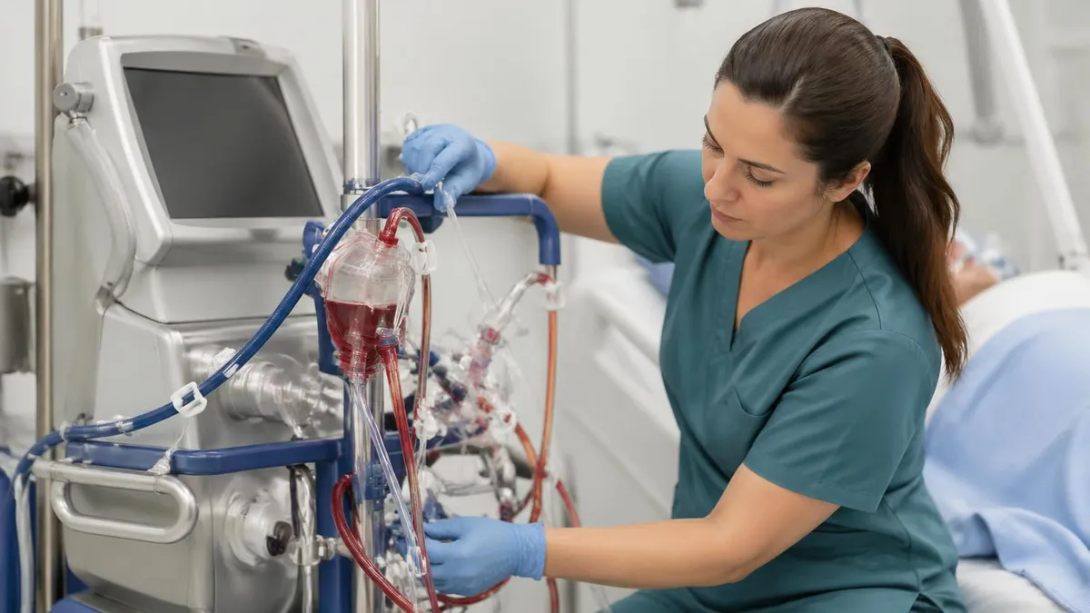
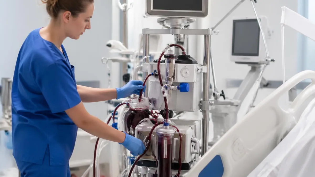
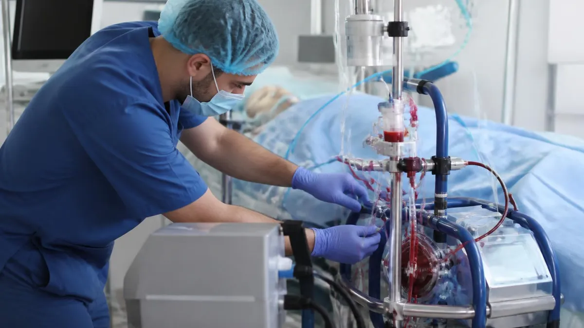
The ECMO circuit has five core components. The pump (centrifugal or roller) generates the blood flow that drives the circuit. The oxygenator — a hollow fiber membrane device — adds oxygen to blood and removes CO2, performing the work of the lungs. The heat exchanger maintains blood temperature, since passage through external tubing causes significant heat loss. Cannulas are the large-bore tubes inserted directly into the patient's veins or arteries to access the circulation. Connecting all of these is the circuit tubing — high-performance polyvinyl chloride or polyurethane lines coated to reduce thrombogenicity.
Centrifugal pumps (the dominant modern design) use a magnetic coupling to spin an impeller that generates flow proportional to rotational speed and inversely proportional to circuit resistance. They're gentler on red blood cells than roller pumps and handle circuit obstructions (a kinked line or thrombus) by reducing flow rather than generating dangerous pressure spikes. Roller pumps (older technology, still used in some centers) compress a segment of tubing to advance blood — they maintain flow regardless of resistance, which is useful in some pediatric applications but risks circuit rupture if downstream obstruction occurs.
The membrane oxygenator is the functional heart of the ECMO machine. Modern oxygenators use hollow fiber technology — thousands of microporous fibers through which blood flows on the outside while oxygen sweeps through the lumen. Gas exchange occurs across the fiber membrane: O2 diffuses into blood, CO2 diffuses out. The sweep gas (typically 100% oxygen or oxygen/CO2 blend) flows countercurrent to blood for optimal efficiency. Oxygenator performance degrades over days to weeks as plasma infiltrates the fibers and as thrombus accumulates — a failing oxygenator requires emergency exchange without interrupting patient support.
Venoarterial ECMO (VA-ECMO) drains blood from the venous system and returns it to the arterial system, bypassing both the heart and lungs. VA-ECMO provides both cardiac and respiratory support — it's the configuration used for cardiogenic shock, cardiac arrest (ECPR — ECMO-assisted CPR), and post-cardiac surgery failure. The major trade-off is that VA-ECMO returns oxygenated blood in a retrograde direction against the aorta, which can create Harlequin syndrome (differential hypoxia) if the native heart is ejecting deoxygenated blood into the proximal aorta while ECMO blood enters from below.
Venovenous ECMO (VV-ECMO) drains venous blood, oxygenates it, and returns it to the venous system — the lung is bypassed but the heart must still pump. VV-ECMO is used exclusively for respiratory failure (ARDS, severe pneumonia, COVID-19 ARDS) when cardiac function is preserved. Because both cannulas are in the venous system, VV-ECMO is generally safer and technically simpler than VA-ECMO, with lower rates of limb ischemia and stroke. The oxygenated blood returned to the right atrium mixes with deoxygenated venous return before being pumped by the native heart through the lungs and aorta.
Recirculation is the main flow efficiency problem in VV-ECMO: some of the oxygenated blood returned to the venous system is immediately re-drained by the drainage cannula before it reaches the right atrium, reducing effective DO2. Recirculation fraction depends on cannula positioning (the drainage and return cannulas must be positioned so their flow fields don't significantly overlap), flow rates, and the patient's own cardiac output. The clinical sign of excessive recirculation is a return saturation that's much lower than expected given the pump flow and oxygenator performance.
ECMO Machine: Three Critical Management Areas
ECMO circuit monitoring involves continuous assessment of flows (pump rpm and flow rate), pressures (pre- and post-pump, pre- and post-oxygenator transmembrane pressure), and circuit appearance (color of blood in tubing, fibrin deposition on circuit surfaces, clot formation in the bladder or oxygenator). An increasing transmembrane pressure gradient across the oxygenator indicates fibrin buildup or thrombus and predicts impending oxygenator failure. Circuit checks should be standardized and documented at least hourly.
Arterial and venous blood gas monitoring tells you whether the ECMO circuit is performing its intended function. The post-oxygenator (post-membrane) blood gas should show near-normal or supranormal PaO2 and a CO2 level determined by your sweep gas settings. The delivered oxygen to the patient (SaO2 in the return limb) and the mixed venous saturation (SvO2 from the drainage limb) together tell you whether the circuit is meeting the patient's oxygen demand and whether your flow rate is adequate for their metabolic state.
Cannulation strategy determines whether ECMO can be placed at the bedside or requires an operating room. Peripheral cannulation — inserting cannulas into the femoral vessels (VA-ECMO) or the right internal jugular and femoral vein (VV-ECMO) — is typically done by intensivists, surgeons, or interventionalists at the ICU bedside using Seldinger technique and ultrasound guidance. Percutaneous femoral VA-ECMO can be placed in under 15 minutes in an experienced center during cardiac arrest (ECPR), which is one of its primary advantages over central cannulation.
Central cannulation — placing cannulas directly into the right atrium and aorta — is an operating room procedure requiring sternotomy and is typically used in postcardiotomy patients who cannot be weaned from bypass or who deteriorate after cardiac surgery. Central cannulation allows higher flows with lower resistance than peripheral cannulation and eliminates the risk of limb ischemia associated with femoral arterial cannulas in VA-ECMO, but it requires a surgeon and an open chest environment that limits mobility and increases infection risk.
Limb ischemia is the most dreaded complication of peripheral femoral VA-ECMO: the arterial return cannula occupies much of the femoral artery's lumen, reducing distal perfusion to the leg. Standard practice is to place a distal perfusion catheter (DPC) — a small antegrade cannula inserted into the superficial femoral artery — to maintain limb perfusion. Hourly distal limb assessment (Doppler of pedal pulses, near-infrared spectroscopy of calf muscle oxygenation) is standard of care. Limb ischemia progressing to compartment syndrome requires emergency fasciotomy or cannula revision.

Circuit priming removes air from the tubing and fills it with fluid before connection to the patient. In adults, crystalloid prime (normal saline or Lactated Ringer's) is standard — the hemodilution effect is manageable. In neonates and small children, the prime volume represents a significant fraction of blood volume, requiring blood prime (packed red blood cells and fresh frozen plasma) to prevent severe hemodilution and its consequences: severe anemia, coagulopathy, and hypothermia. Blood-primed circuits must be carefully checked for compatibility and temperature before connection.
The ECMO bladder (a small reservoir in the venous drainage limb) acts as a volume sensor and safety device. When drainage is insufficient — due to hypovolemia, cardiac tamponade, or cannula malposition — the bladder collapses and triggers an automatic pump-off response, preventing high negative pressure that would cause hemolysis. The bladder collapsing is a critical clinical sign: it tells you that venous return to the circuit is inadequate and prompts immediate evaluation of volume status, cardiac function, and cannula position.
Hemolysis is a circuit-related complication that occurs when mechanical shear forces (especially in the pump or at high-velocity orifices in connectors and clamps) destroy red blood cells. Plasma free hemoglobin accumulates, causing renal injury, pulmonary vasoconstriction, and coagulopathy. Monitoring plasma free hemoglobin or LDH trends allows early detection. Hemolysis refractory to flow adjustments and circuit inspection typically requires circuit change. It's especially common at very high pump speeds in small-caliber pediatric circuits.
ECMO Machine: Clinical Benefits and Inherent Risks
- +Bridges patients with reversible heart or lung failure when no other therapy can sustain life
- +VV-ECMO allows lung-protective ventilation (near-apneic) that reduces ventilator-induced lung injury
- +ECPR (ECMO-assisted CPR) enables resuscitation of refractory cardiac arrest patients in specialized centers
- +Heat exchanger enables deliberate therapeutic hypothermia or emergency rewarming in hypothermic arrest
- +Allows time for clinical decisions — transplant evaluation, implantable device planning, family discussions
- +ELSO registry and growing clinical experience have substantially improved protocolized management over two decades
- −Systemic anticoagulation required throughout creates constant bleeding vs. thrombosis tension
- −Limb ischemia in peripheral femoral VA-ECMO requires vigilant monitoring and often a distal perfusion catheter
- −Drug pharmacokinetics are profoundly altered — standard dosing is often inadequate for sedation and antibiotics
- −Neurological injury (stroke, intracranial hemorrhage) is a major complication, particularly in neonates
- −Cost exceeds $100,000 per run and requires 24/7 specialized nursing and perfusion or ECMO specialist coverage
- −Oxygenator failure, pump thrombosis, or cannula dislodgement can be immediately life-threatening without skilled response
Neonatal ECMO has the highest survival rates of any ECMO population — approximately 75% for meconium aspiration syndrome (MAS), the most common indication. Other neonatal indications include persistent pulmonary hypertension of the newborn (PPHN), congenital diaphragmatic hernia (CDH), sepsis with cardiac failure, and post-cardiac surgery failure. Neonates present unique circuit challenges: small blood volumes (250–400 mL in a term neonate) mean the prime volume matters enormously, flow rates are low (100–200 mL/kg/min compared to 60–80 mL/kg/min in adults), and cannula sizing is technically challenging in vessels measured in millimeters.
The right internal jugular vein and right common carotid artery are the traditional cannulation sites for neonatal VA-ECMO — both are surgically ligated after ECMO support, which was once considered a permanent sacrifice of the right internal carotid. Right carotid ligation in neonates has been associated with subtle neurodevelopmental differences in follow-up studies, though the relationship isn't fully established. Newer techniques attempt to restore carotid flow after decannulation, and some centers use femoral or umbilical cannulation approaches for very small neonates to preserve the carotid.
Intracranial hemorrhage (ICH) is the most feared neonatal ECMO complication and a common reason for ECMO discontinuation. The combination of systemic anticoagulation, altered cerebral autoregulation, and hemodynamic instability at ECMO initiation creates significant stroke risk. Head ultrasound is performed before ECMO initiation to identify pre-existing hemorrhage and repeated serially (typically at 24 and 48 hours). Grade III or IV IVH is generally considered a contraindication to continuing ECMO — the anticoagulation required to maintain the circuit will extend a hemorrhage that is already progressing.
ECMO Machine Daily Management: 10 Essential Checks
- ✓Check pump flows and rpm every hour — document trends, as progressive flow reduction at constant rpm suggests circuit resistance is increasing.
- ✓Monitor transmembrane pressure (TMP) across the oxygenator — rising TMP gradient indicates fibrin buildup and predicts oxygenator failure.
- ✓Draw post-oxygenator blood gas to verify oxygenator gas exchange performance and adjust sweep gas accordingly.
- ✓Assess distal limb perfusion hourly in femoral VA-ECMO — Doppler pedal pulses, capillary refill, limb temperature, and NIRS oximetry.
- ✓Review anticoagulation targets and current monitoring results (ACT, anti-Xa, or TEG) — adjust heparin infusion per protocol.
- ✓Inspect all circuit surfaces and connectors for fibrin strands, clot formation, and discoloration indicating thrombus development.
- ✓Verify cannula position and security — check insertion depth marks at the skin, confirm suture integrity, and document any movement.
- ✓Assess patient for ECMO-related complications: hemolysis (urine color, plasma free Hgb), neurological status, surgical site bleeding.
- ✓Review drug dosing for ECMO pharmacokinetic effects — sedation depth, antibiotic levels (trough if available), vasopressor requirements.
- ✓Confirm emergency response readiness — circuit change supplies at bedside, hand crank for pump failure, clamp pair immediately accessible.
ECMO pharmacology is one of the most practically challenging aspects of machine management for bedside nurses and physicians. Fentanyl, a lipophilic opioid, has a volume of distribution that increases 3–5 fold during ECMO — meaning a dose that would produce adequate analgesia pre-ECMO may provide virtually no effect once the circuit is running. Midazolam, also highly lipophilic, is similarly sequestered. Propofol infusion causes lipid loading into the oxygenator membrane and is generally avoided in ECMO. Most centers shift to dexmedetomidine or ketamine-based sedation regimens that are less affected by circuit sequestration.
Vancomycin is the antibiotic most commonly impacted by ECMO. Its distribution volume increases substantially, requiring higher loading doses and more frequent therapeutic drug monitoring. Beta-lactam antibiotics are less affected. Antimycotics (fluconazole, voriconazole) are highly protein-bound and may have unpredictable pharmacokinetics during ECMO. When managing septic ECMO patients, pharmacist consultation for ECMO-adjusted dosing protocols is not optional — it's standard of care at specialized centers.
Weaning from ECMO begins when the native organ is showing recovery. For VV-ECMO, signs of pulmonary recovery include improving lung compliance, decreasing ventilator support requirements, and improving gas exchange at reduced ECMO flows. The sweep gas is progressively weaned (reducing CO2 clearance via ECMO, forcing the native lung to do more) and then a formal VV-ECMO trial off is performed with the circuit clamped and running to prevent thrombosis. For VA-ECMO, echo evidence of recovering myocardial function and ability to tolerate progressive flow reduction (in 0.5 L/min decrements) guide weaning timing and pace.
Anticoagulation monitoring in ECMO requires more than a single test. ACT alone misses low-molecular-weight heparin effects and doesn't distinguish between thrombin inhibition and platelet dysfunction. Anti-Xa levels provide a direct measure of heparin's effect on factor Xa but require careful interpretation when antithrombin III is depleted (common in ECMO patients). Viscoelastic tests (TEG or ROTEM) provide a global assessment of clot formation, clot strength, and fibrinolysis — allowing targeted interventions (clotting factor replacement, antifibrinolytic therapy, platelet transfusion) rather than reflex fresh frozen plasma. Ideally, all three monitoring modalities are used in combination for complex ECMO patients.
Hemostasis in ECMO is complicated by multiple simultaneous perturbations: heparin causes inhibition of thrombin and factor Xa; contact activation on circuit surfaces consumes platelets and coagulation factors; the membrane oxygenator generates high shear stress that destroys von Willebrand factor; and the continuous anticoagulation combined with hemodilution creates diffuse coagulopathy. Acquired von Willebrand syndrome (aVWS) in VA-ECMO and high-flow axial pump VADs causes gastrointestinal bleeding from angiodysplasia — a complication increasingly recognized as ECMO runs extend beyond 1–2 weeks.
The post-ECMO period requires equally careful management. After decannulation, anticoagulation is typically stopped or switched to antiplatelet therapy. The insertion sites need monitoring for bleeding and thrombosis. Neurological assessment is performed systematically, since subclinical strokes may become apparent when sedation is lightened. The kidneys, which were often protected by ECMO flows during the acute illness, may declare injury as support is withdrawn. Full recovery from an ECMO run often takes months — this isn't a technology that fixes the illness, it's one that provides time for the body to fix itself.
ECPR — ECMO-assisted cardiopulmonary resuscitation — represents one of the most time-critical applications of the ECMO machine. In refractory cardiac arrest (no ROSC after 20+ minutes of conventional CPR), rapid institution of ECMO can restore circulation and allow treatment of the underlying cause (acute coronary syndrome, pulmonary embolism, hypothermia). ECPR outcomes are best when arrest is witnessed, response time is short (less than 20 minutes from arrest to ECMO flow), and the underlying cause is potentially reversible.
ECPR programs require 24/7 cannulation capacity, pre-assembled circuits, and trained teams able to respond immediately — logistics that currently limit ECPR to specialized cardiac centers.
The ELSO guidelines for ECMO provide the reference framework for clinical decision-making, anticoagulation targets, circuit management, and monitoring. They're updated periodically to reflect evolving evidence and are available on the ELSO website. Most ECMO centers develop institution-specific protocols layered on top of ELSO guidance — the anticoagulation targets, weaning protocols, and circuit change triggers may vary between centers based on local experience, equipment, and patient population. When rotating through a new ECMO center, always review their specific protocols rather than assuming another center's approach applies.
The ECMO specialist role — typically a perfusionist, respiratory therapist, or specially trained nurse who manages the circuit — has formalized training pathways through the American Board of Cardiovascular Perfusion and institution-specific competency programs. ELSO's ECMO specialist training course provides a framework. Proficiency requires both classroom knowledge (circuit components, pharmacology, anticoagulation, hemodynamics) and hands-on simulation and proctored clinical experience. The ECMO specialist is the person most immediately responsible for circuit integrity — their continuous vigilance is what prevents circuit emergencies from becoming patient fatalities.
ECMO Practice Test Questions
Prepare for the ECMO - Extracorporeal Membrane Oxygenation exam with our free practice test modules. Each quiz covers key topics to help you pass on your first try.
ECMO in Neonatal and Pediatric Populations
ECMO Exam Questions covering ECMO in Neonatal and Pediatric Populations. Master ECMO Test concepts for certification prep.
ECMO Pharmacology and Drug Management
Free ECMO Practice Test featuring ECMO Pharmacology and Drug Management. Improve your ECMO Exam score with mock test prep.
ECMO - Extracorporeal Membrane Oxygenation...
ECMO Mock Exam on - Extracorporeal Membrane Oxygenation Anticoagulation and Hemostasis. ECMO Study Guide questions to pass on your first try.
ECMO - Extracorporeal Membrane Oxygenation...
ECMO Test Prep for - Extracorporeal Membrane Oxygenation Cannulation Techniques and Strategies. Practice ECMO Quiz questions and boost your score.
ECMO - Extracorporeal Membrane Oxygenation...
ECMO Questions and Answers on - Extracorporeal Membrane Oxygenation Circuit Components and Management. Free ECMO practice for exam readiness.
ECMO - Extracorporeal Membrane Oxygenation...
ECMO Mock Test covering - Extracorporeal Membrane Oxygenation Complications and Troubleshooting. Online ECMO Test practice with instant feedback.
ECMO - Extracorporeal Membrane Oxygenation...
Free ECMO Quiz on - Extracorporeal Membrane Oxygenation ECMO Physiology and Principles. ECMO Exam prep questions with detailed explanations.
ECMO - Extracorporeal Membrane Oxygenation...
ECMO Practice Questions for - Extracorporeal Membrane Oxygenation ECPR and Special Populations. Build confidence for your ECMO certification exam.
ECMO - Extracorporeal Membrane Oxygenation...
ECMO Test Online for - Extracorporeal Membrane Oxygenation Patient Monitoring and Assessment. Free practice with instant results and feedback.
ECMO - Extracorporeal Membrane Oxygenation...
ECMO Study Material on - Extracorporeal Membrane Oxygenation Veno-Arterial (VA) ECMO Management. Prepare effectively with real exam-style questions.
ECMO - Extracorporeal Membrane Oxygenation...
Free ECMO Test covering - Extracorporeal Membrane Oxygenation Veno-Venous (VV) ECMO Management. Practice and track your ECMO exam readiness.
ECMO - Extracorporeal Membrane Oxygenation...
ECMO Exam Questions covering - Extracorporeal Membrane Oxygenation Weaning and Decannulation. Master ECMO Test concepts for certification prep.
Long-term ECMO (beyond 4 weeks) is uncommon but increasingly attempted in bridge-to-transplant or bridge-to-decision patients who cannot tolerate implantable ventricular assist devices. Chronic circuit management involves progressive oxygenator replacement (before failure occurs, typically every 1–3 weeks depending on circuit), intensive infection prevention, and physiotherapy to prevent the profound muscle wasting that occurs during prolonged ICU stays. Ambulatory ECMO — keeping a patient awake, breathing, and mobile on VV-ECMO while awaiting lung transplant — has been reported at specialized centers and may improve transplant outcomes by preserving physical conditioning.
Neonatal and pediatric ECMO differ from adult practice in circuit scale, flow rates, acceptable anticoagulation ranges, and developmental considerations that affect both indications and outcomes assessment. ELSO maintains separate pediatric and neonatal registries with separate reporting standards. The timing of neurodevelopmental follow-up after neonatal ECMO is standardized — at 1, 2, 5, and 8 years in ELSO protocols — because subtle cognitive and motor differences may not manifest until school age. Centers that cannulate neonates are expected to participate in longitudinal follow-up programs to track outcomes beyond the initial hospitalization.
For clinicians preparing for ECMO specialist certification, fellowship examinations, or board reviews, the combination of circuit mechanics, anticoagulation pharmacology, cannulation anatomy, and disease-specific management creates a dense content area that benefits from structured practice. The quiz practice sets on this site cover the major domains — neonatal/pediatric populations, pharmacology and drug management, anticoagulation and hemostasis, and cannulation techniques — systematically, allowing you to identify knowledge gaps and focus study time where it will have the greatest impact on exam performance and, ultimately, on patient safety.
ECMO Questions and Answers


About the Author
Educational Psychologist & Academic Test Preparation Expert
Columbia University Teachers CollegeDr. Lisa Patel holds a Doctorate in Education from Columbia University Teachers College and has spent 17 years researching standardized test design and academic assessment. She has developed preparation programs for SAT, ACT, GRE, LSAT, UCAT, and numerous professional licensing exams, helping students of all backgrounds achieve their target scores.
Join the Discussion
Connect with other students preparing for this exam. Share tips, ask questions, and get advice from people who have been there.
View discussion (4 replies)