ECMO Circuit: How Extracorporeal Membrane Oxygenation Works, Components, and Clinical Use
🎯 Learn how the ECMO circuit works, its key components, and clinical use in neonates, adults, and COVID-19 patients. Free practice questions included.

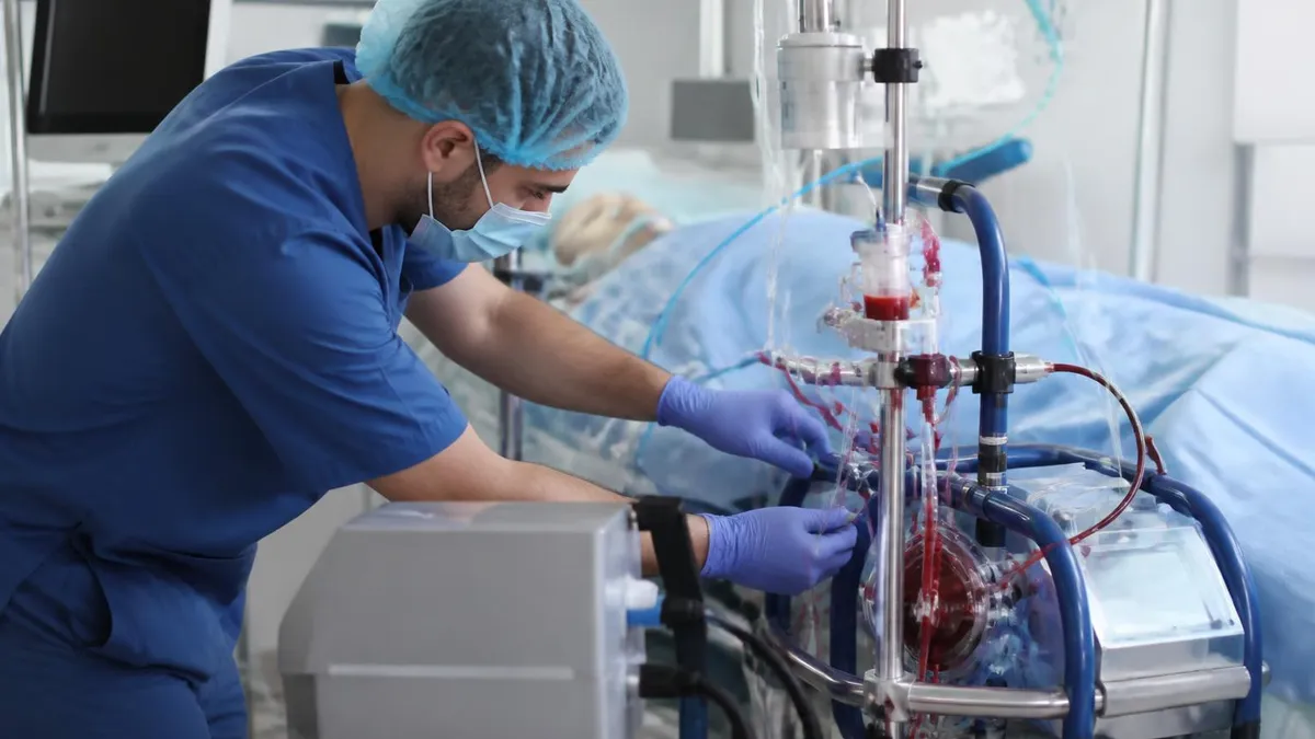
Extracorporeal membrane oxygenation in neonates represents one of the most life-saving applications of modern critical care technology. The ecmo circuit — the physical assembly of tubing, pump, oxygenator, and heat exchanger — is the mechanical backbone that makes ECMO possible. Understanding how each component functions, how they integrate, and how clinicians troubleshoot problems is essential knowledge for anyone preparing for ECMO certification or working in a neonatal or adult intensive care setting. Mastery of circuit physiology separates competent ECMO specialists from truly expert practitioners.
The extracorporeal membrane oxygenation circuit operates on a deceptively simple principle: blood is drained from the patient, pumped through an artificial lung that adds oxygen and removes carbon dioxide, warmed back to body temperature, and returned to the patient. In practice, managing this circuit requires vigilance over dozens of variables simultaneously — flow rates, sweep gas composition, anticoagulation levels, pressures across the membrane, and the patient's own hemodynamic response. Every element of the circuit interacts with every other element, and a change in one parameter will almost always affect several others.
The extracorporeal membrane oxygenation procedure begins long before the first milliliter of blood enters the circuit. Specialists must prime the circuit with crystalloid or blood prime, de-air all components, confirm all connections are secure, and verify that the backup pump and emergency hand-crank are immediately accessible. This priming process typically takes 30 to 60 minutes for an experienced team and must be completed flawlessly, because circuit errors discovered after cannulation can be catastrophic and time-consuming to correct while a critically ill patient is on support.
Circuit configuration differs depending on whether venovenous extracorporeal membrane oxygenation or venoarterial ECMO is chosen for a given patient. In venovenous ECMO, both the drainage and return cannulas are placed in veins, making it appropriate for patients with isolated respiratory failure whose cardiac function remains adequate. Venoarterial ECMO, by contrast, drains venous blood and returns oxygenated blood to the arterial system, providing both cardiac and respiratory support simultaneously. The circuit hardware is largely the same between configurations, but cannula sizing, flow targets, and monitoring priorities differ substantially.
Extracorporeal membrane oxygenation treatment has expanded significantly beyond its neonatal origins. Adults with refractory acute respiratory distress syndrome, cardiogenic shock, cardiac arrest, and even severe extracorporeal membrane oxygenation COVID complications are now routinely managed on ECMO in specialized centers. This expansion has driven innovation in circuit design — modern centrifugal pumps, polymethylpentene membrane oxygenators, and heparin-bonded circuits have all reduced complications compared to the roller pump circuits used in early ECMO programs. Understanding the evolution of circuit technology helps clinicians appreciate why certain protocols and monitoring strategies exist.
For clinicians preparing for certification exams, the ECMO circuit represents a high-yield content area. Questions about oxygenator function, pump mechanics, pressure monitoring interpretation, and troubleshooting scenarios appear frequently in ELSO-aligned assessments. Understanding the extracorporeal membrane oxygenation diagram — the schematic showing drainage cannula, pump, oxygenator, heat exchanger, and return cannula — provides a cognitive scaffold onto which all clinical knowledge can be organized. Visual familiarity with the circuit diagram makes it easier to reason through questions about what happens upstream and downstream of any given component when a problem develops.
This article provides a comprehensive review of ECMO circuit anatomy, physiology, troubleshooting, and clinical management across neonatal, pediatric, and adult populations. Whether you are a respiratory therapist, perfusionist, intensivist, or neonatal nurse practitioner entering ECMO practice, the material here will strengthen your foundation. Readers are encouraged to test their knowledge with the free practice questions embedded throughout the article, as active recall significantly improves retention of complex technical material compared to passive reading alone.
ECMO Circuit by the Numbers

How the ECMO Circuit Works: Step-by-Step Flow
Blood Drainage
Pump Propulsion
Oxygenation and CO2 Removal
Heat Exchange
Blood Return
Continuous Monitoring
The oxygenator is the most critical component of the extracorporeal membrane oxygenation circuit, and understanding its function is essential for any clinician managing ECMO patients. Modern oxygenators use polymethylpentene (PMP) hollow-fiber membranes that allow gas exchange while maintaining a true membrane barrier between blood and gas phases.
This design virtually eliminates the plasma leakage that plagued older silicone membrane oxygenators during prolonged runs, making current devices far more reliable for support lasting days to weeks. The PMP fiber bundles are arranged to maximize surface area — typically 1.5 to 2.5 square meters in adult devices — within a compact housing that minimizes prime volume and foreign surface exposure.
Gas exchange in the oxygenator follows the same principles as the natural lung but with important differences in how it is controlled. Oxygen delivery to the blood is primarily determined by the fraction of inspired oxygen (FiO2) in the sweep gas, which can be dialed from 0.21 to 1.0. Carbon dioxide removal is primarily controlled by sweep gas flow rate — increasing sweep flow increases the CO2 gradient across the membrane and accelerates removal, while decreasing sweep flow allows CO2 to rise.
This independent control of oxygenation and ventilation is one of the most powerful features of ECMO and allows clinicians to apply lung-protective strategies with minimal native ventilator settings.
The pressure drop across the oxygenator is monitored continuously as an index of membrane integrity and clot burden. A normal trans-membrane pressure gradient is typically 20 to 40 mmHg; rising gradients suggest progressive fibrin deposition within the fibers, which reduces gas exchange efficiency and increases hemolysis risk. When the gradient reaches 50 to 80 mmHg or gas transfer measurably deteriorates, oxygenator changeout is planned. Emergency changeout must be performed within minutes if the membrane fails catastrophically, requiring the team to have a primed backup circuit or oxygenator immediately available at the bedside at all times.
The heat exchanger integrated into most modern oxygenators uses countercurrent water flow to warm blood efficiently. Water temperature is maintained by a servo-controlled heater-cooler unit set 1 to 2 degrees above the target patient temperature to account for heat loss along the return tubing. In neonatal circuits, which have shorter tubing lengths and lower priming volumes, temperature management is more precise. Conversely, in adult circuits with long tubing runs to femoral cannulas, significant heat loss can occur, and clinicians must account for ambient temperature effects, particularly in operating rooms where room temperature may be kept low for the surgical team.
Anticoagulation management is intimately linked to circuit function. Unfractionated heparin remains the anticoagulant of choice for most ECMO runs, dosed to maintain an activated clotting time (ACT) of 160 to 220 seconds or an anti-Xa level of 0.3 to 0.7 IU/mL depending on institutional protocol. Subtherapeutic anticoagulation leads to circuit thrombosis, particularly in areas of low flow and blood stasis such as oxygenator fiber interstices and stagnant connector segments. Supratherapeutic anticoagulation increases bleeding complications, which can be devastating in post-surgical neonates or adults with intracranial pathology. Balancing these competing risks is one of the most nuanced aspects of ECMO management.
Hemolysis is an important circuit-related complication that deserves careful monitoring. Mechanical shear stress from the pump, turbulence at cannula tips, and high circuit pressures all contribute to red cell membrane disruption. Plasma-free hemoglobin levels above 50 mg/dL indicate significant hemolysis and warrant circuit inspection for thrombus at high-shear areas. Rising lactate dehydrogenase, falling haptoglobin, and pink-tinged plasma in circuit tubing are early warning signs. Adjusting pump speed to the lowest effective flow rate, correcting hypovolemia to improve preload, and ensuring cannula positions are optimized all help minimize circuit-induced hemolysis during a prolonged ECMO run.
Blood prime versus crystalloid prime for circuit priming is a critical decision, especially in neonates. Because neonatal blood volumes are small — typically 80 to 90 mL/kg — a crystalloid-primed circuit will acutely dilute the patient's blood on initiation of ECMO, potentially causing severe anemia and dilutional coagulopathy.
Most neonatal programs use packed red blood cells to prime the circuit, adjusting the prime to achieve a target hemoglobin of 12 to 14 g/dL. Fresh frozen plasma is often added to provide clotting factors. The circuit prime is carefully crossmatched and irradiated to prevent transfusion reactions and graft-versus-host disease in immunocompromised neonatal patients.
Venovenous Extracorporeal Membrane Oxygenation vs Venoarterial: Key Differences
Venovenous extracorporeal membrane oxygenation is indicated for isolated respiratory failure with preserved cardiac function. Both the drainage and return cannulas are placed in the venous circulation — most commonly the right internal jugular vein for return and the femoral vein for drainage in adults, or a dual-lumen bicaval cannula via the right internal jugular vein. VV ECMO does not provide direct hemodynamic support; cardiac output remains entirely dependent on native heart function. Target ECMO flows are typically 60 to 80 percent of calculated cardiac output to achieve adequate oxygenation.
One critical concept in VV ECMO management is recirculation — the phenomenon where freshly oxygenated blood from the return cannula is immediately suctioned back into the drainage cannula before reaching the patient's right heart. Recirculation fractions of 20 to 30 percent are common and reduce circuit efficiency. Optimizing cannula position to maximize the distance between return and drainage ports, ensuring adequate circuit flows, and avoiding excessive pump speeds that create vigorous suction all help minimize recirculation. Clinicians monitor recirculation by comparing pre-oxygenator and post-oxygenator saturations against patient SaO2 trends.

ECMO Circuit: Benefits and Limitations
- +Provides complete cardiac and/or respiratory support for patients with otherwise fatal organ failure
- +Allows lung-protective ventilation by offloading oxygenation and CO2 removal from the native lungs
- +Modern PMP oxygenators support runs of 2–4 weeks with minimal plasma leakage
- +Centrifugal pumps reduce hemolysis and offer pulsatile-flow mimicry compared to older roller pumps
- +Heparin-bonded circuits reduce systemic anticoagulation requirements and hemorrhagic complications
- +Serves as a bridge to recovery, transplant, or durable mechanical circulatory support device implantation
- −Requires continuous bedside specialist management — not a set-and-forget therapy
- −Major bleeding complications occur in 20–40% of patients, including intracranial hemorrhage in neonates
- −Circuit thrombosis and oxygenator failure can require emergent changeout under pressure
- −Significant cost — daily circuit consumables, ECMO specialist staffing, and ICU care exceed $5,000–$10,000 per day
- −Hemolysis, complement activation, and inflammatory response worsen with longer circuit runs
- −Extracorporeal membrane oxygenation machine price and infrastructure requirements limit availability to tertiary centers
ECMO Circuit Daily Management Checklist
- ✓Inspect all circuit tubing for visible clots, fibrin strands, or color changes every 2 hours
- ✓Document pre- and post-oxygenator pressures and calculate trans-membrane pressure gradient every hour
- ✓Verify oxygenator gas exchange efficiency by comparing pre-pump and post-oxygenator blood gas values
- ✓Confirm sweep gas flow rate and FiO2 settings match physician orders and patient targets
- ✓Check all circuit connections for security, especially at stopcocks, bridge, and cannula connectors
- ✓Verify heater-cooler water temperature and circuit blood temperature are within 2°C of target
- ✓Document pump speed (RPM), flow rate (L/min), and verify flow alarm limits are active and appropriate
- ✓Review current ACT or anti-Xa level and adjust heparin infusion per protocol if outside therapeutic range
- ✓Perform bladder box or flow sensor alarm test and confirm emergency hand-crank is present at bedside
- ✓Assess cannula insertion sites for bleeding, hematoma, or signs of infection and document findings
The Oxygenation Index Threshold for ECMO Initiation
An oxygenation index (OI) above 40, calculated as (MAP × FiO2 × 100) / PaO2, predicts greater than 80% mortality with conventional ventilation in term neonates. Most neonatal ECMO programs use OI ≥ 40 on two consecutive blood gases as a trigger for ECMO cannulation, while some centers initiate at OI ≥ 25 for congenital diaphragmatic hernia given its unique pathophysiology. Knowing this threshold and its calculation is a high-yield item on ECMO certification examinations.
Extracorporeal membrane oxygenation for adults has become increasingly common at major academic medical centers, driven by evidence supporting its use in refractory ARDS and cardiogenic shock. The landmark CESAR trial published in 2009 demonstrated improved survival and disability-free survival for adults with severe ARDS transferred to ECMO-capable centers, and while the subsequent EOLIA trial showed a non-significant mortality benefit at 60 days, a Bayesian reanalysis strongly supported ECMO efficacy. These trials established the foundation for current ELSO guidelines and shaped how clinicians think about patient selection, timing of ECMO initiation, and management goals throughout the run.
Patient selection for adult ECMO requires careful assessment of reversibility. ECMO is appropriate for patients with a potentially reversible cause of cardiorespiratory failure — such as viral pneumonia, post-cardiotomy shock, drug toxicity, or status asthmaticus — where the underlying disease process can resolve with time and organ support.
It is generally contraindicated in patients with severe multi-organ failure unrelated to the primary indication, advanced malignancy with a prognosis of less than one year, irreversible neurological injury, or extreme obesity that prevents adequate cannulation. These criteria require honest multidisciplinary conversations about goals of care before ECMO is initiated, ideally with the patient and family participating whenever feasible.
The extracorporeal membrane oxygenation COVID application emerged rapidly during the 2020–2021 pandemic, with case series from China, Europe, and North America reporting ECMO use in patients with COVID-19-associated ARDS refractory to prone positioning and optimized ventilatory management.
ELSO published emergency guidance recommending ECMO be considered for COVID patients under 65 years of age with an Murray lung injury score ≥ 3 or PaO2/FiO2 ratio below 80 mmHg on optimized ventilation. Early data showed survival rates of approximately 50 to 60 percent in COVID ECMO patients, comparable to outcomes in non-COVID ARDS, though resource constraints during surge periods severely limited access at many hospitals.
Managing ECMO patients in the context of COVID-19 introduced several circuit-specific challenges. COVID-associated coagulopathy — characterized by elevated D-dimer, fibrinogen, and a prothrombotic state — significantly increased circuit thrombosis rates and oxygenator clotting. Many centers increased their anticoagulation targets during COVID ECMO runs and performed more frequent circuit inspections. The cytokine storm associated with severe COVID-19 also accelerated complement activation at circuit surfaces, worsening systemic inflammation. Some programs explored anticoagulation strategies beyond heparin, including argatroban or bivalirudin for patients with heparin-induced thrombocytopenia who needed ECMO during COVID illness.
Weaning from ECMO is a structured process that assesses the patient's readiness to sustain adequate gas exchange and hemodynamics independently of circuit support. For VV ECMO weaning, the sweep gas is progressively reduced — often in decrements of 1 L/min every 30 to 60 minutes — while native ventilator support is gradually reintroduced.
If blood gases remain acceptable at minimal sweep flows, a trial of capping or removing sweep gas entirely tests the patient's respiratory function. VA ECMO weaning additionally requires assessment of cardiac recovery through echocardiography, hemodynamic monitoring, and sometimes a brief trial at reduced pump flows before the decision to decannulate is made.
Decannulation involves removing the ECMO cannulas and achieving hemostasis at the cannulation sites. For peripheral femoral cannulas in adults, this is typically accomplished at the bedside with direct manual pressure or surgical closure of the vessels. Neonatal carotid decannulation requires a formal surgical procedure in the operating room or a well-equipped intensive care bay.
Carotid reconstruction — using the internal jugular vein as a patch angioplasty to restore carotid continuity — is attempted in neonates at many centers, with studies showing patency rates of approximately 70 to 80 percent at one year. Post-decannulation care includes monitoring for rebound pulmonary hypertension in neonates, cardiac function assessment in cardiac ECMO patients, and imaging to evaluate for cannula site vascular complications.
Long-term outcomes after ECMO vary substantially by indication and population. Neonatal survivors of ECMO for meconium aspiration syndrome or persistent pulmonary hypertension have generally excellent long-term outcomes, with the majority entering school age with normal cognitive function. Neonatal congenital diaphragmatic hernia patients have more variable outcomes related to their underlying anatomical severity and pulmonary hypoplasia.
Adult ECMO survivors face a significant burden of post-intensive care syndrome including cognitive impairment, muscle weakness, and post-traumatic stress disorder, as well as the physical rehabilitation challenges from prolonged immobility. Dedicated ECMO survivor follow-up programs at leading centers have demonstrated that structured rehabilitation and psychological support improve functional recovery substantially.

Air entrainment into the ECMO circuit is an immediately life-threatening emergency requiring instant response. If air bubbles are visualized in the arterial limb of the circuit, clamp the return cannula immediately, alert the team, and begin manual ventilation and cardiac compressions if the patient decompensates. Do not attempt to push air through the circuit — place the patient in Trendelenburg position and aspirate air from the highest point in the circuit. Review all circuit connections for the source of air entry and confirm they are secure before resuming ECMO flow.
Troubleshooting the ECMO circuit requires a systematic approach organized around the anatomy of the circuit itself. When a problem is identified — whether a pressure alarm, a flow discrepancy, a visible clot, or an unexpected change in patient oxygenation — the clinician should mentally trace blood flow from the drainage cannula through each component to the return cannula, asking at each point whether that component could be responsible for the observed finding.
This systematic anatomical approach prevents anchoring bias and ensures that upstream causes of downstream problems are not overlooked. A low flow alarm, for example, might reflect poor venous drainage, pump inlet obstruction, hypovolemia, tension pneumothorax, or a kinked drainage cannula — all require different interventions.
Chatter — visible vibration and intermittent collapse of the drainage line tubing — is a characteristic sign of inadequate venous drainage relative to the pump's demand for blood. The pump is attempting to pull more blood than the venous system can supply, creating cyclic suction pressure that collapses the flexible tubing.
Addressing chatter requires identifying its cause: hypovolemia should prompt a fluid bolus; high intrathoracic pressure from aggressive ventilation should prompt ventilator adjustment; positional obstruction of the drainage cannula may require repositioning under fluoroscopic or echocardiographic guidance. Temporarily reducing pump speed to eliminate chatter while the cause is identified prevents hemolysis from repetitive tubing collapse and suction trauma to red blood cells at the cannula tip.
Oxygenator failure manifests as progressive deterioration in gas exchange efficiency despite unchanged sweep gas settings, rising trans-membrane pressure gradient, visible clot or discoloration in the fiber bundle, or plasma leakage — identified as pink or bloody fluid in the sweep gas exhaust line. When oxygenator failure is recognized, planning for changeout should begin immediately, with a primed replacement oxygenator or full circuit being prepared while the failing device is still functional.
Emergency changeout in a decompensating patient is extremely high-risk and requires flawless teamwork, clear role assignments, and a pre-established protocol that the team has rehearsed during simulation training. Most experienced ECMO programs conduct quarterly circuit changeout simulations to maintain readiness.
Pump failure is the most immediately life-threatening circuit emergency because loss of pump function means loss of all circuit flow within seconds. Modern centrifugal pumps have excellent reliability records, but power failures, motor bearing failures, and console malfunctions can occur. The emergency response to pump failure begins with immediate hand-cranking of a backup roller pump or manual circuit clamping to prevent backflow of venous blood through a non-spinning centrifugal pump, which would act as a passive drain.
The hand-crank must be immediately accessible, physically attached to the circuit console, and its location must be known to every team member. Power backup via uninterruptible power supply protects against electrical failures, and ECMO circuits should always be connected to emergency power outlets.
Cannula complications are among the most common reasons for unexpected ECMO circuit problems. Venous drainage cannulas can migrate out of optimal position — typically defined as positioning the drainage cannula tip at the right atrial-inferior vena cava junction under echocardiographic confirmation. Migration can reduce drainage flows by placing the cannula tip in a high-resistance position within a small vessel.
Return cannulas in VA ECMO can migrate distally in the femoral artery, increasing the risk of limb ischemia downstream of the cannula insertion point. Monitoring distal limb perfusion via pulse oximetry on the affected extremity and maintaining a low threshold for placing a distal perfusion catheter are standard of care in peripheral VA ECMO programs.
Documentation requirements for ECMO circuit management are extensive and serve multiple purposes: clinical communication between bedside specialists, quality assurance monitoring, and medicolegal record-keeping. Most programs use dedicated ECMO flow sheets documenting circuit parameters, patient vital signs, laboratory values, interventions, and specialist assessments on an hourly basis during the acute phase of an ECMO run. Electronic health records increasingly integrate ECMO-specific documentation templates that capture required data points without excessive free-text narrative. Understanding what needs to be documented, why each parameter matters clinically, and how documentation errors can compromise patient safety are all important competencies for ECMO specialists entering practice.
Simulation-based training has become a cornerstone of ECMO education and competency maintenance. High-fidelity circuit simulators allow trainees to practice emergency responses — air embolism management, pump failure response, circuit changeout — in a controlled environment without risk to real patients. Studies from multiple ECMO programs have demonstrated that simulation training reduces response time to circuit emergencies and improves team communication during crises. The ELSO training curriculum recommends that ECMO specialists complete both didactic training and simulation competency assessment before independent practice, and that annual recertification include simulation scenarios to maintain emergency response proficiency throughout a practitioner's career.
Preparing for ECMO certification requires a strategic approach that goes beyond memorizing circuit components. The ELSO certification examination tests not just factual knowledge but clinical reasoning — the ability to interpret a scenario, identify the most likely cause of a problem, and select the most appropriate immediate intervention.
Reading the ELSO Red Book (ECMO Specialist Training Manual) cover to cover provides the foundational knowledge base, but active practice with case-based questions is what develops the reasoning skills needed to perform well on examination day. Candidates should aim for at least 200 practice questions in the weeks leading up to their exam, reviewing not just correct answers but the explanations for why incorrect options are wrong.
Time management during ECMO certification examinations is a skill that requires deliberate practice. Many candidates spend too long on difficult questions early in the exam and run short of time for questions they could have answered correctly later. A practical strategy is to answer all questions you are confident about first, marking uncertain questions for review, and then returning to difficult items with remaining time.
This ensures that knowledge you possess translates into correct answers rather than being lost to time pressure. Most ECMO certification exams allow sufficient time for a complete pass plus a full review cycle if pace is managed appropriately from the start.
Understanding the extracorporeal membrane oxygenation diagram — the schematic representation of circuit components and flow direction — is not just an academic exercise. Being able to mentally visualize blood traveling through each component helps clinicians troubleshoot problems in real time. When a downstream oxygenator shows unexpectedly low post-membrane saturation, can you trace the differential diagnosis upstream?
When drainage pressures are suddenly more negative than expected, which component or patient factors are most likely responsible? The circuit diagram is a clinical thinking tool, and practitioners who have internalized it respond faster and more accurately to circuit emergencies than those who must consciously reconstruct the anatomy under pressure.
Pharmacokinetic changes during ECMO alter how medications behave in ECMO patients, making drug management a critical knowledge domain for certification. The ECMO circuit sequesters lipophilic drugs — particularly fentanyl, midazolam, and many antibiotics — into circuit tubing and oxygenator surfaces, dramatically reducing effective blood levels.
Standard dosing that would be adequate in non-ECMO patients is often insufficient during ECMO, leading to inadequate sedation, pain control, or antibiotic exposure. ECMO specialists must understand which drug classes are most affected, how to monitor therapeutic drug levels during ECMO, and how to adjust dosing to account for both circuit sequestration and the volume of distribution changes caused by the hemodilution inherent in circuit priming.
Nutrition management during ECMO is increasingly recognized as a factor that influences outcomes. Early enteral nutrition — initiated within 24 to 48 hours of ECMO cannulation when hemodynamics permit — is associated with better gut mucosal integrity and reduced infectious complications. Calculating caloric needs in ECMO patients is complicated by the fact that the extracorporeal circuit alters normal metabolic rate measurement, making standard predictive equations potentially inaccurate.
Some centers use indirect calorimetry adapted for ECMO to guide nutrition prescriptions. Avoiding both underfeeding and overfeeding is important: underfeeding accelerates muscle wasting while overfeeding increases CO2 production and complicates ventilator weaning in VV ECMO patients with residual lung disease.
Family communication and psychosocial support represent dimensions of ECMO care that are not tested on certification exams but profoundly affect the experience of patients and families. Seeing a loved one connected to a complex mechanical circuit with multiple alarms, large-bore cannulas in major vessels, and a continuous hum of pump machinery is a deeply frightening experience.
ECMO specialists play a crucial role in demystifying the circuit for families — explaining in plain language what each component does, why alarms sound, and what changes in circuit parameters mean for the patient's progress. Families who understand the ECMO circuit as a support tool rather than a source of fear are better prepared to participate in goals-of-care discussions and to cope with the inevitable uncertainties of a prolonged ECMO run.
Continuous quality improvement in ECMO programs depends on systematic data collection and analysis. ELSO maintains an international registry to which member centers contribute circuit event data, complications, and outcomes. Participating centers use this benchmarking data to identify areas where their outcomes deviate from peer institutions and to implement evidence-based practice improvements.
Common quality metrics include circuit prime-to-cannulation time, circuit changeout frequency, major bleeding rates, nosocomial infection rates, and survival to hospital discharge stratified by indication. ECMO specialists who understand how their program's performance is measured and how individual circuit management decisions contribute to program-level outcomes are more engaged, more accountable, and ultimately more effective practitioners.
ECMO Questions and Answers

VA ECMO: Veno-Arterial Configuration, Indications, and Cannulation Guide

ECMO Machine: Components, How It Works, VA vs VV Types, and Clinical Management
ECMO Extracorporeal Membrane Oxygenation Practice Test PDF (Free Printable 2026)

ECMO: How Extracorporeal Membrane Oxygenation Works and Who Needs It
ECMO Procedure
About the Author

Educational Psychologist & Academic Test Preparation Expert
Columbia University Teachers CollegeDr. Lisa Patel holds a Doctorate in Education from Columbia University Teachers College and has spent 17 years researching standardized test design and academic assessment. She has developed preparation programs for SAT, ACT, GRE, LSAT, UCAT, and numerous professional licensing exams, helping students of all backgrounds achieve their target scores.
Join the Discussion
Connect with other students preparing for this exam. Share tips, ask questions, and get advice from people who have been there.
View discussion (7 replies)