
ECMO Cannulation Techniques: A Complete Guide to Extracorporeal Membrane Oxygenation in Neonates and Adults
Master ECMO cannulation techniques for neonates and adults. Learn circuit setup, cannula types, and venovenous ECMO procedures. ✅ Complete 2026 October guide.

Extracorporeal membrane oxygenation in neonates represents one of the most technically demanding applications of life-support medicine, requiring precise cannulation to establish adequate blood flow through an external circuit. The lava ecmo cannula and similar specialized access devices have transformed how clinicians approach vascular access for ECMO, allowing for safer insertion with reduced vessel trauma. Understanding cannulation fundamentals is essential for any ECMO specialist, as errors during this phase can compromise circuit performance and patient outcomes from the very first moment of support.
The extracorporeal membrane oxygenation procedure begins long before the pump starts spinning. Surgical teams must evaluate vessel anatomy, choose appropriate cannula sizes, and decide between open surgical cutdown and percutaneous approaches. Patient size, urgency, and the intended ECMO mode — venovenous versus venoarterial — each influence which vessels are targeted and how cannulas are secured. Neonates present unique challenges because their vessels are tiny, fragile, and highly prone to spasm, making size selection and insertion technique critically important for circuit success.
Modern extracorporeal membrane oxygenation circuits rely on cannulas that provide adequate drainage and return flow while minimizing hemolysis and thrombosis. Cannula design has evolved significantly over the past two decades, with improvements in tip geometry, flow characteristics, and biocompatible coatings reducing circuit-related complications. Teams that understand these engineering principles can make better intraoperative decisions and troubleshoot flow problems more effectively when they arise during a run that may last days or even weeks.
Cannulation site selection varies by patient population and clinical indication. In neonatal cardiac ECMO, the right internal jugular vein and right common carotid artery remain the traditional access points, providing reliable flow without the risks associated with femoral approaches in small patients. For venovenous extracorporeal membrane oxygenation in older children and adults with respiratory failure, the right internal jugular vein combined with the femoral vein — or a single dual-lumen cannula in the jugular — offers an effective and increasingly preferred configuration that avoids arterial cannulation entirely.
The extracorporeal membrane oxygenation treatment landscape expanded dramatically during the COVID-19 pandemic, when ECMO centers worldwide placed thousands of adult patients with refractory hypoxemia on venovenous support. This experience accelerated adoption of percutaneous cannulation techniques, ultrasound guidance protocols, and team-based training programs. Lessons learned during the pandemic continue to refine how centers approach cannulation efficiency, sterility maintenance, and rapid deployment in emergency situations where every minute of delay increases the risk of irreversible organ injury.
For learners preparing for the ECMO Specialist certification or studying for competency assessments, mastering cannulation theory is just as important as hands-on procedural training. Knowing why a specific vessel is chosen, how cannula position affects drainage, and what fluoroscopic or echocardiographic landmarks confirm correct placement will help candidates answer complex scenario-based questions confidently. Those seeking structured practice should explore ecmo cannulation techniques to reinforce procedural knowledge with evidence-based clinical context.
This guide covers the full spectrum of ECMO cannulation knowledge — from basic circuit anatomy and cannula selection through site-specific approaches, complication prevention, and emerging technologies. Whether you are a perfusionist, intensivist, neonatal nurse, or respiratory therapist expanding your ECMO knowledge, the information in these sections will build a strong conceptual foundation that supports both clinical practice and examination preparation at every level of experience.
ECMO Cannulation by the Numbers

Step-by-Step ECMO Cannulation Process
Pre-Cannulation Planning
Vascular Access & Vessel Exposure
Cannula Insertion & Positioning
Circuit Connection & Prime
Flow Titration & Monitoring
Cannula Securement & Documentation
Extracorporeal membrane oxygenation in neonates demands the highest level of technical precision because patients may weigh as little as 1.5–2 kg, with vessel diameters barely exceeding 3–4 mm. The right internal jugular vein is the most common venous access site for neonatal ECMO, accommodating an 8–12 French venous drainage cannula in most term infants. The right common carotid artery serves as the arterial return vessel in venoarterial configurations. While carotid ligation was historically accepted, many centers now attempt carotid reconstruction at decannulation to preserve cerebrovascular integrity in long-term survivors.
Cannula size selection for neonatal patients is guided by weight-based nomograms and target flow calculations. A general rule is that drainage cannula size should allow flows of approximately 100–150 mL/kg/min with reasonable negative pressure. Oversized cannulas risk vessel injury, while undersized ones limit flow capacity and may cause excessive negative pressure artifact — a condition called suction events — that can cause circuit chatter, hemolysis, and inadequate oxygen delivery to the patient when demands are highest.
Pediatric ECMO cannulation beyond the neonatal period introduces additional anatomical considerations. Children between 3 and 15 kg may benefit from femoral vessel access when cervical anatomy is unfavorable or when prior cervical surgery has distorted normal landmarks. In older pediatric patients weighing more than 20–25 kg, femoral venous and arterial cannulation becomes technically straightforward and avoids potential neurological risks associated with carotid manipulation. The extracorporeal membrane oxygenation procedure in this age group increasingly mirrors adult protocols.
Dual-lumen cannulas represent a major innovation in pediatric and adult ECMO cannulation. Devices like the Avalon Elite — and similar products — are positioned through the right internal jugular vein and advanced so the tip sits in the inferior vena cava while the return port faces the tricuspid valve. This configuration allows complete venovenous support through a single venous access site, eliminating the need for femoral cannulation and improving patient mobility. Correct positioning requires careful fluoroscopic and echocardiographic guidance, as malposition can cause recirculation or inadequate drainage.
Recirculation is a major concern in VV ECMO and occurs when oxygenated blood exiting the return cannula is immediately drained back into the circuit before reaching the pulmonary circulation. In dual-lumen cannula configurations, return flow must be directed toward the tricuspid valve rather than toward the drainage ports. Clinicians monitor for recirculation by tracking the pre-oxygenator saturation — when drainage saturations are abnormally high, recirculation is likely. Adjusting sweep gas, repositioning the patient, or rotating the cannula can help correct the problem without requiring full recannulation.
Ultrasound guidance has become the standard of care for percutaneous ECMO cannulation in adults and larger pediatric patients. Real-time imaging confirms needle position within the target vessel lumen, reduces the risk of arterial puncture during venous access, and helps advance wires and dilators safely. Transesophageal echocardiography provides excellent views of wire position in the right heart and inferior vena cava, making it particularly valuable during dual-lumen cannula placement. Many centers have adopted dedicated ultrasound training modules as part of their ECMO team credentialing process.
Post-cannulation assessment is as important as the insertion itself. After ECMO flows are established, the team evaluates cannula position with bedside imaging, confirms hemodynamic improvement, and assesses limb perfusion distal to any arterial cannulation site. Femoral arterial ECMO in particular carries risk of limb ischemia, and many centers routinely place a distal perfusion cannula — a small-bore sheath inserted antegrade into the superficial femoral artery — to maintain perfusion to the leg throughout the ECMO run, a practice now widely adopted as standard safety protocol.
Venovenous Extracorporeal Membrane Oxygenation: Approaches, Advantages & Monitoring
Venovenous extracorporeal membrane oxygenation is the preferred configuration for patients with isolated respiratory failure and adequate cardiac function. The most common adult configuration uses a femoral drainage cannula (21–25 Fr) advanced to the right atrium combined with an internal jugular return cannula (17–19 Fr) positioned at the cavoatrial junction. This two-cannula approach provides reliable flow rates of 4–6 L/min in most adult patients and allows the heart to continue pumping oxygenated blood to the systemic circulation without mechanical assistance.
Single-site VV ECMO using dual-lumen cannulas has grown in popularity because it supports earlier patient mobilization and rehabilitation. The Avalon Elite and comparable devices require 27–31 Fr access through the right internal jugular vein. Correct positioning — with the distal drainage tip in the IVC and the return port facing the tricuspid valve — is confirmed by fluoroscopy and echocardiography. When positioned correctly, these cannulas achieve excellent drainage efficiency with minimal recirculation, making them suitable for awake ECMO protocols in cooperative patients awaiting lung transplantation.

Percutaneous vs. Surgical Cutdown ECMO Cannulation: Pros and Cons
- +Faster initiation in emergency settings — percutaneous access can be established in under 15 minutes by experienced teams
- +No surgical incision reduces wound infection risk and simplifies decannulation when ECMO is no longer needed
- +Bedside feasibility allows ECMO initiation in ICU or emergency department without requiring operating room transport
- +Ultrasound guidance improves first-pass success rate and reduces vascular complications during initial access
- +Lower blood loss during insertion because vessel is dilated rather than surgically opened and ligated
- +Easier to teach and standardize across multidisciplinary team members including intensivists and cardiologists
- −Higher risk of malpositioning in small or anatomically complex patients who lack clear ultrasound landmarks
- −Limited to larger vessel access — patients under 10 kg often require surgical cutdown for safe neonatal cannulation
- −Vessel injury from serial dilation can complicate decannulation and require surgical repair at run termination
- −Less direct control of hemorrhage if vessel injury occurs during wire or dilator advancement through the skin
- −Carotid artery reconstruction not possible with percutaneous technique — ligation remains the norm in neonates
- −Fluoroscopy or echo dependence adds equipment requirements that may not be available in all clinical settings
Pros and Cons at a Glance
| Pros | Cons |
|---|---|
| No surgical incision reduces wound infection risk and simplifies decannulation when ECMO is no longer needed | Higher risk of malpositioning in small or anatomically complex patients who lack clear ultrasound landmarks |
| Bedside feasibility allows ECMO initiation in ICU or emergency department without requiring operating room transport | Vessel injury from serial dilation can complicate decannulation and require surgical repair at run termination |
| Ultrasound guidance improves first-pass success rate and reduces vascular complications during initial access | Less direct control of hemorrhage if vessel injury occurs during wire or dilator advancement through the skin |
| Lower blood loss during insertion because vessel is dilated rather than surgically opened and ligated | Carotid artery reconstruction not possible with percutaneous technique — ligation remains the norm in neonates |
| Easier to teach and standardize across multidisciplinary team members including intensivists and cardiologists | Fluoroscopy or echo dependence adds equipment requirements that may not be available in all clinical settings |
ECMO Cannulation Safety Checklist: 10 Critical Steps
- ✓Confirm ECMO mode (VV vs VA) and document rationale in the medical record before starting cannulation.
- ✓Verify cannula sizes against weight-based nomogram and confirm availability of backup sizes in the room.
- ✓Perform pre-procedure ultrasound to assess vessel patency, size, and identify anatomical variants.
- ✓Confirm anticoagulation plan with the team — heparin bolus timing relative to cannula insertion must be agreed upon.
- ✓Ensure the ECMO circuit is fully primed, air-free, and clamps are in correct positions before vessel access.
- ✓Use real-time ultrasound guidance for all percutaneous needle insertions to prevent inadvertent arterial puncture.
- ✓Confirm wire position in the correct vessel with ultrasound or fluoroscopy before advancing dilators.
- ✓Check cannula tip position by echocardiography or chest X-ray before initiating full ECMO flows.
- ✓Place a distal perfusion cannula in the ipsilateral superficial femoral artery when using femoral arterial ECMO.
- ✓Document cannula sizes, insertion depths, site condition, and initial ECMO parameters in a standardized cannulation note.
Tip Position Is the Single Most Modifiable Determinant of ECMO Flow Adequacy
A drainage cannula tip that has migrated even 1–2 cm from its optimal position can reduce ECMO flow by 30–50%, triggering repeated suction alarms and hemolysis. Before attributing poor flows to hypovolemia or cardiac function, always verify cannula depth with bedside echocardiography or chest X-ray. Repositioning — not volume loading — is often the correct first intervention when drainage is inadequate and the patient is euvolemic on clinical examination.
Cannulation-related complications represent a significant source of morbidity in ECMO patients, and understanding their mechanisms is essential for both clinical management and examination preparation. Bleeding at the cannulation site is the most common early complication, occurring in 10–30% of ECMO runs across published series. The combination of systemic anticoagulation, tissue trauma from large-bore cannula insertion, and platelet consumption by the extracorporeal circuit creates a challenging hemostatic environment. Teams must balance bleeding risk against the need for adequate anticoagulation to prevent circuit thrombosis throughout the run.
Limb ischemia distal to a femoral arterial cannula is a serious complication that can lead to compartment syndrome, rhabdomyolysis, and limb loss if not recognized promptly. Clinical monitoring should include hourly assessment of skin color, temperature, capillary refill, and Doppler signals distal to the arterial insertion site. When ischemia is detected, a distal perfusion catheter placed antegrade into the superficial femoral artery — connected to the arterial return limb of the ECMO circuit — restores perfusion in most cases without requiring removal of the main arterial cannula.
Cannula dislodgement is a potentially catastrophic event that can result in massive hemorrhage or acute loss of ECMO support. Prevention relies on secure suture fixation, redundant circuit loops with slack to absorb patient movement, and bed positioning protocols that protect the exit site. All ECMO team members must be trained in emergency clamping procedures and know exactly which clamps to apply first in the event of accidental decannulation. Simulation-based training using task trainers has been shown to reduce response time significantly in controlled studies of ECMO emergency protocols.
Air embolism entering through the cannulation site or circuit connections can cause cardiac dysfunction, pulmonary air lock, or cerebral infarction. Strategies to prevent air entry include maintaining positive pressure during cannula insertion, ensuring all circuit connections are locked and reinforced, and using air detection systems on modern centrifugal pump consoles. When air is detected in the circuit, flows are immediately reduced, the circuit is clamped, and the air is evacuated from the highest point of the circuit before resuming support. Prompt recognition and response are critical skills for the ECMO specialist examination.
Thrombosis within the cannula or circuit is the second most common mechanical complication after bleeding. Inadequate anticoagulation, low flow states, and areas of stagnant blood within the circuit create conditions favorable for clot formation. Clinical signs include rising transmembrane pressure across the oxygenator, dark coloration of the oxygenator membrane, visible fibrin strands in the tubing, and unexplained deterioration in patient oxygenation. Oxygenator replacement or circuit changeout may be required when thrombosis is extensive, a procedure that carries significant risk and requires careful coordination between perfusionist, bedside nurse, and physician.
Cannula-associated infection is a late complication that becomes more significant as ECMO runs extend beyond 7–10 days. Biofilm formation on the cannula surface provides a nidus for bacterial and fungal organisms that are difficult to eradicate without removing the device. Daily assessment of insertion sites, strict sterile dressing protocols, and antimicrobial dressing materials reduce but do not eliminate infection risk. Blood cultures drawn from peripheral sites — rather than from the ECMO circuit — provide the most reliable bacteriological data when infection is suspected during an extended ECMO run.
Neurological complications in neonatal ECMO — including intracranial hemorrhage and ischemic stroke — occur in approximately 10–15% of neonatal ECMO patients and are strongly associated with anticoagulation intensity, carotid ligation, and prior hemodynamic instability. Routine cranial ultrasound surveillance every 24–48 hours is standard practice in neonatal ECMO centers. When hemorrhagic transformation is identified, teams must weigh the risk of anticoagulation reduction against circuit thrombosis, often requiring individualized decision-making by experienced multidisciplinary teams with expertise in neonatal neurocritical care.

Accidental decannulation can result in exsanguination within minutes. Every ECMO bedside must have emergency clamps immediately accessible, and all team members must know exactly which clamp to apply first based on the cannula configuration in use. Do not rely on memory during a crisis — laminated emergency cards at the bedside are a recommended safety practice in all accredited ECMO centers.
The extracorporeal membrane oxygenation machine price and overall cost of ECMO support is a topic that affects health system planning, resource allocation, and patient family counseling. A single ECMO circuit — including the oxygenator, centrifugal pump head, tubing, and initial cannula set — costs approximately $8,000 to $15,000 at acquisition. When daily consumable costs, nursing staffing ratios, pharmacy charges, and laboratory monitoring are factored in, the total cost of a typical 7–10 day ECMO run in the United States ranges from $100,000 to over $500,000 depending on the institution, payer mix, and patient complexity during the hospitalization.
Cannula costs represent a meaningful portion of per-case ECMO expenditure. Standard single-lumen drainage and return cannulas cost $500–$2,000 each, while specialized dual-lumen devices like the Avalon Elite carry price tags of $3,000–$5,000 per unit. The lava ecmo cannula and similar next-generation access devices incorporate advanced tip designs and biocompatible coatings that may reduce hemolytic complications — a factor that, if it shortens ECMO duration or prevents circuit changeouts, can offset the higher upfront device cost through reductions in downstream resource utilization and complication management.
The extracorporeal membrane oxygenation diagram that appears in most training materials illustrates the basic circuit loop: venous blood drains by gravity or centrifugal pump suction from the patient, passes through a pump that generates flow, traverses a membrane oxygenator where oxygen is added and carbon dioxide removed, and returns to the patient via the arterial or venous return cannula. Modern ECMO consoles integrate flow sensors, bubble detectors, pressure transducers, and temperature monitors into a single interface that allows real-time surveillance of every component in the extracorporeal circuit simultaneously.
Oxygenator selection affects both gas transfer efficiency and circuit longevity. Polymethylpentene (PMP) hollow-fiber oxygenators have largely replaced older silicone membrane devices because they provide superior gas exchange, lower resistance, better plasma leak resistance, and longer functional lifespan. In long-term ECMO runs, the oxygenator is often the first component to fail — manifesting as rising carbon dioxide levels despite adequate sweep gas flow or visible plasma weeping from the fiber bundle. Planned oxygenator exchanges every 7–14 days are performed at some centers with high-volume long-term ECMO programs.
Centrifugal pump technology has also advanced considerably, with modern diagonal-flow pumps generating less hemolysis than older radial designs at equivalent flow rates. The Maquet Rotaflow, Thoratec CentriMag, and Medtronic Biopump are examples of centrifugal pumps used in US ECMO centers. Each has specific priming volumes, speed-flow relationships, and afterload sensitivity characteristics that ECMO specialists must understand to optimize circuit management. Roller pumps — the original ECMO pump design — are now used primarily in neonatal ECMO at a small number of centers where institutional experience and protocol support their continued use.
Decannulation is the final procedural step in the ECMO journey and carries its own technical risks and considerations. Before cannulas are removed, the patient must demonstrate adequate cardiopulmonary function during a weaning trial conducted at minimal ECMO support. For surgical cannulation sites, vessel repair or ligation is performed under direct visualization. Percutaneous sites are managed with manual compression or figure-of-eight sutures, followed by pressure dressings for 6–12 hours. Carotid artery reconstruction at neonatal decannulation — when feasible — aims to restore ipsilateral cerebral blood flow and reduce long-term neurodevelopmental consequences.
For clinicians and trainees aiming to deepen their understanding of the full ECMO support continuum, reviewing the broader context of recovery and equipment management is highly valuable. Comprehensive resources on extracorporeal membrane oxygenation treatment outcomes and device-specific guidance can be found in related clinical literature and specialty training programs. Reviewing material on ECMO recovery and patient management alongside cannulation technique knowledge will provide a well-rounded foundation that prepares learners for both board-style examination questions and real-world clinical decision-making during complex ECMO cases.
Preparing effectively for ECMO specialist examinations requires a study strategy that integrates cannulation technique knowledge with physiological principles, equipment familiarity, and clinical management protocols. Begin by mastering the anatomical rationale for each cannulation site — understanding why the right internal jugular is preferred in neonates, why femoral access is favored in adults, and under what circumstances alternative sites are chosen will help you answer both factual recall and clinical reasoning questions correctly. Draw your own ECMO circuit diagrams repeatedly until the flow of blood from patient to circuit and back becomes second nature.
Focus special attention on flow physiology, as this is heavily tested on ECMO certification examinations. Understand how changes in preload, afterload, sweep gas, and rotational speed affect circuit flows and patient oxygen delivery. Practice calculating oxygen delivery using the Fick equation, and know the normal ranges for key hemodynamic parameters during both VV and VA ECMO. Questions that ask you to interpret a scenario where ECMO flows are dropping require you to reason through drainage problems, circuit problems, and patient physiology simultaneously — a multi-step analytical process that benefits greatly from systematic study.
Anticoagulation management during ECMO is closely linked to cannulation success and run duration, making it an area you cannot afford to ignore during your preparation. Know the typical heparin dosing ranges, target aPTT and anti-Xa levels used at most US centers, and the indications for alternative anticoagulants like bivalirudin. Understand how thrombocytopenia, HIT suspicion, and active bleeding each modify the anticoagulation approach. The pharmacology of anticoagulants in the context of ECMO is a high-yield topic that bridges multiple examination domains.
Use case-based practice questions to develop your clinical reasoning under time pressure. ECMO examinations typically present clinical vignettes describing a deteriorating patient on ECMO support and ask what intervention is most appropriate. Train yourself to immediately categorize the problem — is it a circuit issue, a cannula position issue, a patient physiology issue, or a pharmacological issue? Structured problem-solving using this framework reduces cognitive load during the examination and improves both speed and accuracy on scenario-based items that reward systematic thinking.
Hands-on simulation training complements written study by building procedural fluency and crisis response skills. Most accredited ECMO training programs require a minimum number of simulated cannulation exercises, circuit setup drills, and emergency scenario rehearsals before a candidate is considered clinically competent to manage patients independently. If your institution has an ECMO task trainer or high-fidelity simulation lab, take every opportunity to practice — particularly for low-frequency, high-stakes events like air embolism management, accidental decannulation response, and oxygenator changeout procedures that may arise only a few times per year in clinical practice.
Review the ELSO Guidelines for ECMO Centers and the specific population-based guidelines (neonatal, pediatric, adult respiratory, adult cardiac) as primary study references. These documents represent the consensus of international ECMO experts and form the foundation of most certification examination content. Pay particular attention to the sections on patient selection criteria, contraindications, circuit management standards, and weaning protocols, as these areas generate the highest density of examination questions across all ECMO specialty tracks currently offered by credentialing organizations.
Finally, use the practice quizzes and resources available on PracticeTestGeeks to identify your weak areas early in your preparation timeline. Simulated examinations provide immediate feedback on which domains need more attention and help you build test-taking stamina for the lengthy certification examination format. Reviewing explanations for both correct and incorrect answers deepens conceptual understanding far more effectively than simply re-reading textbooks, making active practice the most efficient use of your limited study time as you approach your certification target date.
Sample ECMO - Extracorporeal Membrane Oxygenation Practice Questions
Try these questions from our free ECMO - Extracorporeal Membrane Oxygenation practice tests. The correct answer and an explanation follow each question.
What is the primary advantage of a percutaneous over a surgical cutdown approach for ECMO cannulation in most adults?
- A. Larger cannulas can be placed percutaneously
- B. Faster access, lower bleeding risk at the site, and no surgical wound
- C. Surgical cutdown provides worse visualization of vessel anatomy
- D. Percutaneous cannulas are easier to remove at decannulation
Answer: B. Faster access, lower bleeding risk at the site, and no surgical wound
Percutaneous Seldinger-based cannulation is faster in an emergency, avoids a surgical incision, and is associated with lower local complication rates in experienced hands.
What is the most common cannulation site for peripheral VA ECMO in adults?
- A. Right internal jugular vein and subclavian artery
- B. Femoral vein and femoral artery
- C. Femoral vein and axillary artery
- D. Femoral vein and radial artery
Answer: B. Femoral vein and femoral artery
Femoral vein to femoral artery cannulation is the most common approach for peripheral VA ECMO due to accessibility and speed of insertion.
Which cannulation strategy is preferred when ECPR is performed during active CPR in a pediatric patient under 10 kg?
- A. Femoral-femoral VA-ECMO
- B. Neck (carotid-jugular) cannulation
- C. Subclavian artery cannulation
- D. Transcaval approach
Answer: B. Neck (carotid-jugular) cannulation
Carotid artery and internal jugular vein cannulation is the preferred approach in small pediatric patients due to vessel size limitations at the femoral site.
A key advantage of a percutaneous 'Z-stitch' purse-string suture placed at the time of cannulation is that it:
- A. Prevents cannula migration during ECMO support
- B. Allows rapid hemostasis at decannulation without surgical exploration
- C. Reduces the risk of cannula-related thrombosis
- D. Secures the cannula to the skin to prevent accidental dislodgement
Answer: B. Allows rapid hemostasis at decannulation without surgical exploration
The pre-placed purse-string suture can be tightened and tied at the moment the cannula is withdrawn, achieving immediate hemostasis percutaneously.
Take the full ECMO - Extracorporeal Membrane Oxygenation practice test
ECMO Questions and Answers
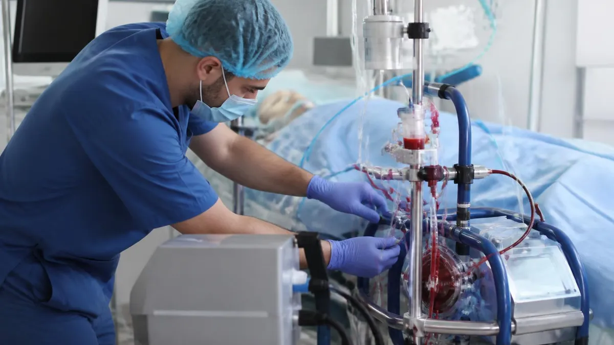
ECMO Procedure

ECMO Recovery: What to Expect After Extracorporeal Membrane Oxygenation Treatment

ECMO Devices: A Complete Guide to Extracorporeal Membrane Oxygenation Equipment

ECMO Patient Guide: What to Expect, Care, Recovery & Outcomes

ECMO Specialist: Career Path, Duties, Salary, and Certification Guide
About the Author

Educational Psychologist & Academic Test Preparation Expert
Columbia University Teachers CollegeDr. Lisa Patel holds a Doctorate in Education from Columbia University Teachers College and has spent 17 years researching standardized test design and academic assessment. She has developed preparation programs for SAT, ACT, GRE, LSAT, UCAT, and numerous professional licensing exams, helping students of all backgrounds achieve their target scores.
Join the Discussion
Connect with other students preparing for this exam. Share tips, ask questions, and get advice from people who have been there.
View discussion (9 replies)