
VA ECMO vs VV ECMO: Configurations, Indications, Outcomes 2026 October
⏳ VA ECMO vs VV ECMO compared: cannulation sites, indications, hemodynamic support, complications, and real-world survival outcomes.
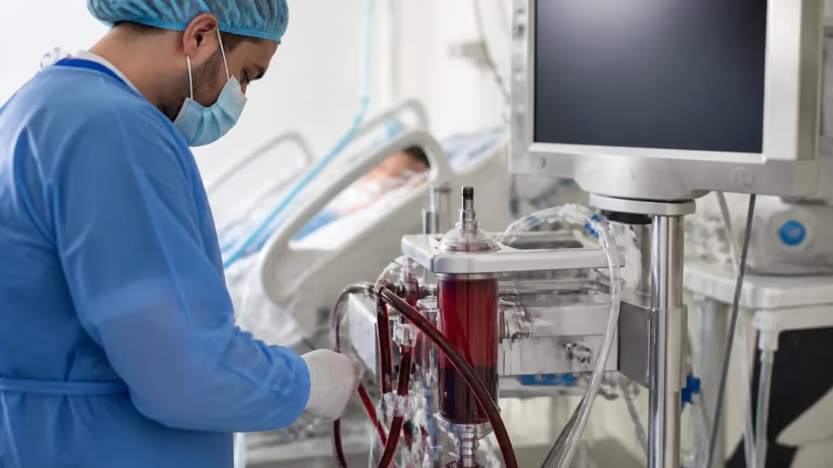
VA ECMO vs VV ECMO at the bedside
Walk into a cardiothoracic ICU and the patient on ECMO is the one with the loud centrifugal pump churning by the bed, two thick cannulas snaking out from the groin, and a perfusionist in a chair watching pre- and post-oxygenator pressures.
ECMO — extracorporeal membrane oxygenation — pulls deoxygenated blood out of the body, runs it through a centrifugal pump and a hollow-fiber oxygenator that adds oxygen and strips carbon dioxide, and returns warm, well-oxygenated blood back to the patient. The whole circuit lives outside the chest. It is, in plain language, a temporary lung, and depending on where you return the blood, a temporary heart too.
That return-site choice is what splits ECMO into its two big configurations. Veno-venous ECMO — VV ECMO — drains from a vein and returns to a vein, so the patient's own heart still has to push the blood around. Veno-arterial ECMO, VA ECMO, drains from a vein but returns to a large artery, bypassing the heart entirely and delivering flow into the systemic circulation.
One supports lungs. The other supports lungs and circulation. The cannulation is different, the indications are different, the complications are different, and the survival numbers are different. Mixing them up at the bedside is dangerous, and on board exams it is one of the cleanest ways to lose points.
This piece walks through both modes the way the bedside team actually thinks about them: what each one does, who needs which, where the cannulas go, what tends to go wrong, and what the outcome data look like once the dust settles. The aim is a clear mental model you can take into a CCRN review, an ECMO specialist course, or the next handover at the start of a night shift.
VA ECMO vs VV ECMO at a glance
VV ECMO: drains and returns to veins. Supports lungs only. Used for severe ARDS, COVID pneumonia, drowning, status asthmaticus. Cardiac output stays with the patient's heart. Typical SpO2 reads 85–92 percent. VA ECMO: drains from vein, returns to artery. Supports heart and lungs. Used for cardiogenic shock, fulminant myocarditis, ECPR, post-cardiotomy failure. Provides 3–5 L/min of mechanical circulatory support. Watch for: harlequin syndrome and limb ischemia in VA; recirculation in VV. Anticoagulation, oxygenator monitoring, and ELSO-trained specialists at the bedside are mandatory for either mode.
What ECMO actually does — the shared core
Before splitting VA from VV it helps to be precise about the circuit they share. Both start with a drainage cannula that sits in a large vein and pulls dark, deoxygenated blood into plastic tubing. A centrifugal pump — usually magnetically levitated to reduce hemolysis — spins between two and four thousand RPM and generates the negative pressure that drains the vein.
The blood passes through an oxygenator, a cartridge full of hollow polypropylene fibers. Sweep gas (usually pure oxygen, sometimes blended) flows through the inside of the fibers, while blood flows around the outside. Oxygen diffuses into the blood and carbon dioxide diffuses out, the same way it would across an alveolar membrane in healthy lungs.
The oxygenator is also a heat exchanger. A separate water line from a heater-cooler keeps blood at body temperature (or deliberately cool, in some protocols). Anticoagulation is non-negotiable — unfractionated heparin titrated to an ACT or anti-Xa target keeps clot from forming on the plastic and the oxygenator fibers. Without heparin the circuit clots within hours. The blood then leaves the oxygenator through the return cannula and goes back to the patient. That is the whole loop: vein in, pump, oxygenator, somewhere back. Where "somewhere" is, is the entire VA-versus-VV question.
Flow rates run between two and seven liters per minute depending on the patient's body surface area and how much support the team wants the circuit to provide. On a 70-kilogram adult, four to five liters per minute is typical full support — effectively replacing native cardiac output (in VA) or matching it (in VV). The circuit is monitored continuously: pre- and post-oxygenator pressures, sweep gas flow, FiO2 on the blender, blood gases drawn from the post-oxygenator port, plasma-free hemoglobin for hemolysis, and ACT or anti-Xa for anticoagulation.
The ECMO specialist at the bedside is the human alarm system — nurses and perfusionists trained specifically to spot circuit problems before they become circuit emergencies.
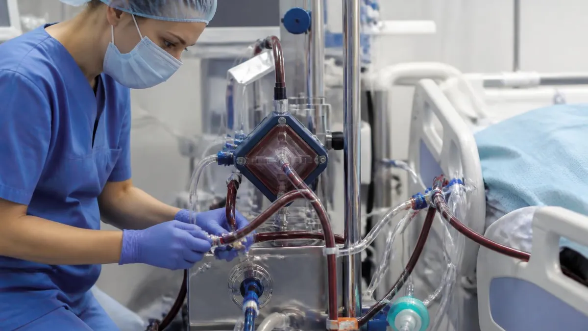
VA vs VV cannulation routes
Drainage cannula in the right common femoral vein with the tip parked at the cavoatrial junction in the inferior vena cava. Return cannula in the right internal jugular vein, tip at the entrance of the right atrium. Two-site configuration is the most common adult setup, used wherever ARDS or other isolated respiratory failure needs full lung rest. Sizes typically run 21 to 25 French drainage and 21 to 23 French return.
Single 27 to 31 French double-lumen catheter placed through the right internal jugular vein under fluoroscopic or echocardiographic guidance. Drainage holes pull from both superior and inferior vena cava. Separate return lumen jets oxygenated blood directly toward the tricuspid valve. Reduces line burden, allows ambulation in awake patients, but is more expensive and demands more precise placement than two-site cannulation.
Drainage cannula in the right common femoral vein, tip at cavoatrial junction. Return cannula in the right or left common femoral artery, threaded retrograde so the tip sits in the abdominal aorta. Fast cannulation, can be done at the bedside under local anesthesia. Risk of limb ischemia distal to the arterial cannula is high enough that most centres also place a small distal perfusion catheter into the superficial femoral artery.
Drainage cannula directly into the right atrium, return cannula directly into the ascending aorta, placed through an open chest. Used most often when the patient cannot wean from cardiopulmonary bypass at the end of cardiac surgery, or in salvage situations where peripheral access is impossible. Excellent drainage and antegrade aortic flow but commits the patient to remaining sedated with chest open until decannulation.
VV ECMO — supporting the lungs alone
VV ECMO is the simpler mode and the more common one in adult medical ICUs. The drainage cannula goes into a large central vein — usually the right common femoral vein, threaded up so the tip sits in the inferior vena cava just below the right atrium. A second cannula returns the oxygenated blood to a different vein, classically the right internal jugular, with its tip aimed at the entrance of the right atrium.
Some centres use a single double-lumen Avalon catheter placed through the right internal jugular: drainage holes pull from both venae cavae and a separate return lumen jets oxygenated blood directly at the tricuspid valve. Single-cannulation reduces line-related complications and lets the patient sit up and ambulate, but it requires fluoroscopic or echo-guided placement and is more expensive.
What VV ECMO does not do is push blood around the body. The patient's own heart still has to pump. So the indication for VV is severe respiratory failure in a patient whose heart is reasonably intact. The textbook case is ARDS that does not respond to lung-protective ventilation, prone positioning, and neuromuscular blockade — a Murray score above three, refractory hypoxemia with PaO2/FiO2 below 80, or hypercapnic respiratory acidosis the ventilator can no longer manage without harming the lungs further.
COVID-19 pneumonia pushed enormous volumes of patients onto VV ECMO between 2020 and 2022. Severe community-acquired pneumonia, influenza, drowning, smoke inhalation, status asthmaticus, pulmonary contusion, and bridge-to-lung-transplant patients also live in VV territory.
Set up correctly, VV ECMO offloads the lungs while the team dials the ventilator down to true rest settings — tidal volumes around three to four millilitres per kilogram of ideal body weight, plateau pressures below twenty-five, PEEP set to keep the lung open rather than to oxygenate. The arterial oxygen saturation on VV typically sits between 85 and 92 percent, which startles teams new to ECMO.
That's normal: the post-oxygenator blood mixes with whatever native cardiac output passes through the diseased lungs, so the systemic SpO2 reflects a blend, not the post-oxygenator number. Tissue oxygen delivery stays adequate because cardiac output is preserved and the hemoglobin is well-oxygenated where it counts.
Indications by mode
Severe ARDS that fails lung-protective ventilation, prone positioning, and neuromuscular blockade. Refractory hypoxemia with PaO2/FiO2 ratio below 80. Refractory hypercapnia with respiratory acidosis the ventilator can no longer manage. COVID-19 pneumonia (the dominant indication 2020 to 2022). Influenza pneumonia, community-acquired pneumonia, drowning, smoke inhalation, status asthmaticus. Pulmonary contusion after trauma. Bridge-to-lung-transplant in awake, ambulatory candidates. Pre-conditions: cardiac function intact on echocardiogram, no severe coagulopathy, no contraindication to systemic anticoagulation, reasonable prognosis if lungs can be rested.
VA ECMO — supporting heart and lungs together
VA ECMO is the bigger intervention. The drainage cannula again goes into a large vein, usually the right common femoral vein with the tip at the cavoatrial junction. But the return cannula goes into a large artery — most often the right common femoral artery, threaded retrograde so the tip sits in the abdominal aorta. Blood comes out of the body venous, gets oxygenated, and is pushed backwards up the aorta. That bypasses the heart and the pulmonary circulation entirely. The pump is now doing what the left ventricle is too weak to do.
That makes VA ECMO the rescue therapy for cardiogenic shock — refractory pump failure where inotropes, vasopressors, and an intra-aortic balloon pump or Impella have not been enough. Acute decompensated heart failure, fulminant myocarditis, massive pulmonary embolism with right-heart failure, post-cardiotomy shock when the patient cannot come off cardiopulmonary bypass, and refractory cardiac arrest all sit in VA territory.
Extracorporeal cardiopulmonary resuscitation — ECPR — is the dramatic edge case: an arresting patient gets cannulated during ongoing CPR, often within thirty to sixty minutes of arrest, in the hope that flow restored through the circuit buys time to find and treat the underlying cause.
Central VA ECMO is a separate beast. Instead of femoral cannulation, the surgeons place cannulas directly into the right atrium and the ascending aorta through an open chest — usually because the patient is already on cardiopulmonary bypass in the OR and cannot be weaned. Central cannulation gives better drainage and antegrade aortic flow but commits the patient to remaining sedated and chest-open until decannulation. Peripheral femoral cannulation is faster and lets the patient stay (slightly) more mobile, but introduces the harlequin and limb-ischemia problems described next.
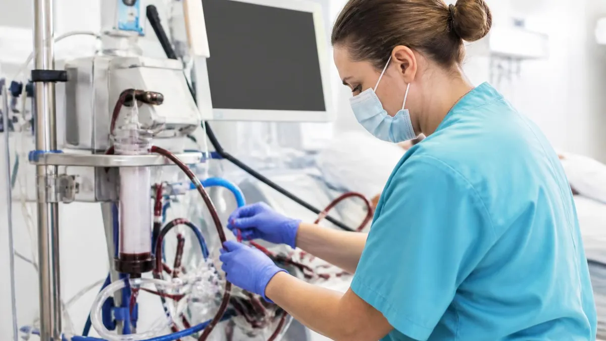
In peripheral VA ECMO, the femoral artery return runs retrograde up the descending aorta. If the patient's own left ventricle is still ejecting poorly-oxygenated blood (sick lungs, normal heart), that blood flows antegrade out the aortic root. Where the two streams collide, in the arch or descending aorta, is the watershed. Coronaries, brain, and right arm sit above it and get the patient's own poorly-oxygenated native flow. Lower limbs and viscera get well-oxygenated circuit blood. Right-hand SpO2 reads 78 percent while the foot reads 99. Standard practice: pulse oximeter on the right hand, arterial line in the right radial, cerebral oximetry on the forehead. Discordance triggers conversion to VAV or escalation of native lung treatment.
The differences that matter at the bedside
The core split is whether the circuit provides circulatory support. VA gives full mechanical circulatory support — the pump can deliver three to five litres per minute against systemic resistance, which is essentially complete replacement of cardiac output. VV gives none. If the heart stops in a VV patient, the circuit keeps oxygenating blood, but nothing pushes it around. Code algorithms run unchanged. In a VA patient, the circuit keeps perfusing the brain and viscera even with no native cardiac output at all — provided drainage and return remain intact.
Mixing in the aorta is the other characteristic VA problem. Femoral arterial return shoots oxygenated blood retrograde up the descending aorta. If the patient's native left ventricle is still ejecting and the lungs are diseased, that ejected blood is poorly oxygenated and flows antegrade down the aortic arch. The two streams meet somewhere in the aortic root or arch — the watershed. Above the watershed (head, right arm, coronaries) the patient gets the patient's own poor-quality blood.
Below it, well-oxygenated circuit blood. The right arm pulse-ox reads low, the left foot reads high. That is harlequin syndrome, sometimes called north-south syndrome. It's not subtle once you look for it: routine practice is to monitor SpO2 on the right hand, draw arterial blood gases from a right radial line, and watch cerebral oximetry on the forehead. Discordant numbers between right arm and femoral are the classic giveaway.
Cannula sizes are bigger on VA than VV, because the artery has to take a return cannula that handles full cardiac-output flow without driving pressures dangerously high. Drainage cannulas run twenty-one to twenty-five French in adults; return cannulas, fifteen to nineteen French arterial or twenty-one to twenty-three venous.
Limb ischemia distal to the femoral arterial cannula — the cannula effectively occludes the artery — is common enough that most centres place a small distal perfusion catheter into the superficial femoral artery at the time of cannulation, fed from a Y off the return limb. Without it, the leg below the cannula has minutes to hours before irreversible damage starts.
Daily ECMO bedside checks
- ✓ACT or anti-Xa heparin level within target band; review trend over preceding 12 hours.
- ✓Pre- and post-oxygenator pressures - rising delta means oxygenator clot or fibre fouling.
- ✓Post-oxygenator blood gas - confirms FiO2 and sweep gas working as set.
- ✓Plasma-free hemoglobin daily - rising number means hemolysis from kink, clot, or pump speed.
- ✓Inspect circuit visually for clot streaks at oxygenator inlet, pump head, and connectors.
- ✓Cannula site dressings clean, dry, no bleeding, no expanding hematoma.
- ✓Limb perfusion check on VA - pulse, colour, capillary refill, Doppler if uncertain.
- ✓Daily echocardiogram - aortic valve opening in VA, RV function in VV.
- ✓Right-hand SpO2 and right radial blood gas in VA - screen for harlequin syndrome.
- ✓Review goals of care daily once duration exceeds five days - recovery, bridge, or withdraw.
Complications — what the team watches for
Both modes share circuit complications: oxygenator failure (rising pre-oxygenator pressure or falling post-oxygenator PaO2), pump-head thrombosis (visible streaks of clot, dropping flows at the same RPM), heparin-induced thrombocytopenia, line-site bleeding, and severe hemolysis when the pump speed climbs to overcome a kinked cannula. Plasma-free hemoglobin above fifty milligrams per decilitre is the warning shot — above a hundred, it usually means a circuit change.
Intracranial hemorrhage is the most feared neurological complication and is more common in patients who arrived already anticoagulated, who had cardiac arrest before cannulation, or who run for many days. Gastrointestinal bleeding, retroperitoneal hematoma at the femoral access site, and surgical-wound bleeding all follow from the same anticoagulation requirement.
VV has one complication essentially unique to it: recirculation. If the drainage cannula and the return cannula tips sit too close to each other inside the right atrium, freshly oxygenated blood from the return cannula gets sucked straight back into the drainage cannula instead of perfusing the body. Recirculation fraction can climb above thirty percent in poor configurations. The signal is well-oxygenated drainage blood (bright red rather than dark) and persistently low systemic SpO2 despite high circuit flows and high sweep. Pulling the drainage cannula back a few centimetres or reorienting an Avalon catheter usually fixes it.
VA has limb ischemia, harlequin, and left-ventricular distension. The third one is subtle but serious: VA ECMO pumps blood backward up the aorta, raising afterload on a left ventricle that may already be stunned and unable to eject against the new pressure. If the LV cannot open the aortic valve to eject the small volume of blood it's still receiving from bronchial circulation, blood pools and the ventricle dilates.
Stagnant blood clots. Pulmonary edema worsens because there's no antegrade outflow. The fix is venting — an Impella, an atrial septostomy, or a direct LV vent. Daily transthoracic or transesophageal echocardiograms looking for aortic valve opening and LV size are routine in any VA program.
Outcomes — the numbers the registry tells us
The Extracorporeal Life Support Organization — ELSO — runs the international registry that anchors most ECMO outcome data. Survival depends heavily on the indication, the patient's pre-cannulation condition, and the centre's volume. Some rough numbers, current as of recent ELSO summary reports:
- VV ECMO for adult respiratory failure (mostly ARDS): roughly 60 percent survival to hospital discharge in well-selected patients. COVID-era cohorts ran lower, dropping to 50 percent or less in the worst surges as candidate selection broadened and durations stretched longer.
- VA ECMO for adult cardiac failure (cardiogenic shock, refractory heart failure): 30 to 50 percent survival to discharge, with patients cannulated for fulminant myocarditis at the upper end and post-cardiotomy failure at the lower end.
- ECPR — VA cannulated during ongoing CPR for refractory cardiac arrest: 20 to 30 percent survival to discharge, with most survivors maintaining good neurological function only if cannulation was achieved within a tight window (low-flow time under sixty minutes).
- Paediatric and neonatal numbers run higher than adult numbers across most indications — congenital diaphragmatic hernia, meconium aspiration, and persistent pulmonary hypertension of the newborn all post survival rates above 75 percent in high-volume neonatal programs.
Volume matters. Centres running more than thirty adult cases a year consistently post better outcomes than centres running fewer than ten, after risk-adjustment. That is the policy argument for regionalising ECMO into level-one trauma and cardiac-surgery centres rather than spreading it thinly across smaller hospitals. Most ECMO programs require ELSO membership and submission of patient-level data to the registry — partly for benchmarking, partly to feed the research base.
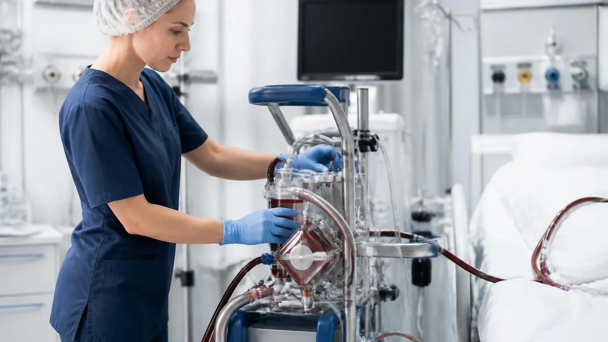
ECMO outcomes - key numbers
Who is at the bedside
Runs the circuit minute to minute. Sets sweep gas and flow targets ordered by the intensivist, draws post-oxygenator gases, watches pre- and post-oxygenator pressures, troubleshoots alarms. Carries ELSO ECMO Specialist certification with annual recertification including water-drill emergencies (air entrainment, oxygenator failure, decannulation). On-call coverage 24/7 in active programs.
Manages the patient as a whole: sedation, fluid balance, antibiotics, vasoactive drugs, ventilator settings, daily decision on goals of care. Orders pump and sweep targets, decides on conversion between modes, leads the multidisciplinary discussion about exit strategy. In most adult programs the intensivist also performs femoral cannulation under ultrasound guidance.
Performs central cannulation when needed, manages limb-ischemia complications (distal perfusion catheter placement, fasciotomy if it progresses), troubleshoots access-site bleeding, performs decannulation. Stays involved in surgical-bridge cases (LVAD implantation, transplant, post-cardiotomy management). In most centres the cardiac surgery service co-manages every VA case.
Daily transthoracic or transesophageal echocardiograms to assess aortic-valve opening and LV size in VA cases, RV function and tricuspid regurgitation in VV cases. Drives the decision to wean or escalate: recovering LV function in VA, falling pulmonary artery pressures in VV. Selects candidates for durable LVAD or transplant when bridge therapy is the plan.
The team behind the circuit
An ECMO program does not run on one specialty alone. Intensivists or cardiothoracic surgeons place the cannulas. Perfusionists or trained ECMO specialists manage the circuit at the bedside — setting sweep and flow, drawing post-oxygenator gases, watching for clot. Critical-care nurses manage the patient, since most of the day-to-day medication titration and tube management is theirs. Cardiology runs daily echocardiograms looking at LV recovery in VA cases or right-heart strain in VV cases. Pharmacy titrates anticoagulation. Physical therapy starts mobility work when stability allows — even ambulating awake VV patients on dual-lumen cannulas, which sounds impossible until you see it.
Training pipelines are formal. The ELSO ECMO Specialist course is a multi-day program with didactics, water drills (running the pump and oxygenator on a saline-filled test loop), and emergency drills (responding to air entrainment, oxygenator failure, decannulation). Annual recertification involves both written testing and hands-on circuit emergencies under instructor supervision. The ECMO practice test PDF walks candidates through the core knowledge domains the exam expects — circuit physics, troubleshooting, anticoagulation targets, and the differences between modes that this article is built around.
Becoming an ECMO specialist usually means at least two years of ICU or CT-ICU bedside experience first, then formal training, then a supervised orientation in the local program before pump independence. Pay scales reflect the responsibility — in most US programs, ECMO specialists carry on-call hours in addition to scheduled shifts and earn premium rates while assigned to active circuits.
VA vs VV ECMO - the trade-off
- +VV: lower cannula complication rates, no harlequin syndrome risk, supports awake ambulation with dual-lumen catheter.
- +VV: lungs can be rested at ultra-low ventilator settings, reducing further injury during recovery.
- +VA: full mechanical circulatory support - flow is preserved even with no native cardiac output.
- +VA: enables ECPR and rescues patients otherwise dying of cardiogenic shock.
- +VA: bridges to durable LVAD, transplant, or recovery in patients with reversible heart failure.
- −VV: no circulatory support - if the heart stops, the circuit cannot push blood around.
- −VV: recirculation problem if cannula tips sit too close inside the atrium.
- −VA: harlequin syndrome from differential oxygenation in the aortic arch.
- −VA: limb ischemia distal to the femoral arterial cannula, requiring distal perfusion catheter.
- −VA: left ventricular distension from increased afterload, sometimes needing Impella or vent.
Pros and Cons at a Glance
| Pros | Cons |
|---|---|
| VV: lower cannula complication rates, no harlequin syndrome risk, supports awake ambulation with dual-lumen catheter. | VV: no circulatory support - if the heart stops, the circuit cannot push blood around. |
| VV: lungs can be rested at ultra-low ventilator settings, reducing further injury during recovery. | VV: recirculation problem if cannula tips sit too close inside the atrium. |
| VA: full mechanical circulatory support - flow is preserved even with no native cardiac output. | VA: harlequin syndrome from differential oxygenation in the aortic arch. |
| VA: enables ECPR and rescues patients otherwise dying of cardiogenic shock. | VA: limb ischemia distal to the femoral arterial cannula, requiring distal perfusion catheter. |
| VA: bridges to durable LVAD, transplant, or recovery in patients with reversible heart failure. | VA: left ventricular distension from increased afterload, sometimes needing Impella or vent. |
Which mode for which patient — the bedside flow
The choice between VA and VV is rarely subtle in real cases. The question the team asks is whether the patient's circulatory failure is driven by the lungs (and therefore correctible by oxygenating the blood and unloading the ventilator) or by the heart (and therefore needing mechanical pump support). A patient with severe ARDS, preserved cardiac function on echocardiogram, and refractory hypoxemia goes on VV.
A patient in cardiogenic shock with poor LV function and rising lactate despite three vasoactive drips goes on VA. A patient with both severe ARDS and a stunned heart sometimes gets VA initially, with the team watching for harlequin signs and considering conversion to VV (or VAV — veno-arterio-venous) if pure cardiac support is no longer needed.
Conversion between modes happens often enough that programs train for it specifically. VA-to-VV conversion when the heart recovers but the lungs lag behind is common after fulminant myocarditis. VV-to-VA conversion when previously normal cardiac function fails partway through a long ARDS run is rarer but real. Each conversion involves placing a new cannula, leaving an old cannula in place temporarily as a bridge, or formal recannulation in the OR — not an outpatient decision.
Duration matters too. VV runs in adults frequently stretch to two or three weeks for severe COVID-era ARDS, with some bridge-to-transplant patients running ninety days or more. VA runs are usually shorter — five to fourteen days — either because the heart recovers, the patient is transitioned to a durable LVAD, listed for transplant, or in honest cases, ECMO is withdrawn because recovery is not coming.
Long VA runs accumulate complications quickly. The decision-making conversation about goals of care while the patient is on a VA circuit is one of the harder conversations in critical care medicine, and most ECMO programs have palliative-care colleagues embedded for that reason.
Exam-prep angle — what board questions test
If you're preparing for the CCRN, the ECMO specialist exam, or a critical-care board, the question stems tend to repeat. They test whether you can pick the configuration from a clinical vignette (cardiogenic shock plus pulmonary edema versus refractory ARDS with preserved EF), whether you recognise harlequin syndrome from a pulse-ox discrepancy, whether you know that recirculation is a VV problem and limb ischemia is a VA problem, and whether you can read a pre-oxygenator pressure trace and tell the difference between drainage trouble and circuit clot.
You'll also be asked about anticoagulation targets, sweep-gas effect on PaCO2 (more sweep means more CO2 removal at the same blood flow), the difference between FiO2 on the blender (post-oxygenator blood) and FiO2 on the ventilator (native lung), and what specifically you change first when a VV patient desaturates — usually sweep gas FiO2, then circuit flow, then recirculation troubleshooting, before you ever touch the ventilator. Take a practice run to get familiar with the question style.
Sample ECMO - Extracorporeal Membrane Oxygenation Practice Questions
Try these questions from our free ECMO - Extracorporeal Membrane Oxygenation practice tests. The correct answer and an explanation follow each question.
A patient on a stable femoro-jugular VV ECMO configuration for severe pneumonia develops acute hypoxemia (SpO2 drops from 95% to 86%). The ECMO circuit is functioning well, with a high post-oxygenator PaO2 and no signs of recirculation. Which patient-related factor is the most likely cause of this deterioration?
- A. A sudden increase in the patient's native cardiac output.
- B. Development of oxygenator membrane failure.
- C. A decrease in the sweep gas flow rate.
- D. Migration of the reinfusion cannula.
Answer: A. A sudden increase in the patient's native cardiac output.
In VV ECMO, the patient's arterial oxygenation is determined by the mixture of oxygenated blood from the ECMO circuit and the deoxygenated blood that passes through the native lungs (shunt). If the patient's cardiac output increases (due to fever, sepsis, agitation, etc.), a larger proportion of blood bypasses the ECMO circuit and goes through the diseased lungs. This larger shunt of deoxygenated blood mixes with the ECMO blood, resulting in a lower systemic arterial oxygen saturation. Oxygenator failure or cannula migration are circuit issues, and a decrease in sweep gas would primarily affect CO2 removal.
A patient on VA-ECMO develops low ECMO flows with high RPM, and the circuit pressure alarm activates. After ruling out hypovolemia, the next assessment should be:
- A. Increase sweep gas flow
- B. Inspect for cannula kinking or positional obstruction
- C. Reduce heparin infusion
- D. Increase ventilator PEEP
Answer: B. Inspect for cannula kinking or positional obstruction
High RPM with low flows and elevated pressures despite adequate volume suggests mechanical obstruction such as cannula kinking, migration, or outflow obstruction requiring immediate inspection.
On VV ECMO, a patient's SaO2 is 88% with ECMO flow at 5 L/min. The native cardiac output is estimated at 8 L/min. What is the approximate recirculation fraction?
- A. 10%
- B. 25%
- C. 37%
- D. 50%
Answer: C. 37%
Recirculation fraction = (ECMO flow – effective flow) / ECMO flow; with total venous return mixed with ECMO flow, approximately 37% recirculation yields this oxygen delivery.
A patient fails multiple VA-ECMO weaning trials over 14 days. The ECMO team should next consider:
- A. Indefinite continuation of ECMO support
- B. Evaluation for durable ventricular assist device implantation or heart transplantation
- C. Switching to VV-ECMO to rest the heart further
- D. Increasing anticoagulation intensity to prevent circuit thrombosis
Answer: B. Evaluation for durable ventricular assist device implantation or heart transplantation
Repeated weaning failure after 2 weeks indicates the heart is unlikely to recover; escalation to a durable VAD or transplant evaluation is the appropriate next step.
Take the full ECMO - Extracorporeal Membrane Oxygenation practice test
ECMO Questions and Answers
About the Author

Educational Psychologist & Academic Test Preparation Expert
Columbia University Teachers CollegeDr. Lisa Patel holds a Doctorate in Education from Columbia University Teachers College and has spent 17 years researching standardized test design and academic assessment. She has developed preparation programs for SAT, ACT, GRE, LSAT, UCAT, and numerous professional licensing exams, helping students of all backgrounds achieve their target scores.
Join the Discussion
Connect with other students preparing for this exam. Share tips, ask questions, and get advice from people who have been there.
View discussion (9 replies)