MRI Full Form — Complete Guide (2026)
MRI full form is Magnetic Resonance Imaging. How it works, what it scans, cost, safety, fMRI, MRA, and how it differs from CT and X-ray (2026).
MRI Full Form — Complete Guide (2026)
Short answer up front: the MRI full form is Magnetic Resonance Imaging. Three words. No hidden meaning, no Latin root — the name describes exactly what the machine does. It uses a magnet, it watches how your body resonates inside that magnet, and it produces an image from the signal.
That's the literal expansion. The interesting question isn't what the letters stand for — it's why a magnetic field can take a picture of soft tissue better than any other tool a hospital owns. The honest answer involves hydrogen, radio waves, and a Nobel Prize. Stick around for the rest of this guide and the machine inside the donut tube stops feeling mysterious.
The word "resonance" is the part most people gloss over. It refers to a physics phenomenon where protons inside your hydrogen atoms tip and recover when hit with a specific radio frequency — and that recovery emits a signal a coil can listen to. No radiation. No needles for most scans. Just magnets and radio. That's why an mri scan can show a torn ligament, a tiny brain lesion, or a slow-growing tumor with detail X-ray can't touch.
This guide walks the full form, the science, the history, the comparisons against CT and X-ray, the common body parts scanned, the specialized variants (fMRI, MRA), safety rules around metal and pacemakers, current 2026 pricing, and what your appointment actually feels like. If you're scheduled for one next week or studying for a tech exam, everything you need is below.
The Full Form, Decoded
MRI = Magnetic Resonance Imaging. A non-invasive scan that uses a powerful magnetic field and pulses of radio waves to create detailed images of soft tissue, organs, and joints.
- M: Magnetic — a superconducting magnet, usually 1.5 Tesla or 3.0 Tesla
- R: Resonance — hydrogen protons in your body align with the magnet, then tip and "resonate" when hit by radio pulses
- I: Imaging — coils detect the signal as protons relax back; software reconstructs cross-sectional images
- No ionizing radiation — unlike X-ray or CT, MRI uses zero radiation
- Best for soft tissue — brain, spinal cord, ligaments, muscles, organs, blood vessels
- Awarded the 2003 Nobel Prize in Medicine to Paul Lauterbur and Sir Peter Mansfield
Breaking Down Each Letter
The scanner's superconducting magnet is the heart of the machine.
- Field strength: Typically 1.5T or 3.0T (Tesla); research scanners reach 7T
- Comparison: Earth's field is ~50 microtesla — a 3T magnet is roughly 60,000x stronger
- Stays on: The magnet is never turned off — it runs 24/7, even between scans
Hydrogen protons align with the magnet, then absorb and re-emit radio energy.
- Target: Hydrogen nuclei (single protons) — abundant in water and fat
- Frequency: Larmor frequency — 63.87 MHz at 1.5T, 127.74 MHz at 3T
- What you hear: The loud knocking sound is the gradient coils switching on and off
A receiver coil captures the signal and software reconstructs slices.
- Output: Cross-sectional slices in axial, sagittal, and coronal planes
- Sequences: T1, T2, FLAIR, DWI, STIR — each highlights different tissue properties
- Resolution: Submillimeter detail in soft tissue — far better than X-ray or ultrasound
How an MRI Actually Works
Your body is roughly 60% water. Water is two hydrogens and one oxygen. Each hydrogen atom has a single proton in its nucleus — and that proton acts like a tiny spinning magnet. Normally those proton magnets point in random directions and cancel out.
Slide into the MRI bore and that randomness ends. The scanner's powerful field forces a small majority of your hydrogen protons to align with the magnet's axis. You feel nothing. The shift is happening at an atomic scale.
The radio pulse
Now the machine sends a radio frequency (RF) pulse at exactly the right frequency — the Larmor frequency — to tip those aligned protons sideways. Think of pushing a child on a swing at exactly the right moment. When the pulse stops, the protons spring back toward alignment with the main magnet. As they relax, they release the energy they absorbed as a radio signal of their own. That signal is what the receiver coil picks up.
Different tissues relax at different rates. Fat relaxes fast. Water relaxes slow. A bruise releases its signal differently than healthy muscle. Software measures these timing differences and translates them into pixel intensity. Bright on T1, dark on T2 — the brightness pattern is what a radiologist reads.
Gradients turn signal into a picture
One radio pulse alone would give you a single number from your whole body — useless. The trick is gradient coils. Three additional smaller magnets switch on and off in pulses, creating tiny variations in the main field along the X, Y, and Z axes. That spatial variation is what tells the scanner whether the signal came from your left knee or your right ear. The knocking sound during a scan? That's the gradient coils physically vibrating as they switch.
Pull all of that together and you get a stack of 2D slices — sometimes hundreds — that a radiologist scrolls through. Modern mri machine models reconstruct full 3D volumes you can rotate on screen. None of it requires a single X-ray. The whole image-making process happens with magnets and radio.
Where MRI Came From: Lauterbur and Mansfield
Magnetic resonance as a physics phenomenon was discovered in 1946 by Felix Bloch and Edward Purcell — they shared the 1952 Nobel Prize in Physics for nuclear magnetic resonance (NMR) spectroscopy. Useful, but it could only analyze test tubes, not living tissue.
The leap to imaging came in 1973. Paul Lauterbur, an American chemist at Stony Brook, published a paper in Nature showing he could use magnetic gradients to encode spatial position into the NMR signal. He called the technique "zeugmatography" — a name that didn't stick. He produced the first 2D image of two water-filled test tubes side by side.
Sir Peter Mansfield, a British physicist at the University of Nottingham, picked up the work and developed mathematical methods to acquire images far faster — fast enough to be clinically useful. He pioneered echo-planar imaging (EPI), the technique still used today for high-speed scans of moving organs like the heart.
The two shared the 2003 Nobel Prize in Physiology or Medicine for their discoveries. By that point MRI was already in every major hospital — about 22,000 scanners worldwide and roughly 60 million scans per year. The Nobel committee called it a "breakthrough in medical diagnostics and research." Hard to overstate. Before MRI, the only way to see soft-tissue detail inside a living person was exploratory surgery.
MRI vs Other Imaging Methods
What it shows: CT excels at bone, lungs, and acute bleeding. MRI excels at soft tissue, brain, spinal cord, ligaments, and tumors.
Radiation: CT uses ionizing X-rays — a single abdominal CT delivers roughly 10 millisieverts, equivalent to about three years of background radiation. MRI uses zero radiation. None. That's the headline difference for pediatric and pregnancy imaging.
Speed: CT wins. A full CT scan takes 30 seconds to a few minutes. MRI typically runs 30–60 minutes depending on the body part. CT is the tool of choice for emergency trauma — fast, broad, finds bleeds quickly.
Cost: CT runs $300–$1,500 typical. MRI runs $1,000–$3,500 typical. See mri vs ct scan for the full breakdown of when each is ordered.
What Doctors Use MRI to Scan
MRI is the imaging tool of choice anywhere soft tissue matters more than bone. That covers a long list of body parts, but six categories handle most of the volume.
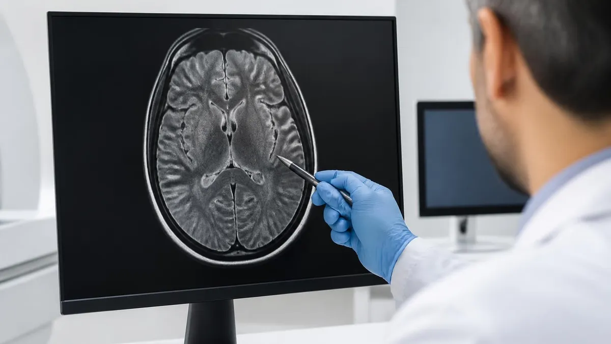
Brain and spine
Brain MRI is the workup for stroke, multiple sclerosis, tumors, dementia, headaches that won't resolve, and unexplained neurological symptoms. A brain mri can detect lesions as small as 2–3 millimeters — far smaller than CT can resolve. MS plaques, micro-bleeds, and early tumors often show up only on MRI. Spinal MRI is the gold standard for herniated discs, spinal stenosis, cord compression, and nerve root impingement. If you've ever been sent for a back MRI after a few weeks of leg pain, this is why.
Joints and musculoskeletal
Orthopedists order MRI for ligament tears, meniscus injuries, rotator cuff problems, labral tears in the shoulder and hip, stress fractures, and bone marrow edema. Plain X-ray shows bone position but cannot show the soft tissue holding it together. A knee mri after a sports injury distinguishes an ACL sprain from a complete tear — which decides whether you operate.
Heart, breast, and prostate
Cardiac MRI evaluates heart muscle damage after a heart attack, congenital defects, cardiomyopathy, and the cause of arrhythmias. A cardiac mri gives functional information no other imaging tool delivers as well — ejection fraction, scar tissue location, viability of damaged muscle. breast mri is used for high-risk screening (BRCA carriers), evaluating known cancers before surgery, and looking at silicone implants. Prostate MRI guides biopsies and stages cancer once detected.
Abdomen and pelvis
Liver lesions, pancreatic masses, kidney tumors, adrenal nodules, uterine fibroids, ovarian cysts, and pelvic floor disorders all fall under MRI's domain. MRCP — magnetic resonance cholangiopancreatography — is a non-invasive way to image the bile ducts and pancreatic duct, replacing the older, riskier ERCP for diagnosis.
MRI by the Numbers
Specialized MRI: fMRI and MRA
Plain MRI maps anatomy. Two important variants map something more interesting — brain activity and blood vessels — using the same hardware with different acquisition sequences.
fMRI — Functional MRI
Functional MRI doesn't show structure. It shows which parts of your brain are working harder right now. The technique relies on a quirk called BOLD — Blood Oxygen Level Dependent contrast. When a brain region activates, local blood flow increases more than oxygen demand, so the ratio of oxygenated to deoxygenated hemoglobin shifts. That shift changes the magnetic properties of blood, which fMRI detects.
The result: a colored heat map laid over brain anatomy showing which regions light up when you wiggle your fingers, listen to music, read words, or imagine a face. Neurosurgeons use functional mri to map language and motor areas before tumor surgery so they can avoid cutting critical functional tissue. Researchers use it for everything from addiction studies to pain mapping. fMRI scans take 30–90 minutes and require the patient to perform tasks inside the bore.
MRA — Magnetic Resonance Angiography
MRA images blood vessels — arteries, veins, and the cerebral circulation. Two methods exist. Time-of-flight MRA uses the natural signal of moving blood to create contrast without injecting anything. Contrast-enhanced MRA injects gadolinium and captures the arterial phase as the contrast travels through.
Common uses: detecting aneurysms in the brain, screening carotid arteries for stenosis, evaluating the renal arteries for fibromuscular dysplasia, and mapping vascular malformations. MRA replaces conventional catheter angiography for many diagnostic questions — no arterial puncture, no radiation, and the patient walks out 45 minutes later. For arterial procedures that need treatment in the same session, catheter angiography is still the tool of choice. For pure diagnosis, MRA usually wins.
MRI Safety: Metal, Implants, and Claustrophobia
An MRI is one of the safer scans in medicine — no radiation, low risk for most patients. But the magnet is always on, which means the safety rules are about what comes near the magnet, not the scan itself.
The metal rule
Ferromagnetic metal — iron, nickel, certain steels — flies toward the magnet at high speed if it enters the room. Hospital incidents involving oxygen tanks, scissors, and even floor buffers becoming projectiles are documented. Patients are screened with a detailed metal questionnaire before every scan. Removed metal: jewelry, watches, hairpins, hearing aids, dentures with metal parts, transdermal patches with metal foil, makeup containing iron oxides.
Implanted metal is more nuanced. Modern surgical hardware — titanium plates, most orthopedic screws, dental implants — is generally MRI-compatible at 1.5T and 3T. Older implants need verification. mri safety protocols require checking the manufacturer's documentation for any implant. Most cardiac pacemakers and defibrillators were once absolute contraindications; newer "MR Conditional" devices allow scans under specific protocols.
Other contraindications
Cochlear implants — usually contraindicated, though newer designs are conditional. Aneurysm clips placed before 1995 — risk of dislodgement; post-1995 titanium clips are safe. Bullet fragments — case-by-case. Insulin pumps — must be removed and left outside the room. Tattoos with old metallic inks can heat up; modern pigments are safer but flag the area on the questionnaire anyway.
Claustrophobia
Roughly 4–10% of patients experience claustrophobia inside the bore. The tube is narrow, the magnet is loud, and you can't move for 30–60 minutes. Options: low-dose anti-anxiety medication before the scan, a blindfold or eye covering, music through MRI-compatible headphones, or an open mri with a wider, more open design (though usually at a lower field strength, which means slightly lower image quality). For severe cases, sedation or even general anesthesia is available.
MRI: Strengths and Drawbacks
- +Zero ionizing radiation — safe to repeat as often as needed and the default choice for pediatric and pregnancy imaging
- +Best-in-class soft-tissue resolution — sees brain, cord, ligaments, organs better than any other tool
- +Multiple imaging sequences (T1, T2, FLAIR, DWI) reveal different tissue properties from a single scan
- +Non-invasive — no needles required for plain MRI, and the patient walks out the same day
- +Specialized variants (fMRI, MRA, MRS) extend the same hardware to brain activity, blood vessels, and metabolic chemistry
- +Multiplanar reconstruction — slice the body in any plane after a single acquisition
- −Expensive — typical US cost runs $1,000–$3,500 per scan, far above CT or X-ray
- −Slow — 30 to 60 minutes per scan limits emergency use compared to CT
- −Loud — gradient coil noise reaches 100+ dB, requiring earplugs or headphones
- −Claustrophobia is a real barrier for 4–10% of patients
- −Contraindicated with many older implants, certain pacemakers, and ferromagnetic foreign bodies
- −Less effective for bone fractures, lung tissue, and acute hemorrhage — CT wins for these
MRI Cost in 2026: What You Actually Pay
Sticker prices look terrifying. Hospital list prices for an MRI can hit $5,000 or more. What you actually pay depends on the body part, the facility, your insurance, and whether contrast is used. Real numbers for 2026:
Typical cash price ranges
Without insurance, plain MRI runs roughly $1,000–$3,500. Common ranges: brain $1,200–$3,200, knee or shoulder $700–$2,500, lumbar spine $1,000–$3,000, abdominal $1,500–$3,500, breast $1,800–$4,500. Contrast adds $100–$500 to any of these. The mri cost reference page has the full breakdown by body part and state.
Independent imaging centers — not hospital-based — usually run 30–60% cheaper than hospital outpatient departments for the same scan. Same equipment, same radiologist tier, dramatically different bill. Cash-pay deals are common at imaging-center chains; some publish flat rates of $400–$700 for a knee or shoulder.
With insurance
If you have insurance, your responsibility depends on your deductible, copay, and coinsurance. Common scenarios: you've met your deductible and pay 20% coinsurance — that's $200–$700 on a typical scan. You haven't met your deductible — you may pay the full negotiated rate (often $400–$1,500 even with insurance) until you hit it. High-deductible plans frequently mean MRIs are out-of-pocket events.
Medicare covers MRI when ordered for medical necessity. You pay 20% of the Medicare-approved amount after meeting Part B deductible. Medicare Advantage and supplemental plans further reduce out-of-pocket. Workers' comp and personal injury claims usually cover 100% with no patient cost.
How to lower the bill
Three things move the price most. First — ask for an independent imaging center referral rather than the hospital. Second — request the cash price before they bill insurance; sometimes cash is cheaper than your insurance's negotiated rate. Third — ask whether contrast is truly necessary, since omitting it saves a couple hundred dollars and avoids gadolinium concerns.
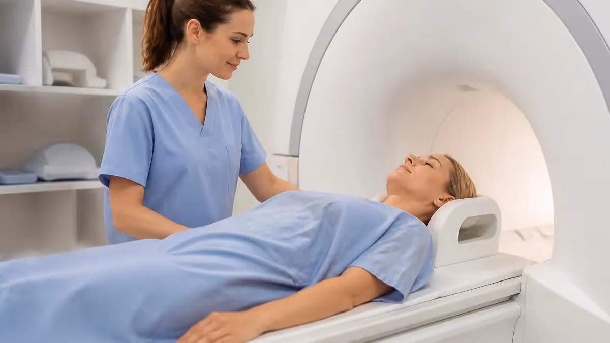
What Your MRI Appointment Actually Looks Like
Most people have never had one. Here's the play-by-play.
Before the scan
You arrive 15–30 minutes early. The tech runs the metal-and-implant questionnaire. You change into a gown — no metal zippers, no underwire, no jewelry, no watch, no phone. They double-check tattoos, piercings, dental work, and previous surgeries. If contrast is ordered, they place an IV.
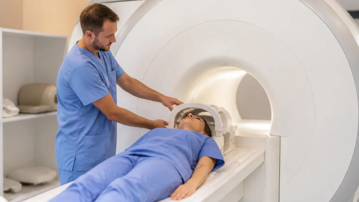
On the table
The tech positions you on a sliding table, head- or feet-first depending on the body part scanned. Foam wedges keep you still. They'll give you earplugs and often padded headphones — the gradient coils are loud, around 100 dB, like a jackhammer. A panic squeeze ball goes in your hand. Press it and the scan stops immediately.
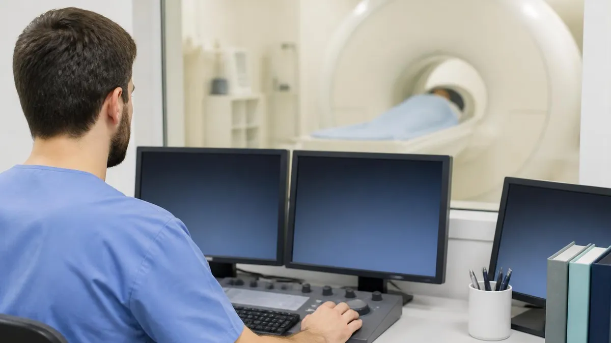
The table slides into the bore. You hear knocking and buzzing in patterns — different sounds for different sequences. The tech speaks to you through the intercom between scans, telling you what's next and how long each sequence will last. Most scans run 30 seconds to 4 minutes per sequence, with 4–10 sequences total.
Staying still matters
Even small movements blur the image. For brain and spine, hold completely still. For chest and abdomen, you'll get breath-hold instructions — "breathe in, hold, breathe out" — synchronized with the acquisitions. The scan ends when the tech says "that's the last one." You slide out, the IV comes out if you had contrast, and you change back into your clothes.
After
You drive yourself home unless you had sedation. The radiologist reads the images over the next 1–3 business days, sometimes faster for urgent cases. Results go to the ordering doctor, who shares them with you at a follow-up visit or through the patient portal.
MRI Appointment Prep Checklist
- ✓Bring a list of every surgery, implant, and medical device — manufacturer and date if possible
- ✓Remove all metal — jewelry, watch, hairpins, glasses, dentures with metal, hearing aids
- ✓No makeup or hairspray with metallic glitter on the day of scan
- ✓Wear easy-to-remove clothing without zippers, underwire, or metal buttons
- ✓Eat and drink normally unless told otherwise (some abdominal MRIs require fasting)
- ✓Bring earplugs if you're sensitive to loud noise — the facility provides them but extras are fine
- ✓If claustrophobic, ask about anti-anxiety medication or an open MRI option in advance
- ✓Stay hydrated if contrast is planned — helps your kidneys clear gadolinium afterward