
How Does an MRI Machine Work? — Complete Guide (2026 September)
🏆 How does MRI work? Magnetic field aligns hydrogen protons, RF pulse disrupts them, relaxation emits signals — computer builds the image. No
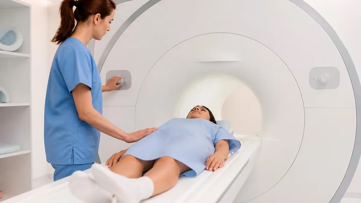
Walk into any hospital radiology department and you'll find one of medicine's most powerful diagnostic tools — a machine that sees inside the human body without a single dose of radiation. That's the MRI scanner. Short for Magnetic Resonance Imaging, it produces stunningly detailed images of soft tissue, organs, joints, and brain structures that X-ray and CT simply can't match.
So how does it actually work? The core principle sounds almost counterintuitive: an MRI machine uses a powerful magnetic field and radio waves — nothing more — to generate images. No ionizing radiation. No nuclear material. Just physics.
Here's the key insight: your body is mostly water. And water molecules contain hydrogen atoms. Each hydrogen atom has a single proton at its nucleus, and that proton behaves like a tiny compass needle — it has a magnetic moment, meaning it naturally wants to align with an external magnetic field.
When you slide into the bore of an MRI machines and that powerful magnet switches on, billions of hydrogen protons throughout your body align along the field. Then a precisely tuned pulse of radio waves knocks them out of alignment. When the pulse stops, those protons snap back — and as they do, they emit a faint radio signal. The machine captures that signal, and a computer reconstructs it into an image.
It sounds simple. In practice, the engineering is extraordinary. The magnet must maintain a field thousands of times stronger than Earth's. The radiofrequency system must pulse at exact frequencies. Gradient coils must shift the magnetic field rapidly to encode spatial information. And the reconstruction algorithms turn raw signal data into the crisp anatomical images radiologists use to diagnose strokes, torn ligaments, brain tumors, and hundreds of other conditions.
This guide breaks down the complete physics of MRI imaging — from the superconducting magnet to T1 and T2 weighting, Tesla strengths, contrast agents, and what all that noise is about. Whether you're studying for an MRI registry exam or just want to understand what happens during a scan, you're in the right place.
MRI's advantage over other modalities is stark when it comes to soft tissue. An X-ray shows bone beautifully — dense calcium attenuates the beam, producing bright white shadows on the detector. But soft tissue is nearly invisible. A CT scan improves on this with cross-sectional anatomy and faster acquisition, but soft-tissue contrast is still limited compared to what MRI achieves.
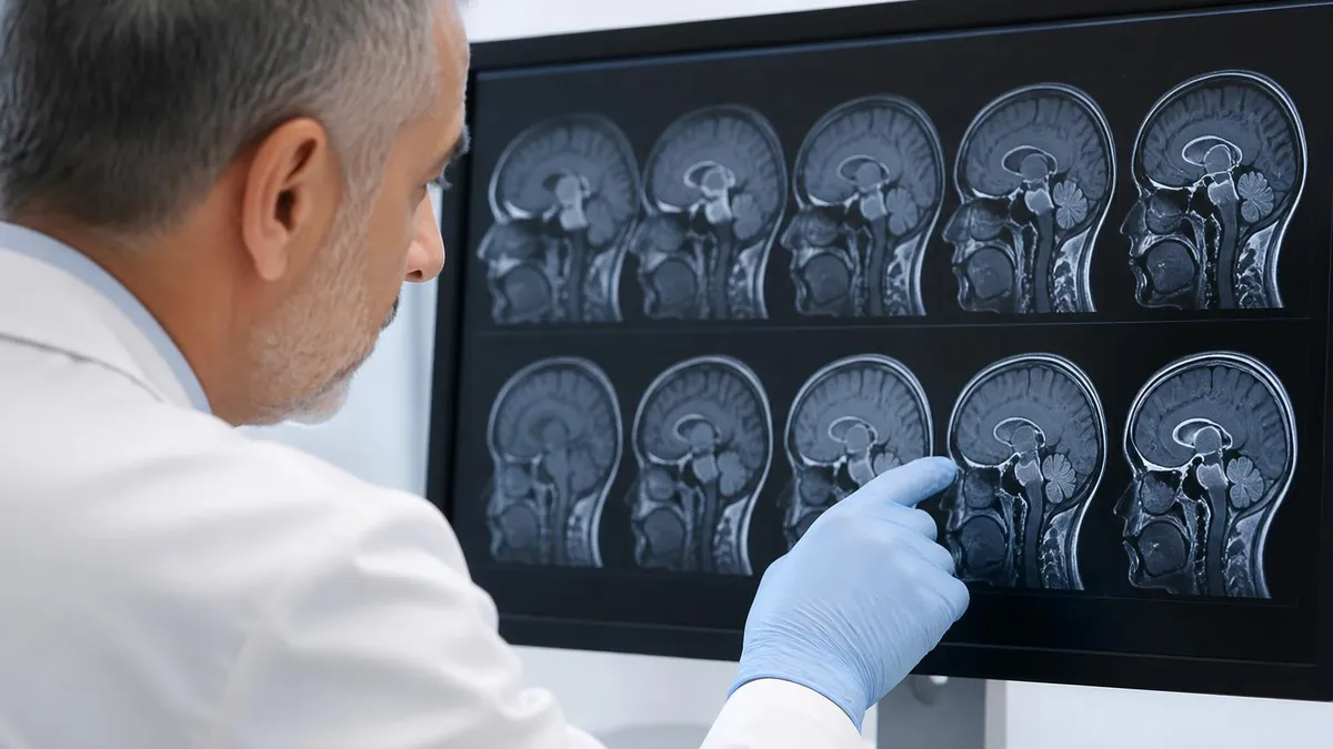
An MRI scanner, by exploiting the different magnetic properties of tissues, can distinguish white matter from gray matter in the brain, identify a torn ACL from intact ligament fibers, and detect subtle edema in a joint weeks before it would show on any other imaging modality.
The machine itself is a marvel of engineering — a doughnut-shaped structure roughly 1.5–2 meters in diameter and 1.5–2 meters long, dominated by a cylindrical bore through which the patient passes. Inside the walls of that bore lies a superconducting electromagnet wound from niobium-titanium wire, cooled by liquid helium to just a few degrees above absolute zero. Surrounding that are the gradient coils, radiofrequency coils, and the computer-controlled electronics that orchestrate the entire imaging sequence. Understanding each component reveals why MRI is both so powerful and so demanding to operate correctly.
MRI Key Facts
MRI uses a magnetic field and radio waves — no ionizing radiation. Standard clinical field strength: 1.5T. Higher field (3T) = better resolution but more noise and cost. The magnetic field is always on, even when no scan is running.
A superconducting electromagnet cooled to near absolute zero generates the static magnetic field that aligns hydrogen protons in the body.
- Temperature: ~4 Kelvin (−269°C) using liquid helium
- Field strength: 1.5T clinical standard; 3T for neuroimaging
- Always on: The field never turns off — even between patients
A radiofrequency pulse at the Larmor frequency tips protons out of alignment. When it stops, protons relax back, emitting a detectable signal.
- Larmor frequency: 63.87 MHz at 1.5T; 127.74 MHz at 3T
- T1 relaxation: Longitudinal — fat bright, fluid dark
- T2 relaxation: Transverse — fluid bright, good for pathology
Three sets of gradient coils (X, Y, Z) rapidly vary the magnetic field across the patient's body to encode spatial location of each signal.
- Function: Slice selection, frequency encoding, phase encoding
- Switching speed: Rapid on/off switching causes the loud banging noise
- Resolution: Faster, stronger gradients = higher spatial resolution
Raw signal data fills k-space — a mathematical grid. A Fourier transform converts k-space data into the final anatomical image.
- Data format: k-space (frequency domain)
- Transform: Inverse 2D/3D Fourier transform
- Output: T1, T2, FLAIR, DWI, MRA sequences and more
Step-by-Step: The Physics of MRI Imaging
Understanding MRI means following a proton through the entire scan process. Here's exactly what happens, from the moment you enter the bore to the moment the image appears on the radiologist's screen.
Step 1 — Magnetic Field Aligns the Protons
The scanner's superconducting magnet creates a powerful, uniform static magnetic field (B0) running along the length of the bore. Hydrogen protons in your body — in water, fat, and organic molecules — naturally have spin angular momentum. In the presence of B0, they precess around the field direction like tiny spinning tops. Most align parallel to the field, creating a net magnetization pointing along B0. The speed of that precession is the Larmor frequency, which depends directly on field strength. At 1.5T, it's about 63.87 MHz. At 3T, it doubles to roughly 127.74 MHz.
Why hydrogen? Because it's the most abundant element in the human body — predominantly in water (H₂O) and fat. Its single proton gives it a strong magnetic moment relative to other nuclei. You could theoretically image phosphorus or sodium, and some research systems do exactly that. But for clinical imaging, hydrogen is unmatched in signal strength and practical utility.
Step 2 — RF Pulse Disrupts Alignment
A radiofrequency (RF) transmitter coil fires a brief pulse of electromagnetic energy at exactly the Larmor frequency — this is the resonance in "magnetic resonance." The pulse tips the net magnetization away from B0. A 90° pulse tips it fully into the transverse plane. A 180° pulse flips it entirely. Once tipped, the protons precess in the transverse plane, generating a rotating magnetic field that induces a measurable voltage in receiver coils — the raw MRI signal.
The precision required here is remarkable. The RF pulse must match the Larmor frequency within a very narrow band — essentially a targeted resonance. Protons not at that frequency (at different field strengths) won't respond. This selectivity is what allows the scanner to image one slice at a time: apply a gradient to make the field slightly different at each position, then pulse at the frequency corresponding to the target slice's field strength. Only protons in that slice resonate.
T1 (longitudinal relaxation): How fast protons re-align with B0 after the RF pulse. Short T1 = fat. Long T1 = water. Short TR/TE pulse sequences emphasize T1.
T2 (transverse relaxation): How fast protons lose phase coherence with each other. Short T2 = muscle. Long T2 = fluid. Long TR/TE sequences emphasize T2.
Step 3 — Proton Relaxation Emits the Signal
After the RF pulse ends, protons begin relaxing back toward equilibrium. Two relaxation processes happen simultaneously. T1 relaxation (longitudinal) describes how quickly the magnetization recovers along B0. T2 relaxation (transverse) describes how quickly the transverse magnetization decays as protons lose phase coherence with each other. Different tissues have different T1 and T2 values — that's what gives MRI its extraordinary soft-tissue contrast.
Fat has a short T1 (relaxes quickly back to B0) and a short T2. Free water — like cerebrospinal fluid — has a long T1 and a long T2. Muscle, brain gray matter, and white matter each have characteristic values. By choosing the repetition time (TR) and echo time (TE) of the pulse sequence, the radiographer selects which tissue properties dominate the image contrast. Short TR/short TE emphasizes T1. Long TR/long TE emphasizes T2.
Step 4 — Gradient Coils Encode Spatial Position
Here's the clever part. A single RF pulse would generate one combined signal from the entire patient. Gradient coils — three orthogonal sets running in X, Y, and Z directions — superimpose small, controlled variations on the main field. Because Larmor frequency depends on field strength, protons at different positions now precess at slightly different frequencies. By systematically varying the gradients, the system performs slice selection (choose a plane), frequency encoding (encode position along one axis), and phase encoding (encode position along the perpendicular axis). The raw data fills a grid called k-space.
k-space is not an image — it's raw frequency-domain data. Each point in k-space holds information about the entire image. The outer regions of k-space contain high-frequency information (fine detail, edges). The center of k-space holds low-frequency information (overall contrast and brightness). Advanced techniques like parallel imaging (SENSE, GRAPPA) and compressed sensing exploit this structure to undersample k-space and reconstruct images faster — dramatically cutting scan times.
Step 5 — Computer Reconstructs the Image
An inverse Fourier transform converts k-space data from the frequency domain into image space — the familiar anatomical picture. The computer applies filters, adjusts windowing, and outputs the image in whatever weighting was selected (T1, T2, FLAIR, DWI, etc.). This entire cycle repeats many times per slice, per sequence. A complete brain MRI might involve tens of thousands of RF pulses and gradient switches. The history of how this technology evolved — from Raymond Damadian's first whole-body scanner in 1977 to today's 7T research machines — is traced in detail in the MRI machine history.
Modern MRI reconstruction increasingly uses AI and deep learning. Vendors now offer neural network-based denoising and acceleration (Siemens Deep Resolve, GE AIR Recon DL, Philips SmartSpeed) — these can reconstruct images from highly undersampled k-space data with quality that rivals or exceeds conventional reconstruction from fully sampled data. The result: scans that used to take 6 minutes now take under 2, with equal or better image quality. MRI physics keeps evolving.
T1-weighted images use a short repetition time (TR) and short echo time (TE). Fat appears bright (hyperintense); fluid appears dark (hypointense). T1 images are excellent for anatomy, post-contrast enhancement, and evaluating fatty tissue. Think of T1 as showing structure. T2-weighted images use a long TR and long TE. Fluid appears bright; fat and muscle appear darker. T2 is sensitive to edema, inflammation, cysts, and most pathological processes — tumors, multiple sclerosis plaques, disc herniations. Think of T2 as showing pathology. FLAIR (Fluid-Attenuated Inversion Recovery) is a T2-weighted sequence with fluid suppressed — so CSF appears dark but periventricular lesions stay bright. It's the go-to sequence for MS and ischemic strokes near the ventricles. DWI (Diffusion-Weighted Imaging) detects restricted water diffusion — critically sensitive for acute ischemic stroke within minutes of onset, where CT would show nothing.
MRI Field Strengths: What Tesla Actually Means
When clinicians and technologists talk about MRI strength, they're measuring in Tesla (T) — the SI unit of magnetic flux density. For context: Earth's magnetic field is about 0.000025 T, or 0.25 Gauss. A 1.5T clinical MRI is roughly 60,000 times stronger than the Earth's field. That's why the scanner room has strict metal safety protocols.
Field strength matters because signal-to-noise ratio (SNR) scales nearly linearly with field strength. Higher field = more signal = sharper images, or shorter scan times for equivalent quality. But higher field also brings trade-offs: susceptibility artifacts, radiofrequency energy deposition limits (SAR), and engineering cost all scale with field strength too.
The 1.5T scanner remains the global standard. It's reliable, well-characterized for all body regions, and supported by decades of clinical validation. Protocols are mature and reproducible. For most abdominal, pelvic, cardiac, and musculoskeletal MRI, 1.5T delivers excellent results. The vast majority of clinical MRI worldwide — in community hospitals, outpatient imaging centers, and regional medical facilities — runs at 1.5T.
Moving up to 3 Tesla MRI roughly doubles the SNR compared to 1.5T. In neuroimaging, that extra signal translates into thinner slices, higher in-plane resolution, and better visualization of subtle white matter changes, cortical architecture, and small vascular lesions. 3T MRI scanners are increasingly standard at academic medical centers and neuroimaging-focused facilities.
The downside: stronger field means stronger susceptibility artifacts (signal dropout near air-tissue interfaces — think sinuses, temporal bones, or bowel gas), more radiofrequency energy deposited in tissues (requiring careful SAR management in patients with implants), and greater scanner cost and shielding requirements. RF coil design is also more complex at 3T, and dielectric padding is sometimes needed for body imaging.
At 7T and above — currently deployed at research institutions and a small number of clinical centers with specific FDA or CE approvals — the imaging possibilities are extraordinary. Submillimeter cortical imaging, direct visualization of the subthalamic nucleus for deep brain stimulation targeting, and high-resolution MR spectroscopy are practical at 7T. Hippocampal subfield segmentation for epilepsy and Alzheimer's research becomes feasible.
But the challenges multiply: RF inhomogeneity creates uneven image brightness across the field of view, dielectric effects can cause artifacts in large patients, specific absorption rate limits are stricter, and the list of contraindicated implants is longer than at lower fields. 7 Tesla MRI is still largely confined to research and specialty clinical indications.
At the low end, 0.5T open scanners sacrifice image quality for patient comfort and accessibility. They're far less claustrophobic, quieter, and can accommodate bariatric patients and those with moderate anxiety. For straightforward extremity imaging or patients who can't tolerate a closed-bore system, they're a practical choice. Some newer wide-bore 1.5T systems (70 cm bore diameter vs the traditional 60 cm) bridge the gap — offering better patient comfort without the image quality penalty of true open systems.
The MRI machine cost reflects these field-strength differences significantly — a 1.5T system might cost $1–2 million installed, while a 3T system runs $2–3 million, and a 7T research unit can exceed $10 million before installation, shielding, and ongoing helium costs. Clinical MRI tesla strength is a major driver of both image quality and operational expense.
- ✓Patient screening form completed — metal implants, pacemakers, tattoos, claustrophobia assessed
- ✓All metal objects removed (jewelry, piercings, hearing aids, watches, belts)
- ✓Earplugs or MRI-compatible headphones provided before entering the bore
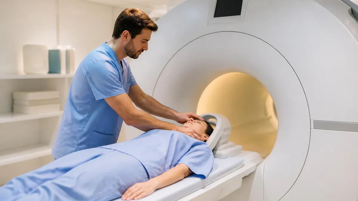
- ✓Patient positioned on the scanner table; appropriate RF coil placed over the body region being scanned
- ✓Table slides into the bore; positioning confirmed by technologist from the control room
- ✓Breath-hold instructions given for specific sequences (abdomen, cardiac) via intercom
- ✓IV contrast (gadolinium) administered mid-scan if ordered by the referring physician
- ✓Multiple sequences acquired — each sequence takes 2–8 minutes; total scan time typically 30–60 minutes
- ✓Patient can communicate with tech via intercom or squeeze bulb at any time
- ✓Table slides out; patient assisted off the table; IV removed if contrast was given
- ✓Images reviewed by radiologist; report typically available within 1–3 business days
MRI Noise, Contrast Agents, and Safety
Why Is MRI So Loud?
If you've ever had an MRI, you remember the noise. Rhythmic banging, thumping, knocking — loud enough that earplugs are mandatory. The culprit is the gradient coil system. Those three sets of coils must switch on and off thousands of times per sequence, carrying enormous electrical currents in a powerful static magnetic field.
The Lorentz force — the same force that drives electric motors — causes the coil windings to physically flex with each pulse. That flexing, transmitted through the structural housing of the scanner, generates the characteristic percussive noise. You can read more in our deep dive on MRI machine noise, including why different sequences have different noise signatures and what manufacturers are doing to quiet modern systems.
Gadolinium Contrast Agents
Many MRI protocols use an intravenous contrast agent to improve visualization of certain structures. The standard MRI contrast is a gadolinium-based agent (GBCA) — a chelated form of gadolinium, a rare earth metal with paramagnetic properties. Gadolinium shortens T1 relaxation times in adjacent tissues, making those areas appear brighter on T1-weighted images. This is particularly useful for tumor characterization (enhancing lesions vs. non-enhancing), blood vessel imaging (MR angiography), and detection of blood-brain barrier breakdown in conditions like multiple sclerosis or meningitis.
Gadolinium is generally well tolerated. Adverse reactions are uncommon — mild reactions like nausea or brief warmth occur in less than 1% of patients. Severe allergic reactions are rare (approximately 0.001–0.01%). The main concern in recent years is gadolinium retention — small amounts of gadolinium can deposit in brain tissue and bone, particularly with linear (non-macrocyclic) chelates and repeated administrations. Macrocyclic agents are more stable and are preferred in patients requiring multiple contrast-enhanced studies. Gadolinium is also contraindicated in severe renal impairment due to the risk of nephrogenic systemic fibrosis (NSF).
MRI Safety — Metal, Pacemakers, and Implants
The static magnetic field never turns off. This makes ferromagnetic objects — items containing iron, nickel, or cobalt — potentially dangerous. Projectile effects: a ferromagnetic object brought into the scan room can become a missile, accelerating toward the magnet bore at dangerous speed. This is why strict screening protocols exist, and why MRI suites are locked zones with 5-Gauss line signage.
For patients with implanted devices, the risks are more nuanced. Traditional cardiac pacemakers are an absolute contraindication for most MRI systems — the magnetic field can interfere with device programming or pacing function, and gradient switching can induce currents in leads. Modern MRI-conditional pacemakers can be scanned under specific conditions. Cochlear implants typically require removal of the external processor and may have magnet-related restrictions.
Cerebral aneurysm clips vary — some older clips are ferromagnetic and absolutely contraindicated; newer titanium clips are MRI-safe. Dental implants, joint replacements, and surgical staples made from titanium or stainless steel are generally safe. Screening every patient with a detailed implant history is non-negotiable. No radiation risk — but that doesn't mean "no risk at all."
Patient Positioned in Bore
Static Field Aligns Protons
RF Pulse Fired
Protons Relax — Signal Emitted
Gradients Encode Spatial Position
k-Space Filled
Fourier Transform Reconstructs Image
Radiologist Reads the Images
- +MRI: superior soft-tissue contrast — brain, spinal cord, joint cartilage, ligaments
- +MRI: no ionizing radiation — preferred for pediatric and repeat imaging
- +MRI: multi-planar imaging without repositioning the patient
- +MRI: functional imaging capabilities (fMRI, perfusion, spectroscopy)
- +MRI: better for pelvic, liver, and biliary tract pathology
- +CT: much faster — full body scan in under 1 minute
- +CT: excellent for bone, calcifications, and lung parenchyma
- +CT: better for acute trauma and hemorrhage (speed critical)
- −MRI: slower (30–60+ min) — not ideal for unstable patients
- −MRI: loud, claustrophobic, more motion artifact in uncooperative patients
- −MRI: contraindicated with many metallic implants and pacemakers
- −MRI: more expensive — scanner cost $1–3M, scan cost typically higher
- −CT: ionizing radiation — not preferred for repeated studies in young patients
- −CT: lower soft-tissue contrast — may miss lesions MRI would detect
- −CT: contrast is iodinated — more nephrotoxic than gadolinium
- −CT: poor for posterior fossa evaluation due to beam hardening artifacts
MRI Questions and Answers
About the Author

Medical Laboratory Scientist & Clinical Certification Expert
Johns Hopkins UniversityDr. Sandra Kim holds a PhD in Clinical Laboratory Science from Johns Hopkins University and is certified as a Medical Technologist (MT) and Medical Laboratory Scientist (MLS) through ASCP. With 16 years of clinical laboratory experience spanning hematology, microbiology, and molecular diagnostics, she prepares candidates for ASCP board exams, MLT, MLS, and specialist certification tests.
Join the Discussion
Connect with other students preparing for this exam. Share tips, ask questions, and get advice from people who have been there.
View discussion (8 replies)