Cardiac MRI: Complete Guide for Patients and MRI Technologists 2026 July
Cardiac MRI (CMR) reveals heart function, ejection fraction, scar tissue, and valve disease — 📝 without radiation. Learn what to expect and how the
Cardiac MRI — you'll also hear it called cardiovascular MRI or CMR — is one of the most comprehensive tests a cardiologist can order. It uses magnetic fields and radio waves to build detailed images of your heart's structure and function, and it does this without a single dose of radiation. That last point matters more than you might think, especially for younger patients who'll need repeated imaging over years or decades.
What separates CMR from most other cardiac tests is tissue characterization. An echocardiogram (ultrasound) can show how the heart moves, but it can't reliably tell you why the walls are moving abnormally. Is it a scar from an old heart attack? Active inflammation from myocarditis? Protein deposits from amyloidosis? Cardiac MRI can distinguish between these — and that distinction drives very different treatment decisions.
Understanding how CMR mri mrcp cpt code it actually measures, and when doctors choose it over other tests is valuable whether you're a patient preparing for your first scan or an MRI technologist expanding into cardiac protocols. This guide covers both angles.
Also called cardiovascular MRI (CMR) — uses magnetic fields and radio waves, zero radiation, 45–75 minutes, requires ECG gating and often IV gadolinium contrast. Gold standard for ejection fraction measurement, myocardial tissue characterization, and congenital heart disease evaluation.
The term "cardiac MRI" covers a family of imaging sequences — not a single scan. A typical CMR protocol layers several different acquisition types. Steady-state free precession (SSFP) cine sequences generate movie-like loops showing the heart contracting and relaxing. Phase-contrast sequences quantify blood flow through valves. Late gadolinium enhancement (LGE) sequences — acquired 10–15 minutes after contrast injection — reveal scar tissue lit up bright white against dark healthy muscle. T1 and T2 mapping sequences produce quantitative tissue values rather than just visual impressions.
Each sequence asks something different of the patient and the technologist. Cine imaging requires 10–15 second breath-holds timed to the cardiac cycle. LGE requires precise timing after contrast injection. T2 mapping needs excellent gating. For a standard MRI scan, breath-holding isn't usually critical — for cardiac MRI, it's fundamental to image quality.
What Cardiac MRI Actually Shows
Ejection fraction is the number most cardiologists want first. It's the percentage of blood the left ventricle pumps out with each beat — normal range is 55–70%. Below 40% is reduced, and below 35% triggers consideration of implantable defibrillators. CMR is the reference standard for EF measurement because it makes no geometric assumptions about heart shape. Echo-based EF calculations assume the ventricle is an ellipsoid; it often isn't, especially in patients with abnormal hearts.
Wall motion analysis uses the AHA's 17-segment model. Each segment gets graded: normal, hypokinetic (reduced movement), akinetic (no movement), or dyskinetic (paradoxical outward movement during systole). This segmental mapping tells the cardiologist which coronary artery territory is affected — key information for planning intervention.
Late gadolinium enhancement is where CMR truly earns its reputation. Gadolinium accumulates in damaged tissue because the normal cell membrane barrier is disrupted in fibrosis and necrosis. On T1-weighted images acquired 10–15 minutes post-injection, scarred areas appear bright. The pattern of enhancement is diagnostic: ischemic scar starts at the subendocardium and extends outward; myocarditis shows mid-wall or epicardial enhancement; cardiac amyloidosis produces a diffuse global pattern.
Valvular disease — regurgitation (leaking) and stenosis (narrowing) — can be assessed quantitatively using phase-contrast sequences. The regurgitant fraction and valve orifice area measurements from CMR are highly reproducible, making them valuable for serial follow-up. Pericardial disease, including constrictive pericarditis, has characteristic MRI findings, and CMR often resolves ambiguous cases that echo or CT couldn't definitively characterize.
Key Cardiac MRI Numbers
When Doctors Order a Cardiac MRI
Post-heart attack assessment is one of the most common indications. After a myocardial infarction, the cardiologist needs to know how much muscle survived and how much became scar. That scar burden — expressed as a percentage of left ventricular mass — predicts recovery of function after revascularization. Segments with extensive transmural scar (above 50% wall thickness) won't recover contractile function even if the blocked artery gets opened. CMR-based viability assessment guides decisions about stenting, bypass surgery, or conservative management.
Unexplained heart failure often lands patients in the CMR suite when echo results are inconclusive. Hypertrophic cardiomyopathy, dilated cardiomyopathy, arrhythmogenic right ventricular cardiomyopathy (ARVC) — all have characteristic CMR patterns that confirm diagnosis and risk-stratify patients. ARVC in particular relies heavily on CMR because the right ventricle is notoriously difficult to image with echo.
Suspected myocarditis is another strong indication. A young patient presents with chest pain and troponin elevation after a viral illness — coronary angiogram is clean. CMR with LGE and T2 mapping can confirm active myocardial inflammation and guide decisions about activity restriction. Athletes with myocarditis need to know before returning to training.
Common Reasons for Cardiac MRI
Quantifies scar tissue burden and determines which segments may recover function after revascularization — guides stenting and bypass decisions.
Identifies cardiomyopathy type (hypertrophic, dilated, ARVC) when echo is inconclusive — characteristic LGE patterns often confirm the diagnosis.
Confirms active myocardial inflammation after viral illness — T2 mapping shows edema, LGE shows injury extent and location.
Evaluates complex anatomy and post-surgical conduits without radiation — ideal for serial follow-up of pediatric and adult congenital patients.
When echocardiography can't answer the clinical question — poor acoustic windows, RV assessment, valvular quantification — CMR steps in.
Baseline functional assessment before surgery and surveillance of repair outcomes, including conduit function and residual shunts.
Cardiac MRI vs Echocardiogram
- +Superior tissue characterization (scar, inflammation, infiltration)
- +No radiation — safe for serial follow-up
- +Gold standard for ejection fraction and RV assessment
- +Excellent for congenital heart disease anatomy
- +Highly reproducible quantitative measurements
- +Visualizes pericardium and great vessels in one exam
- −45–75 minutes — much longer than echo (20–30 min)
- −Significantly more expensive
- −Not portable — can't be done at bedside or in ICU
- −Requires patient cooperation for breath-holds
- −Gadolinium contrast carries NSF risk in severe renal disease
- −Less available — not every hospital has cardiac MRI capability
Cardiac MRI vs Cardiac CT — Different Tools, Different Questions
Cardiac CT is fast. A coronary CT angiogram takes seconds to acquire. The images show coronary artery anatomy with remarkable clarity — stenoses, plaques, stent positions. CT calcium scoring quantifies calcified plaque burden and feeds directly into cardiovascular risk calculators. For coronary anatomy, CT is genuinely superior to MRI. The vessels are small, move constantly, and achieving the spatial and temporal resolution needed requires CT's speed advantage.
Cardiac MRI takes 45–75 minutes and can't reliably image coronary arteries with the clarity CT provides. What it does instead — tissue characterization, flow quantification, comprehensive functional assessment — CT simply can't match. There's also the radiation question: a coronary CTA delivers 2–12 mSv depending on protocol, while CMR delivers none. For patients needing repeated imaging over years, that difference accumulates. Understanding the broader MRI vs CT scan comparison clarifies why both modalities coexist in modern cardiology rather than one replacing the other.
In practice, many cardiology workups use both. A patient with chest pain might get a CT angiogram first to rule out significant coronary stenosis, then a cardiac MRI to characterize an incidentally found wall motion abnormality. The tests are complementary, not competing.
Before Your Cardiac MRI
- ✓Inform your doctor of all metal implants, including pacemakers, ICDs, stents, and surgical clips
- ✓Bring your device card if you have a pacemaker or ICD — the MRI team needs the exact model number
- ✓Tell your care team if you have kidney disease — gadolinium contrast requires adequate renal function
- ✓Avoid caffeine for 24 hours if adenosine stress testing is included in your protocol
- ✓Arrive 15–20 minutes early to complete screening paperwork and change into a gown
- ✓Practice slow, controlled breathing — you'll hold your breath for 10–15 seconds repeatedly
- ✓Bring earplugs or ask for them — cardiac MRI sequences are loud
- ✓Discuss claustrophobia concerns with your ordering physician before the day of the scan
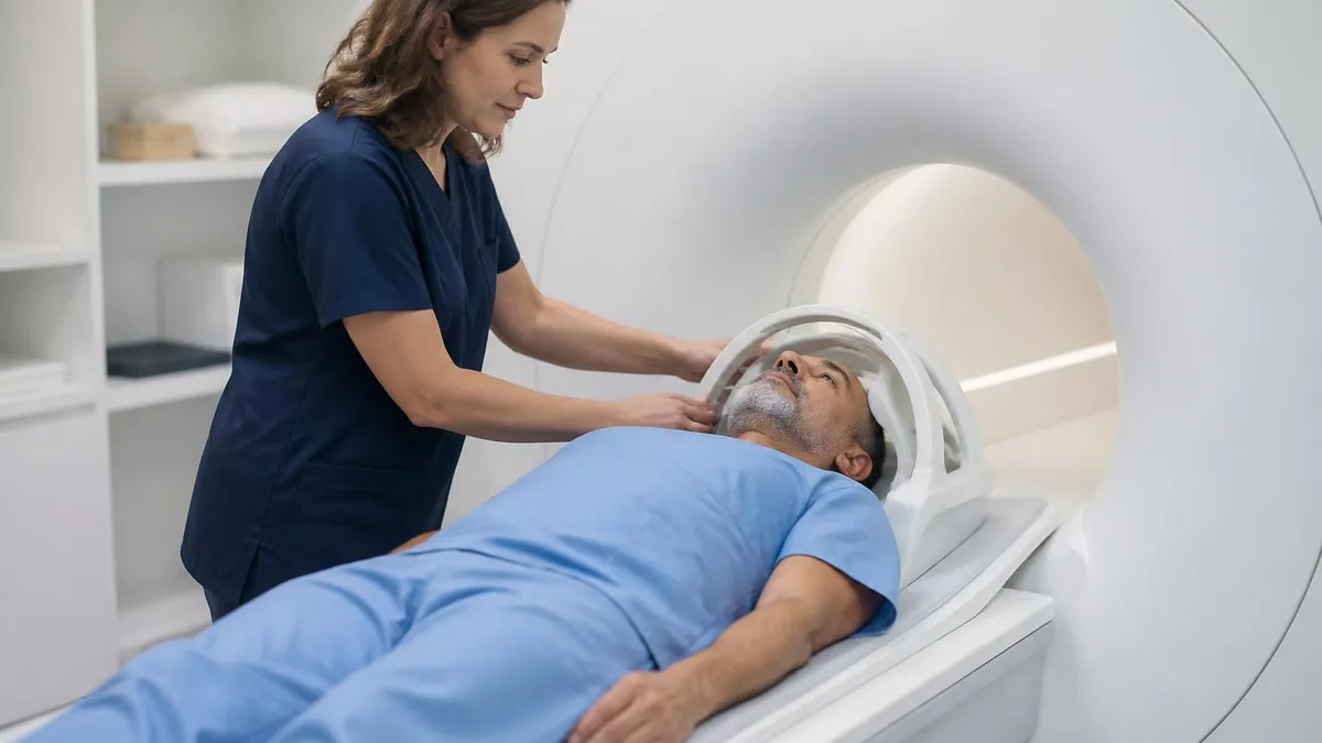
What Happens During the Scan
Plan on 45–75 minutes from the time you're positioned on the table. Some complex protocols — particularly those including stress perfusion imaging — can run longer. Bring earplugs or ask for them at the front desk; different sequences produce very different sounds, from rhythmic banging to high-pitched buzzing.
You'll have EKG leads placed on your chest before entering the bore. These electrodes synchronize image acquisition to your heartbeat — a process called ECG gating. The scanner detects the R-wave peak of each heartbeat and triggers data acquisition at specific phases of the cardiac cycle. Without clean gating, images blur because the heart has moved during acquisition. Irregular rhythms like atrial fibrillation make this challenging; some sequences use retrospective gating algorithms, but image quality is usually reduced.
The technologist will coach you through breath-holds from the control room intercom. "Take a breath in, breathe out halfway, and hold." Then: "Breathe." You'll do this dozens of times across the exam. The half-exhale position is used because it's more reproducible than full inspiration — diaphragm position varies less, keeping the heart in nearly the same position for each acquisition.
If your protocol includes gadolinium contrast, an IV line will be placed before you enter the scanner. Contrast is injected during the exam — usually after the cine sequences are complete. The LGE sequences are acquired 10–15 minutes after injection, giving gadolinium time to wash out of normal myocardium while staying concentrated in scar tissue. Complete MRI safety screening — including the metal implant questionnaire — happens before you ever enter the scan room.
The noise level surprises most first-time patients. Music through MRI-compatible headphones is offered at many sites. You can still hear the technologist's instructions clearly through the intercom or headphone channel — don't be afraid to signal if you need a break or feel uncomfortable.
Gadolinium Contrast in Cardiac MRI
Most cardiac MRI protocols include gadolinium-based contrast agents (GBCAs). The late gadolinium enhancement sequence depends entirely on contrast. Gadolinium is a paramagnetic metal — by itself it's toxic, so it's chelated (chemically bonded to a carrier molecule that keeps it stable). After IV injection, gadolinium distributes through blood and extracellular space, then clears through the kidneys.
In healthy myocardium, it clears quickly. In damaged myocardium — scar, fibrosis, inflammation — it persists because the extracellular space is expanded. T1-weighted sequences acquired 10–15 minutes post-injection show retained gadolinium as bright signal against dark, cleared normal muscle. That's late gadolinium enhancement.
The primary safety concern is nephrogenic systemic fibrosis (NSF), a rare but serious condition occurring almost exclusively in patients with severe renal impairment (eGFR below 30 mL/min/1.73m²). Macrocyclic agents — gadobutrol, gadoteridol, gadoterate — have a substantially better safety profile than older linear agents and are preferred when renal function is reduced.
Your radiologist weighs these considerations against the diagnostic value of contrast before ordering it. For patients with pacemakers undergoing MRI-conditional device scanning, gadolinium protocols follow standard cardiac MRI procedures — the MRI machine type and field strength matter as much as the contrast agent choice.
Cardiac MRI Contraindications
- Non-conditional pacemakers or ICDs — legacy devices not cleared for MRI use
- Certain cochlear implants — device-specific evaluation required
- Ferromagnetic intracranial aneurysm clips — older clip generations are contraindicated
- Metallic foreign bodies near critical structures — intraocular metal, shrapnel near spine or major vessels
For MRI Technologists: Cardiac Protocols and Subspecialty Practice
Cardiac MRI is among the most technically demanding subspecialties in the field. It combines precise MRI physics with real-time physiological coupling (ECG gating), patient coaching skills (breath-holds), and a clinical vocabulary spanning cardiology, cardiac surgery, and electrophysiology. The learning curve is steep. The rewards — both clinical and professional — are substantial.
A standard CMR protocol includes: scout images → cardiac geometry planning → SSFP cine acquisitions in standard planes (4-chamber, 2-chamber, LVOT, short-axis stack) → phase-contrast at the aortic and pulmonary valves → gadolinium injection → first-pass perfusion (if stress testing is included) → T1 mapping → LGE sequences in matching planes to the cines. T2 mapping for edema quantification is increasingly included in myocarditis protocols.
ECG gating quality is your first quality control checkpoint. A clean, tall R-wave with minimal noise gives the scanner a reliable trigger. Lead placement matters — V4-V6 position on a cardiac MRI surface coil setup typically gives the best R-wave amplitude. Gradient fields induce voltages in the EKG leads (the magnetohydrodynamic effect), which can distort the trace inside the scanner. High-quality MRI-compatible leads with good skin contact minimize this artifact.
Breath-hold coaching is a skill that doesn't come from physics textbooks. Different patients have wildly different capacity and compliance. Coaching tone, pacing, and specific instruction phrasing all affect how reproducibly patients hold at mid-expiration. Misregistration between slices from poor breath-holding creates artifacts that look like wall motion abnormalities — false positives from poor technique cause real clinical problems.
The same patient coaching principles that apply during a brain MRI translate to cardiac work, though cardiac patients need more active guidance due to the breath-hold demands. Subspecialty certification paths include the ARRT Advanced Cardiac Imaging credential and the ARMRIT cardiac certification — both require dedicated case volume. Understanding what MRI stands for and the underlying physics is foundational, but cardiac MRI demands additional training in ECG gating, sequence planning, and cardiology anatomy.
MRI Questions and Answers
How Long Does Cardiac MRI Take to Get Results?
Scan acquisition is the fast part. The 45–75 minutes in the scanner collects raw data — but interpreting a full cardiac MRI study takes significantly longer than reading a chest X-ray or even a standard echocardiogram. A board-certified radiologist or cardiologist with specialized CMR training reviews the study, performs post-processing analysis (ejection fraction calculations, LGE quantification, T1/T2 mapping), and writes the report.
In most hospital settings, you'll have results within 24–48 hours for routine studies. Urgent scans — suspected acute myocarditis in a young athlete, post-STEMI viability before emergent bypass — get read and reported within hours. Your ordering cardiologist reviews the report and typically contacts you within a few days to discuss findings and implications for your treatment plan.
Don't expect the technologist to interpret findings. They're responsible for image quality, not diagnosis. Any question about what the images show should wait for your cardiologist or radiologist. You're completely within your rights to ask the technologist whether the scan went well technically — a confident "yes" means the diagnostic information you need is captured. Interpretation comes later, and it's worth discussing every finding — even incidental ones — with your ordering physician before drawing conclusions.
What's Included in a Cardiac MRI Report
- ✓Left ventricular ejection fraction (LVEF) — percentage of blood pumped per beat
- ✓Right ventricular ejection fraction (RVEF) — often 45–50% or above in normal hearts
- ✓End-diastolic and end-systolic volumes — chamber size measurements
- ✓Wall motion analysis by AHA 17-segment model — grades each segment as normal, hypo-, a-, or dyskinetic
- ✓Late gadolinium enhancement (LGE) — presence, location, transmurality, and pattern
- ✓T2 signal / T2 mapping — edema indicating active or recent myocardial injury
- ✓Valvular assessment — regurgitant fraction, valve orifice area if indicated
- ✓Pericardial findings — thickness, effusion, constrictive features
- ✓Incidental extracardiac findings — aortic dimensions, pleural effusions, masses
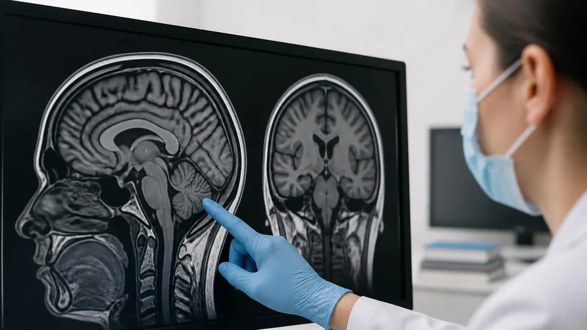
Reading Your Cardiac MRI Report
Radiology reports are written for referring physicians — but knowing the key terms helps you have a more productive conversation with your cardiologist. Left ventricular ejection fraction (LVEF) is almost always listed first, along with end-diastolic and end-systolic volumes. Normal LVEF is 55–70%. Reduced (below 40%) and mildly reduced (40–50%) are the current guideline-defined categories.
Wall motion analysis follows — the 17-segment model grades each LV segment as normal, hypokinetic, akinetic, or dyskinetic. Segments matching a specific coronary artery territory often point to ischemic disease in that vessel.
The LGE section is often the most clinically important: it describes which segments are affected and the transmurality — the percentage of wall thickness enhanced. A 25% transmural infarct has a much better chance of functional recovery with revascularization than a 75% transmural infarct in the same segment. Your cardiologist will focus on this number when deciding whether intervention is worth pursuing.
T2 signal findings indicate edema — active or recent injury rather than chronic scar. Valvular findings, pericardial assessment, and incidental extracardiac findings (aortic dimensions, pleural effusions, masses) appear at the end. The impression section synthesizes everything into primary findings and clinical implications — that's where your cardiologist focuses first. Ask about every finding, even the ones labeled incidental.
About the Author
Medical Laboratory Scientist & Clinical Certification Expert
Johns Hopkins UniversityDr. Sandra Kim holds a PhD in Clinical Laboratory Science from Johns Hopkins University and is certified as a Medical Technologist (MT) and Medical Laboratory Scientist (MLS) through ASCP. With 16 years of clinical laboratory experience spanning hematology, microbiology, and molecular diagnostics, she prepares candidates for ASCP board exams, MLT, MLS, and specialist certification tests.
Join the Discussion
Connect with other students preparing for this exam. Share tips, ask questions, and get advice from people who have been there.
View discussion (6 replies)