Knee MRI: What It Shows, How It Works, and What to Expect 2026 June
Learn about knee MRI scans — 📗 what a knee MRI shows, how to prepare, how long it takes, what conditions it diagnoses, and how to read your results.
What Is a Knee MRI?
A knee MRI is a medical imaging procedure that uses magnetic fields and radio waves to produce detailed images of the soft tissue structures inside and around the knee joint. Unlike X-rays and CT scans, which excel at imaging bone, MRI produces high-resolution images of cartilage, ligaments, tendons, the meniscus, and the synovial lining of the joint — the soft tissue structures that are most commonly injured in knee problems and which X-rays simply cannot visualise. This makes MRI the gold standard diagnostic tool for most knee injuries and disorders.
The knee joint is one of the most complex joints in the body and one of the most frequently injured, particularly among athletes and active individuals. The structures at risk include the anterior cruciate ligament (ACL), posterior cruciate ligament (PCL), medial and lateral collateral ligaments (MCL and LCL), the medial and lateral menisci, articular cartilage, tendons (particularly the patellar tendon and quadriceps tendon), and the bone itself.
When a patient presents with knee pain, swelling, instability, locking, or restricted range of motion, a knee MRI gives the orthopaedic surgeon or sports medicine physician the detailed picture needed to identify which structures are affected and to what degree.
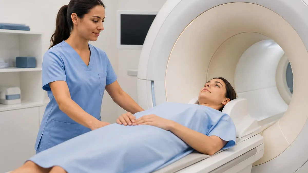
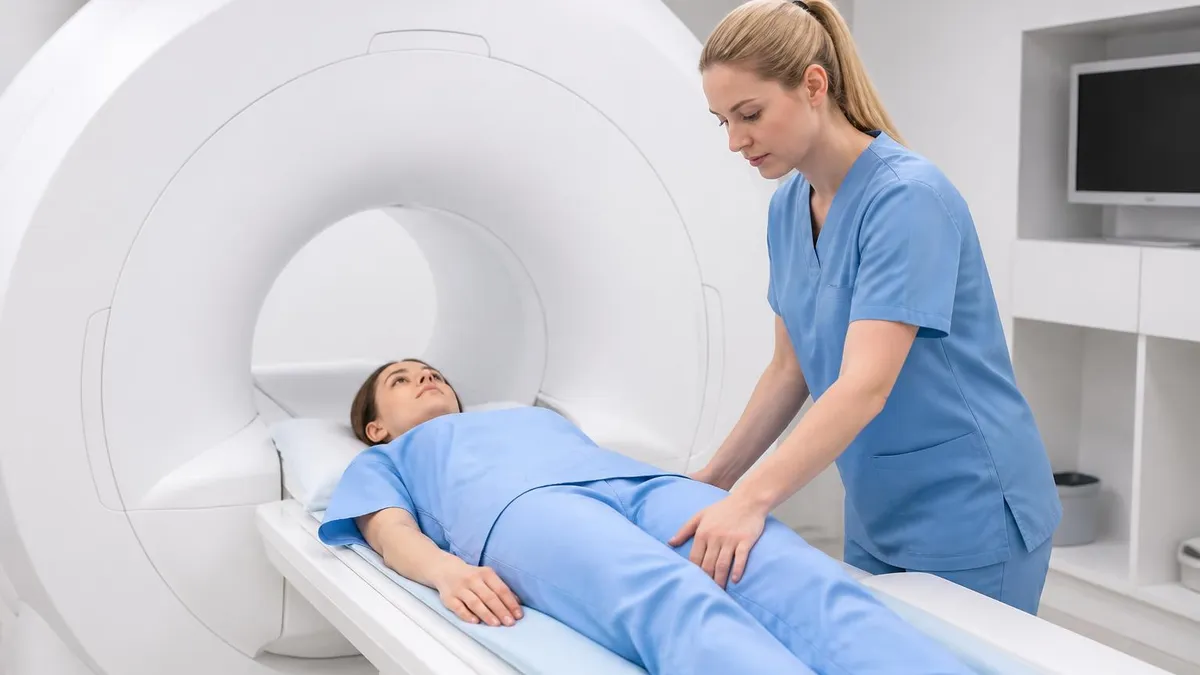
Knee MRI is non-invasive and uses no ionising radiation — unlike X-rays and CT scans which expose patients to radiation. The scan typically takes 30-60 minutes. You lie still in the MRI scanner while the machine captures multiple sequences of images from different angles and orientations, producing a comprehensive three-dimensional picture of the knee's internal structures. The resulting images are interpreted by a radiologist who produces a report that your referring physician uses to guide treatment decisions.
Most knee MRIs are ordered after a clinical examination suggests a soft tissue injury that needs imaging confirmation, before surgical intervention to plan the procedure, or when pain or functional limitation persists despite initial treatment and the cause isn't clear from examination alone. For a foundational understanding of how MRI technology works and what differentiates it from other imaging modalities, the complete MRI guide covers the physics, clinical applications, and what to expect across all MRI body regions.
- What it images: Ligaments (ACL, PCL, MCL, LCL), menisci, cartilage, tendons, bone marrow, and joint fluid — structures X-rays cannot show
- Duration: 30-60 minutes for a standard knee MRI; with contrast, slightly longer
- No radiation: MRI uses magnetic fields and radio waves — no ionising radiation unlike X-rays or CT scans
- Contrast use: Most knee MRIs are performed without contrast; contrast (gadolinium injection) is added when infection, tumour, or post-surgical assessment is needed
- Claustrophobia: Knee MRIs are performed in a feet-first position — most of your body is outside the machine, making claustrophobia less of a concern than head or chest MRIs
- Metal implants: Most modern joint replacements and plates are MRI-compatible — inform your radiographer of all implants before scanning
- Result timing: Radiologist report typically available within 1-7 business days; urgent cases may be reported same day
What Happens During a Knee MRI
Arrival and Safety Screening
Preparation and Positioning
The Scan
After the Scan
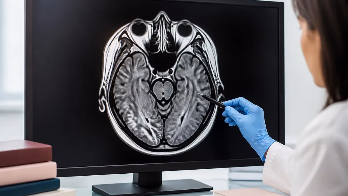
What Does a Knee MRI Show?
Knee MRI produces excellent visualisation of all the major soft tissue and bony structures of the knee joint. The structures that MRI can assess include the four major ligaments, both menisci, articular cartilage, tendons, bursae, the joint capsule, bone marrow (where stress fractures and bone bruising appear), and the synovial membrane. The scan is acquired in multiple planes — axial (top-down), sagittal (side view), and coronal (front view) — giving the radiologist a complete three-dimensional picture of the joint.
Ligament injuries are one of the most common reasons for knee MRI referral. The ACL (anterior cruciate ligament) is the most frequently torn ligament in sports — a complete ACL tear appears on MRI as a disruption or absence of the normally taut dark band of ligament tissue, often accompanied by characteristic bone bruising patterns on the lateral femoral condyle and posterolateral tibial plateau.
Partial ACL tears, which are more difficult to diagnose clinically, are reliably identified on MRI. PCL tears, MCL injuries, and LCL complex injuries are similarly well-visualised. MRI also shows the degree of injury — complete versus partial tear — which directly influences surgical versus non-surgical treatment decisions.
Meniscal tears are another primary indication for knee MRI. The medial and lateral menisci are the C-shaped cartilage pads that act as shock absorbers and joint stabilisers between the femur and tibia. On MRI, a normal meniscus appears dark and homogeneous; a tear is visible as a line of increased signal (brightness) extending to the surface of the meniscus.
MRI can identify the type of tear — horizontal, vertical, radial, complex, or the bucket-handle pattern — which helps the surgeon plan the repair or trimming procedure. The overall sensitivity and specificity of MRI for meniscal tears exceeds 90% in most studies, making it highly reliable for this indication.
Articular cartilage damage — a precursor to osteoarthritis — is well-demonstrated on modern high-field MRI with dedicated cartilage-sensitive sequences. Cartilage defects, thinning, and the characteristic changes of early and established osteoarthritis are all visible. Tendon pathology (patellar tendinitis, quadriceps tendon tears, popliteal tendon injuries) and bursal inflammation are similarly assessable.
Bone pathology — stress fractures, bone contusions from impact, osteonecrosis, and tumours — are detectable on the bone marrow sequences included in a standard knee MRI protocol. See the comprehensive MRI scan guide for how MRI imaging is used across different anatomical regions and what distinguishes knee MRI protocols from other body part protocols.
Common Conditions Diagnosed by Knee MRI
Anterior cruciate ligament tears are the most common ligament injury requiring knee MRI. MRI shows complete or partial disruption of the ACL fibres, associated bone bruising patterns, and any concurrent meniscal or cartilage injuries (commonly present with ACL tears). MRI is essential for surgical planning and assessing whether reconstruction is appropriate.
MRI is the gold standard for diagnosing meniscal tears — horizontal, vertical, radial, and bucket-handle patterns are all identifiable. The tear location (anterior horn, body, posterior horn) and extent determine whether arthroscopic repair or trimming is recommended. MRI sensitivity for meniscal tears exceeds 90%.
Articular cartilage thinning, defects, and subchondral bone changes from osteoarthritis are well-demonstrated on MRI. Cartilage-sensitive sequences show early damage before it's visible on X-ray. MRI also shows joint space narrowing, osteophytes, bone marrow oedema, and synovial thickening in established arthritis.
Bone marrow oedema from impact injuries (bone bruising) and stress fractures are invisible on X-ray but clearly visible on MRI as areas of increased signal on fluid-sensitive sequences. Athletes with persistent knee pain and negative X-rays are frequently found to have stress fractures or bone bruising on MRI.
Knee MRI: Preparation, During, and After
Preparation for a knee MRI is straightforward compared to many other medical procedures.
- Metal screening: Inform the MRI centre of all implants, previous surgeries, and medical devices before your appointment — call ahead if you're unsure about compatibility
- Clothing: Wear comfortable clothing without metal (elasticated waistbands, no underwire bras). You may need to change into a gown
- No fasting required: Unlike CT with contrast, a routine knee MRI (no contrast) requires no dietary restrictions beforehand
- Contrast MRI: If contrast is ordered, a blood test for kidney function (eGFR) is often required first, as gadolinium contrast is cleared by the kidneys
- Medications: Continue all regular medications unless specifically told otherwise
- Anxiety: If you experience claustrophobia, mention it when booking — sedation or anti-anxiety medication can be prescribed if needed, and open MRI may be an option
Knee MRI vs. X-Ray vs. CT Scan
The three primary imaging modalities used for knee problems — X-ray, CT scan, and MRI — have different strengths and are suited to different clinical questions. Understanding what each shows helps you understand why your doctor ordered a particular type of scan and what it can and cannot tell them.
X-rays are the standard first-line imaging for knee pain. They're fast, inexpensive, and excellent for assessing bone structure, joint space width (a proxy for cartilage thickness), and bony abnormalities like fractures, dislocations, and significant arthritis. However, X-rays provide virtually no information about soft tissue structures — ligaments, tendons, menisci, and cartilage are invisible on X-ray. When a patient has a normal X-ray but persistent knee symptoms suggesting a soft tissue injury, MRI is the next appropriate step.
CT scans provide excellent three-dimensional bony detail and are useful for complex fractures where precise bony anatomy matters for surgical planning. CT is faster than MRI and more accessible, but it involves ionising radiation and provides significantly inferior soft tissue resolution compared to MRI. For knee injuries where the primary concern is ligament, meniscal, or cartilage damage, CT adds little over X-ray in soft tissue characterisation. CT is occasionally used in post-surgical assessment to evaluate bone healing when MRI is contraindicated due to metallic implants causing artefact.
MRI is unmatched for soft tissue detail and remains the definitive imaging modality for most knee disorders. Its disadvantages are cost, scan time, availability, and contraindications for patients with certain non-MRI-compatible implants or severe claustrophobia. For a detailed comparison of these modalities and when each is appropriate, the CT scan vs MRI guide covers the key differences in radiation, cost, image quality, and clinical use cases. The guide to MRI scan duration covers how long knee MRI and other body part MRIs typically take, including factors that extend scan time.
Knee MRI Preparation Checklist
- ✓Inform the MRI centre of all metal implants (joint replacements, plates, screws, pins) before your appointment — call ahead if you're uncertain about compatibility
- ✓Tell staff about any pacemaker, cochlear implant, neurostimulator, or other electronic medical device — these may be MRI-incompatible
- ✓Wear comfortable clothing without metal fastenings — elasticated waistbands, no belt buckles or underwire
- ✓Remove all jewellery, watches, piercings, and hair clips before entering the MRI suite
- ✓Tell staff if you experience claustrophobia — knee MRI is feet-first and less enclosed than head MRI, but sedation options are available if needed
- ✓If contrast has been ordered, confirm whether you need a prior kidney function blood test (eGFR) — many facilities require this
- ✓Bring your referral letter and any previous knee imaging (X-rays, prior MRI) for the radiologist to compare
- ✓Allow 45-60 minutes at the facility — the scan itself takes 30-45 minutes
- ✓Plan to remain still during the scan — movement during sequences degrades image quality and may require repeat sequences
- ✓You can drive home after a routine knee MRI (no sedation) — if sedation was given, arrange for someone to collect you
Knee MRI: Advantages and Limitations
- +Unmatched soft tissue detail — ligaments, menisci, cartilage, and tendons are clearly visualised and accurately characterised
- +No ionising radiation — unlike X-rays and CT scans, MRI poses no radiation risk, making it safe for repeated use and for younger patients
- +Multi-planar imaging — the same scan produces images in multiple orientations (axial, sagittal, coronal) giving a complete three-dimensional picture
- +High sensitivity for bone marrow abnormalities — stress fractures and bone bruising invisible on X-ray are clearly shown on MRI
- +Can be performed with or without contrast — most knee MRIs need no contrast injection, making the procedure simpler and faster
- −Longer scan time than X-ray or CT — 30-45 minutes of stillness required, which can be difficult for patients in significant pain
- −More expensive than X-ray and CT — MRI costs significantly more and has longer waiting times on the NHS compared to X-ray
- −Not suitable for patients with certain non-MRI-compatible implants — some older pacemakers and cochlear implants are contraindicated
- −Claustrophobia — some patients cannot tolerate the enclosed scanner; open MRI alternatives exist but typically have lower field strength and image quality
- −Requires referral — unlike X-ray which is often done in emergency departments, MRI typically requires a referral and advance booking
Cost and Access: Getting a Knee MRI
In the UK, knee MRI is available through the NHS (free at point of use) or privately. NHS waiting times for a non-urgent knee MRI referral vary by trust but commonly run from 6 weeks to several months. Urgent referrals (suspected tumour, rapidly progressive symptoms) are prioritised and typically scheduled within 2 weeks. For sports injuries requiring prompt diagnosis to guide treatment decisions, many patients choose private MRI to avoid the wait.
Private knee MRI costs in the UK typically range from £250-£600 depending on the facility, location, and whether contrast is included. Many private imaging centres offer self-referral — you can book and pay directly without a GP or specialist referral. This is particularly useful for sports injuries where athletes want prompt diagnosis. Self-referral results are typically sent to a radiologist who produces a report; you then take this report to your GP or directly to a sports medicine physician or orthopaedic surgeon for treatment planning.
In the United States, knee MRI costs vary significantly based on facility type and insurance status. Without insurance, a knee MRI typically costs $700-$2,000 or more at hospital-based radiology departments; free-standing imaging centres often charge $400-$800 for self-pay patients. Most health insurance plans cover knee MRI when ordered by a physician and deemed medically necessary, though deductibles, copays, and prior authorisation requirements vary by plan.
Understanding your insurance coverage and getting prior authorisation before scheduling prevents unexpected bills. The full breakdown of costs in different settings is covered in the guide to MRI costs including how insurance coverage works and how to reduce out-of-pocket expense.
Regarding safety, a knee MRI is exceptionally safe for the vast majority of patients. The magnetic field itself poses no biological harm at the field strengths used in diagnostic MRI. The primary safety concerns relate to metal implants and medical devices — the magnetic field can exert force on ferromagnetic implants and interfere with electronic devices.
Modern orthopaedic implants are virtually all MRI-conditional (safe under specified conditions) or MRI-safe, meaning knee replacement patients and patients with plates and screws can typically have MRI with clearance. Always bring implant documentation to your appointment. For comprehensive MRI safety information, the MRI safety guide covers all contraindications, screening procedures, and what to tell your radiographer.
Knee MRI: Key Statistics
Claustrophobia and Open MRI for Knee Scans
Claustrophobia is one of the most common concerns patients raise when a knee MRI is ordered. The good news is that knee MRI is substantially less claustrophobia-inducing than brain or chest MRI. Because the knee is positioned at the far end of the body, you enter the scanner feet-first and most of your body — including your head — remains outside the bore of the magnet during the scan. Many patients who cannot tolerate brain MRI manage knee MRI without difficulty for this reason.
For patients who do experience significant anxiety in any enclosed space, several options exist. Sedation or anxiolytic medication (typically a low-dose benzodiazepine) can be prescribed to be taken before the appointment, providing sufficient relaxation to complete the scan. This requires someone to drive you to and from the appointment as you cannot drive under sedation. Sedation should be discussed and prescribed in advance — it cannot typically be arranged on the day of the scan.
Open MRI systems are an alternative for patients with severe claustrophobia or patients who cannot fit into standard cylindrical MRI scanners due to size. Open MRI uses two opposed magnet plates with open sides rather than a cylindrical bore, eliminating the enclosed feeling entirely.
However, most open MRI systems operate at lower magnetic field strengths (0.3-1.0 Tesla) compared to standard closed MRI (1.5-3.0 Tesla), which produces lower resolution images. For most routine knee MRI indications, open MRI at 1.0 Tesla provides diagnostically acceptable image quality. For complex or subtle pathology — early cartilage changes, subtle partial ligament tears — higher field closed MRI is preferred.
Wide-bore MRI systems offer a middle ground — a cylindrical bore with a larger diameter (70cm vs the standard 60cm) that reduces the enclosed feeling without sacrificing field strength. Wide-bore 1.5T and 3T systems are increasingly available at major imaging centres and are suitable for most patients who find standard bore claustrophobic but can tolerate a less enclosed environment. If claustrophobia is a concern, ask your referring doctor or imaging centre about wide-bore availability before defaulting to open MRI, as the image quality difference is significant for detailed knee pathology assessment.
The MRI safety screening process exists to protect you — never enter the MRI suite without completing screening and informing staff of all metal implants, medical devices, and previous surgeries. While most modern orthopaedic implants are MRI-safe, some older pacemakers, cochlear implants, and certain vascular clips are not compatible with MRI and could be dangerous in a strong magnetic field. If you're unsure whether an implant is MRI-compatible, bring any implant documentation you have (implant cards, surgical reports) to the appointment and inform the radiographer. They can verify compatibility with manufacturer data before proceeding. Concealing implants or declining to disclose them puts you at serious risk.
Understanding Your Knee MRI Report
Knee MRI reports are written by radiologists for referring clinicians — they use medical terminology that can seem complex but follows consistent patterns that are worth understanding. The report typically has a header (patient information, scan date, referring clinician), clinical indication (why the scan was ordered), technique (field strength, sequences performed, contrast use), findings (the detailed description of all structures assessed), and impression (summary of the key abnormal findings and their clinical significance).
Common terms you may encounter: 'increased T2 signal' in a ligament or meniscus indicates pathology — fluid or damage in a structure that should appear dark; 'free edge tear' describes a meniscal tear reaching the inner surface of the meniscus; 'grade III signal' in a meniscus typically means a tear reaching the articular surface; 'bone marrow oedema' indicates bone bruising; 'chondral defect' refers to cartilage damage; 'effusion' is fluid in the joint; 'osteophytes' are bony spurs associated with arthritis. Your doctor will explain which of these findings are relevant to your symptoms and treatment.
Not all abnormal MRI findings are clinically significant. Studies of asymptomatic knees in middle-aged and older adults consistently find meniscal changes, cartilage thinning, and other abnormalities that produce no symptoms — these are incidental findings. A good radiologist and clinician contextualise MRI findings against your clinical presentation.
A meniscal finding that matches your symptoms and physical examination is likely the cause of your problem; the same finding in an asymptomatic knee may simply be an age-related change that doesn't require treatment. This is why MRI reports are interpreted by your doctor rather than taken in isolation — imaging findings alone don't determine treatment, clinical context does. An MRI that confirms your suspected diagnosis gives you and your surgeon confidence proceeding to treatment; one that shows unexpected findings may redirect management in an important direction.
If you have questions about your knee MRI findings, your referring doctor or orthopaedic surgeon is the appropriate person to ask. Radiologists who produced the report can sometimes be reached for clarification on technical aspects, though treatment decisions and clinical interpretation always rest with the treating clinician. For broader context on MRI technology, scan preparation, and how MRI findings are used across different specialities, the comprehensive MRI guide provides detailed background on how MRI images are acquired and interpreted.
Knee MRI Questions and Answers
About the Author
Medical Laboratory Scientist & Clinical Certification Expert
Johns Hopkins UniversityDr. Sandra Kim holds a PhD in Clinical Laboratory Science from Johns Hopkins University and is certified as a Medical Technologist (MT) and Medical Laboratory Scientist (MLS) through ASCP. With 16 years of clinical laboratory experience spanning hematology, microbiology, and molecular diagnostics, she prepares candidates for ASCP board exams, MLT, MLS, and specialist certification tests.
Join the Discussion
Connect with other students preparing for this exam. Share tips, ask questions, and get advice from people who have been there.
View discussion (4 replies)