Magnetic Resonance Imaging (MRI): Complete Guide 2026 July
Magnetic resonance imaging explained — 📗 how MRI works, types of scans, safety protocols, costs, and what to expect before your exam.
What Is Magnetic Resonance Imaging?
Magnetic resonance imaging — most people just call it an MRI — is a medical imaging technique that uses powerful magnets and radio waves to create detailed pictures of the inside of your body. No X-rays, no radiation. Just a strong magnetic field, a precisely timed pulse of radio waves, and a computer that turns the resulting signals into cross-sectional images your doctor can read.
The physics behind it are genuinely fascinating. Your body is mostly water. Water contains hydrogen atoms. Those hydrogen atoms have nuclei — protons — that spin like tiny tops. Under normal conditions, those spins point in random directions. The moment you slide into an MRI machine, the scanner's magnet aligns those protons along its field. Then a precisely tuned radio-frequency pulse knocks them out of alignment. When the pulse stops, the protons snap back — and as they do, they emit a faint radio signal. Different tissues snap back at different rates. The scanner captures those signals and a computer reconstructs them into images.
It sounds simple enough in a paragraph, but the engineering required to make it work — superconducting coils, rapidly switching gradient magnets, radiofrequency shielding — represents decades of development. The technique earned Paul Lauterbur and Peter Mansfield the 2003 Nobel Prize in Physiology or Medicine, and it's easy to see why.
A Brief History
The underlying physics — nuclear magnetic resonance, or NMR — was discovered in the 1940s by Felix Bloch and Edward Purcell, who shared the 1952 Nobel Prize in Physics for their work. For years, NMR was purely a chemistry tool. Scientists used it to identify molecular structures in the lab, with no thought of using it to image a living person.
The leap to medical imaging came in the early 1970s. Raymond Damadian demonstrated in 1971 that tumors and normal tissue had different NMR relaxation times — meaning a scanner could, in theory, tell them apart. Paul Lauterbur figured out how to add spatial information using gradient magnetic fields, making it possible to build actual images rather than just measuring averaged tissue properties. Peter Mansfield developed the mathematical framework that made acquisition fast enough to be clinically practical.
The first full-body MRI scanner was used clinically around 1980. By the late 1990s, MRI had become standard equipment in hospitals worldwide. Today, more than 35,000 clinical MRI units operate globally, and demand keeps growing as the technology improves and more indications emerge.
How an MRI Scanner Works
There are three core components inside every MRI scanner, and each one plays a distinct role in producing diagnostic images.
The main magnet gives MRI its defining characteristic. Most clinical scanners use a superconducting electromagnet cooled with liquid helium to near absolute zero. At that temperature, the coil's electrical resistance drops to zero — meaning you can run enormous currents through it without energy loss, generating a stable magnetic field measured in Tesla (T). A 1.5T scanner is the workhorse of most hospitals. A 3T scanner delivers higher resolution and is increasingly common for neurological imaging. Research scanners pushing 7T and above exist at academic medical centers.
Gradient coils sit inside the bore. The main magnet creates a uniform field — necessary for aligning protons, but it doesn't tell you where each returning signal is coming from. Gradient coils add small, rapidly switching magnetic fields that vary in strength along each axis of the scanner. By knowing exactly how the gradient field changes at each point in space, the computer can pinpoint where each signal originated. The rapid switching of these coils is responsible for the loud banging and knocking you hear during a scan.
Radiofrequency coils transmit the excitation pulses and receive the returning signals. The large coil built into the scanner bore handles transmission. Smaller, specialized receiver coils — head coils, knee coils, shoulder coils, cardiac coils — are placed around the body part being imaged to pick up weak return signals with much higher sensitivity. Using the right coil for the right anatomy significantly improves image quality.
Types of MRI Scans
The MRI scan your doctor orders depends on what they're looking for and which part of your body needs imaging. The technique is remarkably versatile — it's used from head to toe, across nearly every medical specialty.
Brain and head MRI is the most frequently ordered exam. It evaluates headaches, seizures, stroke, multiple sclerosis, brain tumors, dementia, and a wide range of other neurological conditions. It can visualize gray matter, white matter tracts, blood vessels, and cerebrospinal fluid in ways no other modality can match. Functional MRI (fMRI) goes further — it maps brain activity in real time by detecting changes in blood oxygenation, which is invaluable for pre-surgical planning near eloquent cortex.
Spine MRI is used to assess herniated discs, spinal stenosis, cord injuries, infections, and tumors. Because MRI shows soft tissue so clearly, it can reveal a disc fragment compressing a nerve root in a way that a plain X-ray simply cannot.
Musculoskeletal MRI is ordered constantly in orthopedic and sports medicine practice. Knee, shoulder, hip, and ankle MRI exams can show acl mri, meniscal injuries, cartilage damage, tendon pathology, and stress fractures that aren't yet visible on X-ray. It's the go-to modality when your orthopedist needs to know exactly what's torn before deciding on surgery.
Cardiac MRI (often called CMR) assesses heart structure, function, perfusion, and scar tissue with remarkable accuracy — without radiation and without the geometric limitations of echocardiography. It's growing rapidly as a tool for evaluating cardiomyopathies, infiltrative diseases, and congenital defects.
Abdominal and pelvic MRI evaluates the liver, kidneys, pancreas, prostate, uterus, and rectum. MRI is the preferred modality for staging rectal cancer and characterizing liver lesions because of its superior soft-tissue contrast. It's also the standard of care for local staging of prostate cancer.
Breast MRI is recommended for high-risk patients as a supplement to mammography. It detects tumors that mammograms miss, especially in dense breast tissue, making it a meaningful adjunct for women with BRCA mutations or a strong family history.
MR angiography (MRA) images blood vessels without invasive catheterization. It can detect aneurysms, arterial stenosis, and vascular malformations — often serving as a screening or problem-solving tool before any interventional decisions are made.
MRI With vs. Without Contrast
Some scans are ordered "with contrast" — meaning a gadolinium-based contrast agent is injected intravenously during the exam. Gadolinium is a rare earth metal that alters the magnetic relaxation properties of nearby water, making tissues with disrupted blood-brain barriers or abnormal vascularity light up brightly on the resulting images.
Contrast is especially useful for detecting tumors (which often have leaky blood vessels), assessing inflammation, and evaluating vascular anatomy. It's not always necessary — many excellent MRI exams are performed without it, particularly for musculoskeletal and routine neurological evaluations where the clinical question is about structure rather than enhancement.
Gadolinium is generally safe, but there are real risks to understand. Patients with severely impaired kidney function are at risk for nephrogenic systemic fibrosis — a rare but serious condition. Gadolinium also deposits in the brain and other tissues with repeated exposures; whether this causes clinical harm is still being studied. Your radiologist weighs benefits against risks on a per-patient basis, so don't assume contrast is automatic just because someone ordered it before.
MRI vs. CT Scan: Which Is Better?
This question comes up constantly. The honest answer is: it depends entirely on the clinical situation. The full MRI vs CT scan comparison covers the differences in detail, but here's the practical breakdown.
MRI wins for soft-tissue contrast. It's the superior choice for brain, spinal cord, joints, liver, and pelvic organs. It doesn't use ionizing radiation, so it's safer for repeat imaging and for imaging children. The trade-offs are real — MRI is slower, more expensive, louder, and claustrophobia-inducing for some patients. Certain metallic implants are contraindicated, which rules it out for a meaningful subset of patients.
CT is faster, cheaper, and better for bone detail, lung imaging, and acute trauma. The emergency department can't wait 45 minutes for a trauma MRI when a patient is crashing — a 5-minute CT is the right tool there. CT does deliver ionizing radiation; a chest CT is roughly equivalent to three years of background radiation exposure.
Neither modality is universally better. The right one depends on the clinical question, the urgency of the situation, what's in the patient's body, and what information each test can realistically provide.
MRI Safety Essentials
MRI is one of the safest imaging modalities available — but the magnetic field creates unique hazards that don't exist with X-ray or ultrasound. MRI safety protocols exist for serious reasons, and they're enforced rigorously in every accredited facility.
The most dramatic risk is projectile hazard. The MRI magnet never turns off — it's always on, always generating an intense field, even when no exam is running. Ferromagnetic objects (those containing iron or steel) experience a powerful pull toward the magnet. A forgotten oxygen tank, a floor polisher, even a set of steel keys can become a dangerous projectile if someone walks past the 5-gauss line without authorization. There have been serious injuries — and at least one fatality — involving MRI projectile incidents. This is why the suite is locked, why everyone entering the room must be screened, and why technologists ask the same screening questions every single time without shortcuts.
Internal implants are the other major concern. Pacemakers, cochlear implants, certain aneurysm clips, and metallic foreign bodies in the eyes can all be contraindicated in an MRI environment. The field has evolved significantly — many modern pacemakers are now "MR conditional," meaning they can be scanned under specific conditions with monitoring. But this must be verified before every scan. Implant manufacturers provide safety designations: MR Safe (no hazard), MR Conditional (safe under defined conditions), or MR Unsafe (must not enter the room). Technologists are trained to look these up using standardized resources.
Claustrophobia is also worth mentioning — not because it's a physical safety risk, but because it affects a meaningful percentage of patients. If you're anxious about small spaces, tell your doctor before the exam. A mild anxiolytic prescribed beforehand can make the difference between a completed, diagnostic-quality scan and an aborted exam. Open-bore scanners exist as an alternative, though at some cost to image resolution.
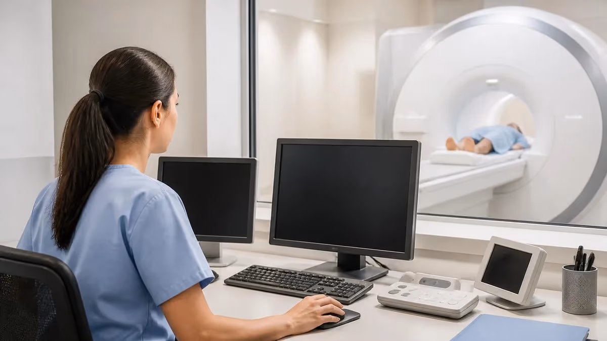
What to Expect During an MRI
If you've never had an MRI, here's what actually happens from arrival to departure.
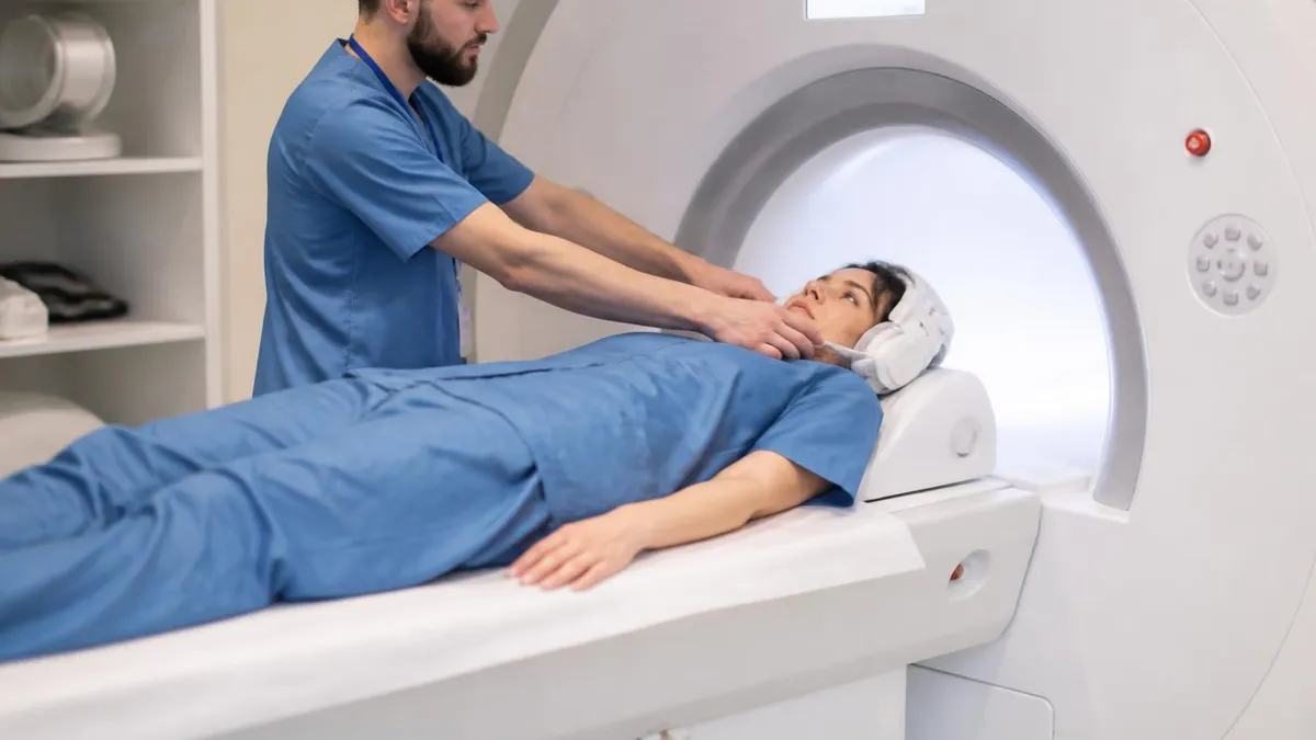
You'll complete a detailed safety screening questionnaire — every item matters, even if a question seems irrelevant to your situation. You'll remove anything metallic: jewelry, piercings, hair clips, glasses, hearing aids, and any clothing with metallic fasteners. The facility usually provides a gown if needed. A locker is available for your belongings.
You'll be positioned on a sliding table. A coil specific to the body part being imaged is placed around or near you. The table moves you into the bore of the magnet — typically about 60–70 cm in diameter and roughly 150 cm deep for a standard closed-bore scanner.
Once scanning begins, you'll hear a series of loud banging, clicking, and buzzing noises. That's normal — it's the gradient coils switching rapidly. Most facilities provide earplugs and headphones so you can listen to music. You need to remain as still as possible during each sequence. Some run 30 seconds; others run three to five minutes. Total scan time ranges from about 20 minutes for a straightforward knee exam to over an hour for a complex cardiac or staging study.
If contrast is needed, a nurse or technologist places an IV line and injects the gadolinium at a specific point during the exam. You may feel a brief cool sensation — the injection is typically painless. After the exam, you can eat, drink, and drive normally. A radiologist reads the images and sends a report to your referring physician, usually within 24–48 hours for routine exams and much faster for urgent cases.
MRI Careers: Becoming an MRI Technologist
Behind every scan is a professional trained in MRI tech school who positions the patient, selects the imaging protocols, monitors safety throughout the exam, and delivers diagnostic-quality images to the radiologist. It's a technically demanding and clinically rewarding career path.
Most MRI technologists begin as radiographers, earn the ARRT RT(R) credential, then add the MRI postprimary credential RT(MR) after completing additional clinical hours and passing the ARRT MRI certification exam. Some programs offer a more direct pathway into MRI. Either way, you'll need an accredited degree, structured clinical training, and a passing score on the certification exam.
Compensation reflects the specialized knowledge required. The MRI tech salary varies significantly by geography, experience level, and setting. Hospital positions, outpatient imaging centers, and travel contracts each have different pay structures — travel MRI techs are particularly well-compensated because demand consistently outstrips supply in many markets. It's one of the few imaging specialties where skilled technologists can command genuinely competitive wages early in their careers.
Emerging Applications and Research
Clinical MRI is remarkable on its own terms. But research applications are pushing the technology into territory that would have seemed implausible not long ago.
Ultra-high-field MRI at 7T and beyond allows researchers to image cortical layers, hippocampal subfields, and individual white matter tracts with submillimeter resolution. That kind of detail is changing what neuroscientists can learn about Alzheimer's disease, schizophrenia, and traumatic brain injury at a structural level that wasn't accessible before.
Diffusion tensor imaging (DTI) maps the movement of water along white matter fiber bundles, producing a kind of wiring diagram for the brain. Surgeons use pre-operative DTI to identify eloquent tracts before resecting brain tumors — reducing the risk of postoperative deficits by knowing exactly where the critical connections run.
MR spectroscopy goes beyond anatomy entirely. It measures the chemical composition of tissue — elevated choline, depressed N-acetylaspartate, the presence of lactate — producing metabolite fingerprints that help characterize tumors and distinguish recurrence from treatment effect without a biopsy.
Quantitative MRI techniques including T1 mapping and T2 mapping generate numerical measurements rather than purely visual images. This makes reliable comparison across time and across scanner manufacturers possible — which matters enormously for clinical trials and long-term patient monitoring.
Artificial intelligence is increasingly being applied to every stage of the MRI workflow — accelerating image acquisition through compressed sensing and deep learning reconstruction, automating anatomical segmentation, and flagging abnormalities for radiologist review. The goal isn't to replace radiologists. It's to let them read more scans, more accurately, in less time.
Understanding Your MRI Results
Most patients receive a written radiology report alongside (or instead of) raw images, though you're entitled to both. Reports follow a standard structure: technique, findings on each sequence, and a conclusions section where the radiologist summarizes what they found and offers a differential diagnosis.
The terminology is dense if you're unfamiliar with it. "Hyperintense" means bright on the specific sequence being described. "Hypointense" means dark. "T1-weighted" and "T2-weighted" refer to different pulse sequences that emphasize different tissue properties — fat is bright on T1; fluid is bright on T2. "No acute intracranial abnormality" means no urgent finding, not that everything is definitively normal. "Clinical correlation recommended" is how radiologists say: I can describe what I see, but your doctor needs to integrate this with the full clinical picture.
If the report leaves you with questions, ask your referring physician to walk you through it. That's what the follow-up appointment is for. Don't try to interpret a radiology report in isolation — context matters enormously in how findings are weighted.
MRI Costs and Access
MRI is expensive compared to X-ray and ultrasound. In the United States, a non-contrast brain MRI at a hospital may list at $1,500–$3,000 without insurance. At a freestanding outpatient imaging center, the same scan might run $400–$900. With insurance, your out-of-pocket cost depends on your deductible and whether the facility is in-network.
If you're uninsured or underinsured, it's worth calling several centers directly and asking about self-pay pricing — freestanding centers often offer substantial cash-pay discounts. Some hospital systems have financial assistance programs. In many other developed countries, MRI is covered by national health insurance and the patient cost is minimal or zero.
The price variation in the U.S. doesn't reflect meaningful differences in diagnostic quality for routine exams. A properly maintained 1.5T scanner at an accredited outpatient center produces clinically equivalent images to a hospital scanner. What matters most is the radiologist's expertise — specifically whether the subspecialty radiologist reading your brain MRI or your musculoskeletal MRI has deep experience with that type of study.
Don't delay a necessary MRI purely because of cost without first exploring your options. Call your insurance company, ask about financial assistance, compare imaging centers. The diagnostic information MRI provides can be irreplaceable — and delaying a scan that was clinically indicated is rarely the money-saving strategy it feels like in the moment.
Practical Tips for Your MRI Appointment
A little preparation makes the whole experience smoother — and helps ensure the images come out diagnostic rather than degraded by movement or incomplete setup.
Before you go, make a list of every implant or device in your body. Pacemakers, neurostimulators, cochlear implants, insulin pumps, stents, joint replacements, surgical clips, shrapnel, even metallic tattoo ink — it all needs to be disclosed. Bring the implant card if you have one. If you don't, knowing the manufacturer name and model number allows staff to look it up. Some implants are MR conditional and can be scanned safely under specific protocols; others are absolute contraindications. The technologist needs this information before the exam starts, not after you're already in the bore.
Wear comfortable, loose-fitting clothing without metallic zippers, snaps, or underwire — or simply plan to change into a gown. Leave jewelry, watches, and hair accessories at home. The locker at the facility is fine for valuables, but leaving metal at home eliminates the possibility of forgetting something in a pocket.
If you're claustrophobic, talk to your referring doctor before your appointment date — not on the day of the exam. There may be time to prescribe a mild anxiolytic, discuss whether an open-bore scanner is appropriate for your exam, or arrange additional support. Knowing what to expect in advance — the sounds, the confined space, the duration — helps many people manage without medication. Others genuinely need pharmacological support, and there's no judgment in that. Your goal is to stay still so the images are actually useful.
Bring prior imaging if you have it — CDs or access to a patient portal with previous MRI or CT studies. Comparing current findings to a prior baseline is often the most useful thing a radiologist can do, especially if you're being monitored for a known lesion, MS plaques, or structural abnormality. A scan without a prior comparison is still valuable, but context changes interpretation significantly.
What MRI Can't Do
It's worth being clear-eyed about the limitations. MRI isn't a perfect test, and understanding what it can't do helps you have realistic conversations with your doctor about what the results actually mean.
MRI doesn't see acute blood well in some sequences. Fresh hemorrhage is actually well-visualized on certain protocols, but the appearance changes rapidly as the blood breaks down — so timing matters, and the type of sequence ordered must match the clinical question. A radiologist who doesn't know the clinical history may misinterpret what they're seeing.
MRI struggles with lung tissue. Air and MRI don't play well together — the air-filled spaces of the lung create severe susceptibility artifacts that obscure detail. CT is far superior for pulmonary imaging. If your doctor is looking for a pulmonary embolism, pneumonia, or a lung nodule, you're getting a CT, not an MRI.
Motion degrades MRI images severely. A patient who can't hold still — whether from pain, anxiety, involuntary movement, or simply being too young — will produce images full of motion artifact that are difficult or impossible to interpret. Children often require sedation for this reason. Adults with severe pain or movement disorders face similar challenges.
And MRI isn't available everywhere. In many low- and middle-income countries, MRI scanners are limited to major urban centers, making access deeply unequal. Even in wealthy countries, wait times can be weeks to months for non-urgent studies. The technology is powerful — but it's not universally accessible.
None of this diminishes what MRI can do when it's the right tool for the right clinical question. It's transformed diagnostic medicine, and for many conditions there's simply no substitute for what it shows. But used thoughtfully — ordered when it's genuinely indicated, interpreted by someone with the right expertise, compared against relevant prior studies — that's when magnetic resonance imaging truly delivers on its potential.
About the Author
Medical Laboratory Scientist & Clinical Certification Expert
Johns Hopkins UniversityDr. Sandra Kim holds a PhD in Clinical Laboratory Science from Johns Hopkins University and is certified as a Medical Technologist (MT) and Medical Laboratory Scientist (MLS) through ASCP. With 16 years of clinical laboratory experience spanning hematology, microbiology, and molecular diagnostics, she prepares candidates for ASCP board exams, MLT, MLS, and specialist certification tests.
Join the Discussion
Connect with other students preparing for this exam. Share tips, ask questions, and get advice from people who have been there.
View discussion (6 replies)