MRI With Contrast: When It's Needed and What to Expect 2026 July
What is an MRI with contrast? Learn how gadolinium contrast dye works, when doctors order contrast MRI, risks, and what to expect during the procedure. 🔎
An MRI with contrast is a magnetic resonance imaging scan that uses an injectable contrast agent — typically a gadolinium-based compound — to make certain tissues and structures appear more clearly on the resulting images. The contrast material doesn't make the MRI machine work differently. Instead, it changes how specific areas of your body appear on the images by altering the magnetic properties of nearby water molecules, which the MRI detects.
The result is sharper differentiation between tissues that might otherwise look similar, making it easier for radiologists to identify tumors, inflammation, infection, damaged blood vessels, and other abnormalities that would be harder to see on a standard scan without contrast.
Not every MRI requires contrast. Many routine scans — musculoskeletal imaging for a torn ligament, a brain MRI to rule out stroke in a patient without suspicious findings, spine imaging for disc problems — are performed without contrast because the standard scan provides all the information the physician needs. The decision to use contrast is made by the ordering physician and the radiologist together, based on the clinical question they're trying to answer. When the physician's question involves vascularity (how well an area is supplied with blood), inflammation, or the presence of a lesion with uncertain boundaries, contrast typically helps.
The contrast agent most commonly used in MRI is gadolinium, a rare earth metal that is chelated — chemically bonded to a carrier molecule — to make it safe for intravenous injection. Gadolinium compounds are not the same as the iodine-based contrast dyes used in CT scans, and patients who have had reactions to CT contrast are not automatically at risk for MRI contrast reactions. However, gadolinium carries its own risk profile that patients and physicians need to evaluate, particularly for people with kidney disease.
Understanding when contrast is used — and when it isn't — helps you prepare for your scan and ask informed questions about your imaging order. If your doctor has ordered an MRI with contrast and you're not sure why, asking is entirely reasonable. Knowing whether the contrast is specifically necessary for your clinical situation, or whether a non-contrast scan might answer the same question, is a conversation worth having.
This guide covers how gadolinium contrast works, when it's indicated, what the procedure involves, and what you should disclose to your imaging team before your appointment. For a broader overview of the MRI procedure, the MRI scan guide covers preparation, what to expect, and what happens after imaging.
It's also worth knowing that contrast and no-contrast MRI are often ordered together as a combined protocol. Many institutions perform an MRI without contrast first, review those images, and then inject gadolinium to acquire contrast-enhanced sequences in the same sitting. This approach provides both the baseline non-contrast images and the contrast-enhanced images that show enhancement patterns.
When your imaging order says "MRI with and without contrast," this is what it means — two sets of sequences in one appointment, not two separate visits. The combined protocol adds time but gives the radiologist maximum information, and it's particularly common for brain, spine, and liver imaging where both baseline and contrast-enhanced findings contribute to the final interpretation.
MRI With Contrast: Key Facts
Gadolinium contrast works by taking advantage of how MRI images are produced. MRI creates images using the magnetic properties of hydrogen atoms in your body's water molecules. When you're placed in the scanner's powerful magnetic field and exposed to radiofrequency pulses, your hydrogen atoms emit signals that the scanner detects and converts into images. The strength and timing of those signals vary depending on the type of tissue — which is how MRI distinguishes between muscle, fat, fluid, and other structures.
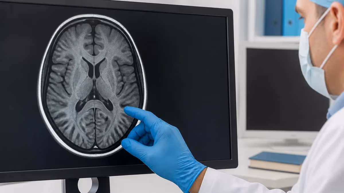
Gadolinium alters this process by shortening the relaxation time of nearby hydrogen atoms, which makes them emit stronger signals faster. Areas where gadolinium has accumulated appear brighter on certain MRI sequences (specifically T1-weighted images) than surrounding tissue. This brightening effect — called enhancement — is what makes contrast useful. Areas with increased blood supply, disrupted blood-brain barriers, active inflammation, or high vascular density accumulate more gadolinium and appear brighter relative to adjacent tissue that accumulates less.
The blood-brain barrier plays a specific role in brain and spine MRI with contrast. Under normal conditions, the blood-brain barrier prevents large molecules — including gadolinium chelates — from crossing from the bloodstream into brain tissue. Most healthy brain tissue doesn't enhance with contrast because the barrier keeps the gadolinium in the blood vessels.
When that barrier is disrupted — as happens with tumors, infections, multiple sclerosis lesions, or acute inflammation — gadolinium can cross into the tissue and causes enhancement. This is why brain MRI with contrast is particularly valuable: enhancement patterns in the brain are highly informative for distinguishing different types of lesions and determining disease activity.
After injection, gadolinium circulates through the bloodstream and is cleared by the kidneys, typically within a few hours in patients with normal kidney function. Most patients don't notice anything during or after the contrast injection other than a mild coolness or warmth at the injection site. Some patients notice a brief metallic taste.
Within 24 hours, virtually all of the gadolinium compound is eliminated through urine in patients with healthy kidneys. For patients with impaired kidney function, clearance takes longer, which is why kidney function is evaluated before contrast is administered. The complete guide to how MRI works explains the underlying physics and imaging principles in more detail for those who want a deeper technical understanding.
Not all gadolinium contrast agents behave identically. There are two structural categories: linear and macrocyclic chelates. Linear gadolinium chelates — including some of the earlier generations of contrast agents — release free gadolinium ions more readily than macrocyclic chelates, which have a more stable ring structure that binds gadolinium more tightly. The difference in stability translates to different rates of tissue deposition. Most major imaging centers now preferentially use macrocyclic gadolinium agents when contrast is clinically indicated, particularly for patients who will need repeated contrast MRI over time, such as those being monitored for multiple sclerosis or cancer treatment response.
Research over the past decade has identified that small amounts of gadolinium can remain deposited in brain tissue and other organs long after a contrast MRI, even in patients with normal kidney function. The clinical significance of this deposition remains under active investigation.
To date, no neurological effects have been definitively linked to gadolinium deposition in patients with normal kidney function, and major radiology organizations continue to endorse gadolinium contrast as safe when clinically indicated. However, the finding has prompted a shift toward using linear gadolinium chelates less frequently in favor of macrocyclic chelates, which have lower deposition rates, when contrast is needed.
Tell your imaging team about any of the following before receiving gadolinium contrast:
- Kidney disease or reduced kidney function — Gadolinium clearance depends on kidney function. Patients with eGFR below 30 mL/min/1.73m² are at elevated risk for nephrogenic systemic fibrosis (NSF), a rare but serious condition. Your team will check your kidney function before proceeding.
- Previous reaction to MRI contrast — Prior adverse reactions to gadolinium should be reported. Pre-medication protocols may be used for patients with previous reactions.
- Pregnancy — Gadolinium use during pregnancy is generally avoided unless the benefit clearly outweighs the risk, as gadolinium crosses the placenta and its effects on fetal development are not fully established.
- Breastfeeding — A very small amount of gadolinium can pass into breast milk. Most guidelines consider it safe to continue breastfeeding after contrast MRI, but some patients choose to pump and discard breast milk for 24 hours after the scan.
- Asthma or multiple drug allergies — Increased risk of contrast reaction; your team may recommend pre-medication.
Doctors order MRI with contrast when the clinical question they're trying to answer is one that a standard non-contrast scan is likely to miss or answer inadequately. The most common indication is evaluating a known or suspected lesion — a mass, a growth, a structural abnormality — where knowing whether it enhances with contrast provides critical diagnostic information. Enhancement indicates vascularity and blood-brain barrier disruption, which helps distinguish between different types of tumors, between active and inactive disease, and between benign and potentially malignant processes.
Brain tumors are one of the clearest indications for contrast MRI. The degree and pattern of enhancement are central to characterizing brain tumors, guiding biopsy targeting, and monitoring treatment response. A glioblastoma, a brain abscess, and a brain metastasis can all produce mass lesions on non-contrast MRI, but their enhancement patterns differ substantially — and distinguishing them changes management completely. Contrast MRI of the brain is also essential for detecting leptomeningeal disease (cancer spread to the fluid surrounding the brain and spinal cord), which often shows minimal findings on non-contrast scans.
Multiple sclerosis is another major indication. Active MS lesions enhance with contrast because demyelination disrupts the blood-brain barrier locally. On a non-abdominal mri cpt code you can see the white matter lesions, but you can't easily tell which ones are new and active versus old and stable. Contrast enhancement identifies active lesions — which is critical for monitoring disease activity, evaluating response to treatment, and making decisions about changing or escalating therapy. The brain MRI guide covers the specific imaging findings associated with MS and other neurological conditions in more detail.
Outside the brain, contrast MRI is commonly ordered for liver lesions (distinguishing hepatocellular carcinoma from benign hemangiomas), soft tissue masses where malignancy is suspected, the spine (where contrast helps identify infection, tumor, or inflammatory disease), the pelvis (for staging gynecological and prostate cancers), and the breast (where contrast-enhanced MRI is used for high-risk screening and surgical planning). Cardiac MRI with contrast evaluates myocardial viability, scar tissue after heart attack, and cardiomyopathy — the cardiac MRI guide covers those specific applications.
Vascular imaging with MRA (magnetic resonance angiography) also often uses gadolinium to visualize blood vessel anatomy, aneurysms, and stenoses more clearly than time-of-flight MRA techniques allow without contrast.
The decision between contrast and no-contrast is sometimes made in real time during the scan. A radiologist may begin a scan without contrast, review preliminary images, and decide that contrast would add important information — in which case the technologist will call you back to the table for an injection and additional sequences. This is a routine part of MRI practice and not a sign that something unexpected was found, though it understandably creates anxiety for patients who weren't told to expect it.
Follow-up imaging is another major context where contrast MRI is ordered. After a tumor has been treated with surgery, radiation, or chemotherapy, monitoring scans with contrast are used to track treatment response and detect recurrence. The enhancement pattern at follow-up can distinguish active tumor from treatment-related changes — a distinction that significantly affects what comes next in a patient's care. For patients in active cancer treatment, contrast MRI may be ordered every few months as part of a structured surveillance protocol.

Common Uses for MRI With Contrast
Enhancement patterns help characterize tumor type, distinguish recurrence from treatment effects, and guide biopsy targeting and surgical planning.
Contrast identifies active, newly demyelinating lesions — critical for monitoring disease activity and evaluating treatment response.
Brain abscesses, spinal epidural abscesses, and other infections show characteristic ring-enhancement patterns with contrast that distinguish them from tumors.
Dynamic contrast-enhanced MRI characterizes liver lesions as hemangioma, hepatocellular carcinoma, metastasis, or other entities based on enhancement timing.
Contrast differentiates active inflammatory disease, tumor, and infection from degenerative changes that can look similar on non-contrast spine MRI.
Contrast MRI evaluates lymph nodes, soft tissue invasion, and vascular involvement in staging pelvis, breast, and other cancers for treatment planning.
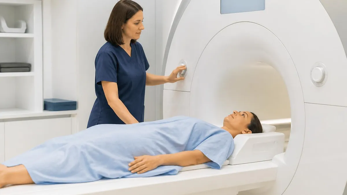
When you arrive for an MRI with contrast, the procedure starts much like any standard MRI. You'll be asked to remove metal-containing items, change into a gown, and complete a safety screening questionnaire about implants, prior surgeries, and medical history. Because contrast will be administered, the technologist will also ask about kidney disease, prior contrast reactions, pregnancy, and allergies. In some facilities, blood is drawn to check kidney function before contrast-eligible patients receive gadolinium — particularly in patients over 60 or with known kidney conditions.
A peripheral IV line is placed, usually in a vein in the arm or hand. The IV is used both to flush the contrast in quickly and to maintain venous access in case any reaction requires treatment. The actual MRI scan begins with non-contrast sequences, which the radiologist will review while you're still in or near the scanner.
When the pre-contrast images are complete, the technologist will inject the gadolinium through the IV — either manually or using an automated power injector — and additional contrast-enhanced sequences are acquired immediately afterward. The timing of imaging after injection matters: some sequences are designed to capture the arterial phase (very shortly after injection), others the venous phase, and others delayed phases — all of which show different enhancement patterns.
Most patients experience little or nothing from the contrast injection itself. A sense of warmth spreading from the injection site through the body is common and normal. Some patients notice a brief metallic or unusual taste. Nausea is less common but can occur. Serious allergic reactions — hives, difficulty breathing, anaphylaxis — are rare, occurring in well under one percent of injections, but imaging centers are equipped with emergency medications and protocols to manage them. If you feel anything unusual during or after the injection, tell the technologist immediately.
The total scan time for an MRI with contrast is typically longer than a non-contrast scan because additional sequences are acquired after the injection. Depending on the body part and the clinical indication, a contrast MRI may run 45 minutes to 90 minutes total. The most important thing during the scan is staying still, especially during the sequences that require the highest image quality.
Movement blurs the images and may require sequences to be repeated. If claustrophobia is a concern, mention it before your scan — mild sedation can often be arranged, or an open MRI may be an option. The open MRI guide explains the differences in image quality and the patient experience compared to traditional closed-bore scanners, and the guide to how long MRIs take breaks down typical scan durations by body part and type.
After the scan, you can typically resume normal activities immediately — there are no restrictions on eating, drinking, or driving after MRI contrast unless you received sedation. Drinking extra fluids after the scan is commonly recommended to help flush gadolinium through your kidneys more quickly, though this is based on general principle rather than specific evidence.
Your imaging results will be interpreted by a radiologist and communicated to your ordering physician, who will discuss the findings with you — typically within a few days for routine studies, or more urgently if the findings require immediate clinical action. If you're being monitored for a known condition, keep a record of each scan date and contrast type used, as cumulative gadolinium exposure may be relevant information for your care team over time.
What to Tell Your Doctor Before MRI With Contrast
- ✓Any history of kidney disease, reduced kidney function, or dialysis — gadolinium clearance depends on kidney function
- ✓Any previous reactions to MRI contrast or CT contrast dye
- ✓Current pregnancy or the possibility of pregnancy
- ✓Whether you are breastfeeding
- ✓All current medications, particularly metformin (for diabetes) — some protocols require temporary discontinuation around contrast administration
- ✓Any implanted devices (pacemakers, neurostimulators, cochlear implants, surgical clips) — some are not MRI-compatible
- ✓History of asthma or multiple drug allergies, which may warrant pre-medication before contrast
MRI With vs. Without Contrast by Situation
With contrast: Tumor characterization, active MS lesions, brain abscess or meningitis, leptomeningeal disease, spinal infection or tumor, post-surgical evaluation to distinguish scar from recurrent tumor.
Without contrast: Initial workup for headache, stroke evaluation, degenerative disc disease, routine spinal stenosis assessment, most first-time brain MRI screening where no lesion is known.
Key principle: If a lesion is already identified on a prior scan, contrast usually adds meaningful information about its current status and activity. Initial screening scans often start without contrast and add it if findings warrant.
MRI With Contrast vs. Without
- +Reveals active inflammation, disrupted blood-brain barrier, and lesion vascularity not visible on standard scans
- +Essential for tumor characterization — enhancement patterns distinguish different tumor types
- +Identifies which MS lesions are newly active versus old and stable
- +Enables vascular imaging (MRA) with sharper vessel definition than non-contrast techniques
- +Low adverse reaction rate — serious reactions occur in well under 1% of administrations
- −Requires IV placement and injection, adding discomfort and time to the procedure
- −Contraindicated or requires precaution in patients with severe kidney disease (NSF risk)
- −Small amounts of gadolinium may be retained in tissue — clinical significance still under investigation
- −Allergic reactions, though rare, can occur and require monitoring for 30 minutes post-injection
- −Adds cost and scan time compared to non-contrast MRI
MRI Questions and Answers
About the Author

Medical Laboratory Scientist & Clinical Certification Expert
Johns Hopkins UniversityDr. Sandra Kim holds a PhD in Clinical Laboratory Science from Johns Hopkins University and is certified as a Medical Technologist (MT) and Medical Laboratory Scientist (MLS) through ASCP. With 16 years of clinical laboratory experience spanning hematology, microbiology, and molecular diagnostics, she prepares candidates for ASCP board exams, MLT, MLS, and specialist certification tests.
Join the Discussion
Connect with other students preparing for this exam. Share tips, ask questions, and get advice from people who have been there.
View discussion (6 replies)