MRI Scan Guide 2026 June: Types, Preparation & What to Expect
Free MRI Scan Guide 2026 June: Types, Preparation practice test with questions and answer explanations. Prepare for the 2026 June exam with instant scoring. 🧠
MRI Scan: Complete Guide for 2026
An mri scan — short for magnetic resonance imaging — uses powerful magnetic fields and radio waves to produce detailed images of your organs, tissues, and bones. Unlike X-rays, it emits no radiation, making it one of the safest diagnostic tools in medicine. Doctors order MRI scans to investigate everything from unexplained headaches to joint pain, tumors, and spinal injuries.
If you've been told you need one, you're probably wondering what it's like. It's louder than most people expect — the machine clicks and thumps during imaging — but the procedure itself is painless. Most scans take 30–60 minutes, and you can go home the same day. Understanding mri meaning and what the scan actually measures can ease a lot of the anxiety that comes with the unknown.
This guide covers everything: the different scan types, how to prepare, what the scanner feels like, costs, and tips for getting the clearest results. Whether you're scheduled for a brain scan, a hip MRI, or a cardiac workup, you'll walk in ready.
How an MRI Scan Works
Your body is roughly 60% water. Water molecules contain hydrogen atoms — and hydrogen nuclei respond to magnetic fields. An MRI machine creates a magnetic field up to 60,000 times stronger than Earth's own field, aligning the hydrogen atoms in your body. A radio wave pulse then knocks those atoms out of alignment; when they snap back, they release a signal. The scanner detects that signal and a computer transforms it into cross-sectional images called slices.
That's why there's no radiation. The process is entirely electromagnetic — magnets and radio waves, nothing ionizing. It's also why metal implants, pacemakers, and certain medical devices are safety concerns inside the scanner room. Before your appointment, review mri safety guidelines with your care team, especially if you have any implanted metal devices.
Two key scan variations exist: with and without contrast dye. Contrast MRI uses gadolinium — a metallic element injected into a vein — to highlight blood vessels, tumors, and areas of inflammation more sharply. Your doctor decides which you need based on what they're looking for.
Types of MRI Scans by Body Part
MRI is flexible — it can image virtually any body region. Different body areas require different coils and sequences. Here's what you need to know about the most common types.
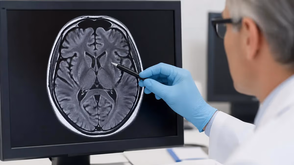
Brain and Head MRI
A head brain mri is ordered for suspected strokes, tumors, multiple sclerosis, seizure disorders, and unexplained cognitive changes. It takes 45–60 minutes and usually requires contrast. Some head scans include a head mri cage — a helmet-shaped coil that surrounds your skull to boost signal strength. Don't be alarmed by it; it doesn't touch your head.
Spine MRI
Spinal MRI is one of the most common orders. A herniated disc in lower back mri shows bulging discs pressing on nerve roots in high resolution — far better than X-ray or CT for soft tissue detail. Cervical (neck), thoracic (mid-back), and lumbar (lower back) scans are ordered separately because each region requires its own coil and protocol. Lumbar scans are typically 30–40 minutes.
Hip and Knee MRI
Hip labrum tear on mri is a frequent finding in active adults with groin pain and clicking. Hip mri with contrast (hip mri contrast) produces sharper labral detail. A hip mri scan typically runs 40–50 minutes. Knee MRI is shorter — around 30 minutes — and is excellent for identifying ACL tears, meniscus injuries, and cartilage wear.
Heart MRI
Cardiac MRI (CMR) assesses heart muscle function, valve disease, and myocardial scarring. Heart mri without contrast works for structural assessment; contrast is added when evaluating for cardiomyopathy or prior heart attack. This is the most technically demanding MRI type — it takes 60–90 minutes and requires the scanner to synchronize imaging with your heartbeat.
Pelvis and Abdomen
A pelvis mri scan evaluates reproductive organs, bladder, prostate, and pelvic floor disorders. It's routinely ordered for endometriosis staging, prostate cancer assessment, and uterine fibroid mapping. abdominal mri cpt code images the liver, kidneys, and pancreas. Both require breath-hold sequences — you'll be asked to hold your breath for 15–20 seconds at a time, several times during the scan.
Femur and Extremity MRI
Femur mri detects stress fractures, avascular necrosis, bone tumors, and soft tissue masses around the thigh. It's particularly useful when X-rays come back normal but symptoms persist. Extremity MRI — wrist, elbow, ankle, foot — can be done in an open or extremity-only scanner, which feels far less claustrophobic than a full-body tube.
Thinking about working in MRI? Learn about mri tech school programs and what the licensing path looks like, or explore the mri tech salary ranges to see if the career fits your goals.
Preparation by Scan Type
No special diet prep is required for most brain MRI scans. Remove all metal — earrings, hair clips, dental retainers if removable. Let your technologist know if you have any metal implants in your skull. Contrast brain MRI: avoid eating 2 hours before if you have a history of nausea with injections. A facial mri scan (for sinus disease, orbit issues, or parotid gland) follows the same protocol but uses a different coil positioned near your face.
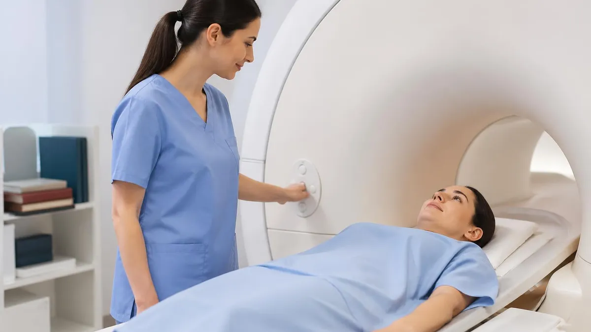
How to Prepare for Your MRI Scan
Preparation is simpler than most people expect — but a few missteps can delay or cancel your scan. Here's what actually matters.
Metal and Implants
The #1 safety rule: tell your MRI team about every piece of metal in or on your body. Implanted devices — pacemakers, cochlear implants, spinal cord stimulators, certain aneurysm clips — may be MRI-incompatible. Bring your implant card if you have one; the technologist verifies compatibility with MRI-Conditional databases before you enter the room. Jewelry, body piercings, and underwire bras must come off. Tattoos with metallic ink can heat up — mention them.
Clothing and Valuables
Wear comfortable, loose clothing with no metal zippers or buttons. Sweatpants and a cotton shirt are ideal. You'll likely change into a gown anyway, but minimal metal reduces the hassle. Leave your wallet, keys, and credit cards in the locker outside the MRI suite — the magnetic field can wipe magnetic stripes and damage certain cards.
Claustrophobia
Roughly 5–10% of patients experience anxiety in the scanner. Don't suffer through it silently. Tell your doctor before your appointment — they can prescribe a mild sedative (typically lorazepam or diazepam) taken 30–60 minutes before. You'll need a driver if you take sedation. Alternatively, some facilities have open MRI scanners that reduce the enclosed feeling, though image quality is lower than closed-bore machines.
Contrast Dye Considerations
If your scan involves gadolinium contrast, tell your doctor if you have kidney disease. Gadolinium is generally safe but can cause complications in patients with severely impaired kidney function (GFR below 30). Drink plenty of water after a contrast scan — it helps flush the dye. Allergic reactions to gadolinium are rare but possible; mention any prior contrast reactions.
Reading Your MRI Results
Your MRI report uses clinical language that can feel intimidating. A few terms you are likely to see: hyperintense means bright on the image; hypointense means dark. Mass effect describes tissue being pushed by a lesion. T1-weighted and T2-weighted are two standard image sequences — T2 lights up fluid, making it useful for spotting inflammation and edema. Do not google your report in isolation. Wait for your doctor to interpret findings in the context of your symptoms and history. Most incidental findings — things spotted that were not the reason for the scan — are benign and require nothing more than a follow-up MRI in 6 to 12 months.
What to Do After Your MRI
Most people resume normal activities immediately. If you received sedation, arrange a ride. If contrast was used, stay hydrated. Your radiologist's report goes to your referring physician — expect to hear back within 24–72 hours. If you haven't received results after 72 hours, call your doctor's office; don't assume no news is good news.
- ✓Confirm your exam appointment and location
- ✓Bring required identification documents
- ✓Arrive 30 minutes early to check in
- ✓Read each question carefully before answering
- ✓Flag difficult questions and return to them later
- ✓Manage your time — don't spend too long on one question
- ✓Review flagged questions before submitting

MRI Pros and Cons
- +Structured MRI study guides organize all required content in exam-aligned order, reducing time spent identifying what to study
- +Combining review guides with practice questions provides both content knowledge and test-taking fluency
- +Focused study plans allow candidates to allocate more time to weak areas rather than reviewing already-mastered content
- +Free and low-cost study resources mean comprehensive preparation is accessible at any budget level
- +Spaced repetition techniques (Anki, regular review sessions) significantly improve long-term retention of tested facts
- −No single study guide covers all tested content optimally — most candidates need 2–3 resources for complete preparation
- −Study guides can become outdated quickly when exam content is updated; verify edition currency before purchasing
- −Self-study requires self-discipline; candidates without structured external accountability often underallocate preparation time
- −Coverage breadth in comprehensive guides can create false confidence — recognizing content is not the same as answering questions correctly under timed conditions
- −Study time estimates in guides often assume ideal conditions; real preparation time is typically 30–50% longer due to life disruptions
About the Author
Medical Laboratory Scientist & Clinical Certification Expert
Johns Hopkins UniversityDr. Sandra Kim holds a PhD in Clinical Laboratory Science from Johns Hopkins University and is certified as a Medical Technologist (MT) and Medical Laboratory Scientist (MLS) through ASCP. With 16 years of clinical laboratory experience spanning hematology, microbiology, and molecular diagnostics, she prepares candidates for ASCP board exams, MLT, MLS, and specialist certification tests.
Join the Discussion
Connect with other students preparing for this exam. Share tips, ask questions, and get advice from people who have been there.
View discussion (4 replies)