Brain MRI: What It Shows, How It Works & What to Expect 2026 June
Brain MRI explained: what it detects, how the scan works, prep tips, and what results mean. Learn what a brain MRI can and can't show. 🏆
What Is a Brain MRI?
A brain MRI—short for magnetic resonance imaging of the brain—is one of the most detailed diagnostic tools available to neurologists, neurosurgeons, and radiologists today. Unlike X-rays or CT scans, it doesn't use ionizing radiation. Instead, it uses powerful magnetic fields and radio waves to produce highly detailed images of the brain's structure, tissue, and blood vessels.
You'll hear the terms "brain MRI" and "head MRI" used almost interchangeably, though technically a head MRI can include facial bones and sinuses while a brain MRI focuses specifically on brain tissue and the surrounding structures. Either way, if a doctor orders one, they're trying to get a clear picture of what's happening inside your skull—and that's a good thing.
Brain MRIs are used to diagnose an enormous range of conditions: strokes, tumors, multiple sclerosis, epilepsy, dementia, brain injuries, infections, and more. They're also used to monitor known conditions, plan surgical procedures, and evaluate symptoms that don't have an obvious cause—like persistent headaches, memory problems, or unexplained dizziness.
How Does a Brain MRI Work?
Here's the basic physics without getting too deep into the weeds. Your body is mostly water, and water contains hydrogen atoms. The MRI machine generates a powerful magnetic field that temporarily aligns the hydrogen protons in your body. When radio waves are pulsed through, those protons absorb energy and then release it as they return to alignment. The scanner detects that released energy and uses it to build cross-sectional images of the brain.
Different tissues release energy at different rates, which is why the MRI can distinguish between gray matter, white matter, cerebrospinal fluid, blood vessels, tumors, and areas of inflammation or injury. The contrast is remarkable—far sharper than what a CT scan can produce for soft tissue.
There are several MRI sequences used for brain imaging, and the radiologist will often choose a combination depending on what they're looking for:
- T1-weighted images — show anatomy clearly; fat appears bright, fluid appears dark
- T2-weighted images — fluid appears bright; great for detecting edema and lesions
- FLAIR (Fluid-Attenuated Inversion Recovery) — suppresses cerebrospinal fluid signal to make lesions near ventricles more visible
- DWI (Diffusion-Weighted Imaging) — critical for detecting acute stroke within minutes of onset
- MRA (MR Angiography) — images blood vessels without traditional contrast dye
- fMRI (Functional MRI) — maps brain activity by detecting blood flow changes; used before brain surgery
Most routine brain MRIs take between 30 and 60 minutes. If your doctor orders a contrast MRI, a technologist will inject gadolinium—a contrast agent—through an IV partway through the scan. Gadolinium makes certain structures, like tumors or inflamed tissue, show up more vividly on the images.
What a Brain MRI Can Detect
The range of conditions a brain MRI can identify is genuinely wide. It's one of the reasons neurologists rely on it so heavily—it's not a one-trick diagnostic tool. Here's a breakdown of major categories:
Strokes and Vascular Disease
When brain tissue is deprived of blood flow, it changes in detectable ways—fast. Diffusion-weighted MRI can spot an ischemic stroke within minutes of it happening, which is critical for treatment decisions. Brain MRI also detects hemorrhagic strokes, chronic small vessel disease, and transient ischemic attacks (TIAs) that left traces.
Brain Tumors
Both primary brain tumors (like glioblastoma or meningioma) and metastatic tumors that spread from cancers elsewhere in the body show up clearly on MRI—especially with contrast. The scan can reveal tumor size, location, and often give clues about whether it's likely to be benign or malignant before a biopsy is performed.
Multiple Sclerosis
MS causes demyelinating lesions in the brain and spinal cord. MRI is the gold standard for MS diagnosis and monitoring. The number, location, and activity of lesions (some enhance with contrast, indicating active inflammation) directly inform treatment decisions. It's hard to overstate how central MRI scan technology is to MS care.
Epilepsy and Seizures
For someone with epilepsy, brain MRI helps identify a structural cause—scar tissue, cortical dysplasia, hippocampal sclerosis, or a tumor. This matters hugely when surgery is being considered. If MRI shows a resectable lesion causing seizures, surgery can sometimes eliminate them entirely.
Dementia and Neurodegenerative Disease
In Alzheimer's disease, brain MRI reveals characteristic patterns of atrophy, particularly in the hippocampus. It can help distinguish Alzheimer's from other dementias like Lewy body dementia or frontotemporal dementia—which look different on imaging. Magnetic resonance imaging is increasingly used to track disease progression and evaluate participants in clinical trials.
Brain Infections and Inflammation
Encephalitis, meningitis, abscesses, and autoimmune encephalopathies all produce characteristic MRI findings. When someone presents with altered consciousness, seizures, or neurological symptoms and an infectious or inflammatory cause is suspected, brain MRI is often one of the first tests ordered.
Traumatic Brain Injury
While CT is often done first in acute head trauma (it's faster and better for detecting fresh blood), MRI provides far more detail about diffuse axonal injury and subtle white matter damage that CT misses. In patients who seem "more injured than their CT suggests," MRI often reveals the full picture.
Headaches and Unexplained Neurological Symptoms
Not every headache warrants a brain MRI—but certain red flags do: sudden severe headache, new neurological deficits, headaches that wake you from sleep, or headaches that progressively worsen over weeks. Your doctor weighs symptoms carefully before ordering one.
Preparing for a Brain MRI
Preparation is usually straightforward, but there are a few things you need to know ahead of time.
The most important is metal. The MRI machine uses an extremely powerful magnet—strong enough to pull metal objects across the room at speed. Before entering the scanner room, you'll be asked detailed questions about any metal implants, devices, or foreign bodies. This includes:
- Cardiac pacemakers and defibrillators (most older models are contraindicated; some newer ones are MRI-conditional)
- Cochlear implants
- Certain aneurysm clips
- Metal fragments in the eyes (relevant for people who've worked with metal without eye protection)
- Deep brain stimulators
Most modern orthopedic implants—hip replacements, knee replacements, titanium screws from spine surgery—are MRI-safe and won't cause issues. But you should always tell the MRI team about any implants or surgeries.
You'll remove jewelry, piercings, hearing aids, glasses, and any clothing with metal fasteners before the scan. A locker will be provided for your belongings.
Contrast scans require additional preparation. If you have kidney disease, your doctor will check your kidney function before ordering gadolinium contrast, since impaired kidneys can't clear it properly. You may also be asked if you have any gadolinium allergies or history of prior contrast reactions.
If you're claustrophobic, tell your doctor before the scan—not after. Open MRI machines are available (though lower field strength) and some facilities have wide-bore scanners that feel less enclosed. Oral sedation or anti-anxiety medication can be prescribed if needed. The scanner is loud—you'll be given earplugs or headphones—and you'll need to lie still, which is harder than it sounds for some people.
There's no specific diet restriction for a standard brain MRI without contrast. For contrast scans, some centers ask you to avoid eating for a few hours beforehand, though this varies.
What Happens During the Scan
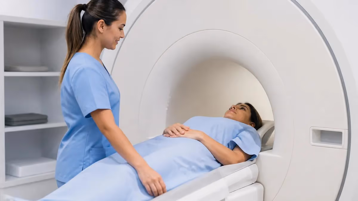
On the day of your scan, you'll check in at the imaging center or hospital radiology department. A technologist—an MRI tech—will go through a safety screening questionnaire with you and verify there are no contraindications. This is thorough, and for good reason.
You'll change into a gown, secure your belongings, and be positioned on the scanner table, typically lying on your back. For brain imaging, a specialized coil—essentially a helmet-shaped receiver—is placed around your head. It doesn't touch you painfully, but it does feel snug. This coil dramatically improves image quality by capturing the radio frequency signals more efficiently.
The table slides into the bore of the magnet. For a standard closed MRI, the bore is roughly 60-70cm in diameter. You'll hear a series of loud knocking, buzzing, and thumping sounds—this is completely normal and expected. The sounds come from gradient coils within the machine rapidly switching on and off. Earplugs significantly reduce this noise.
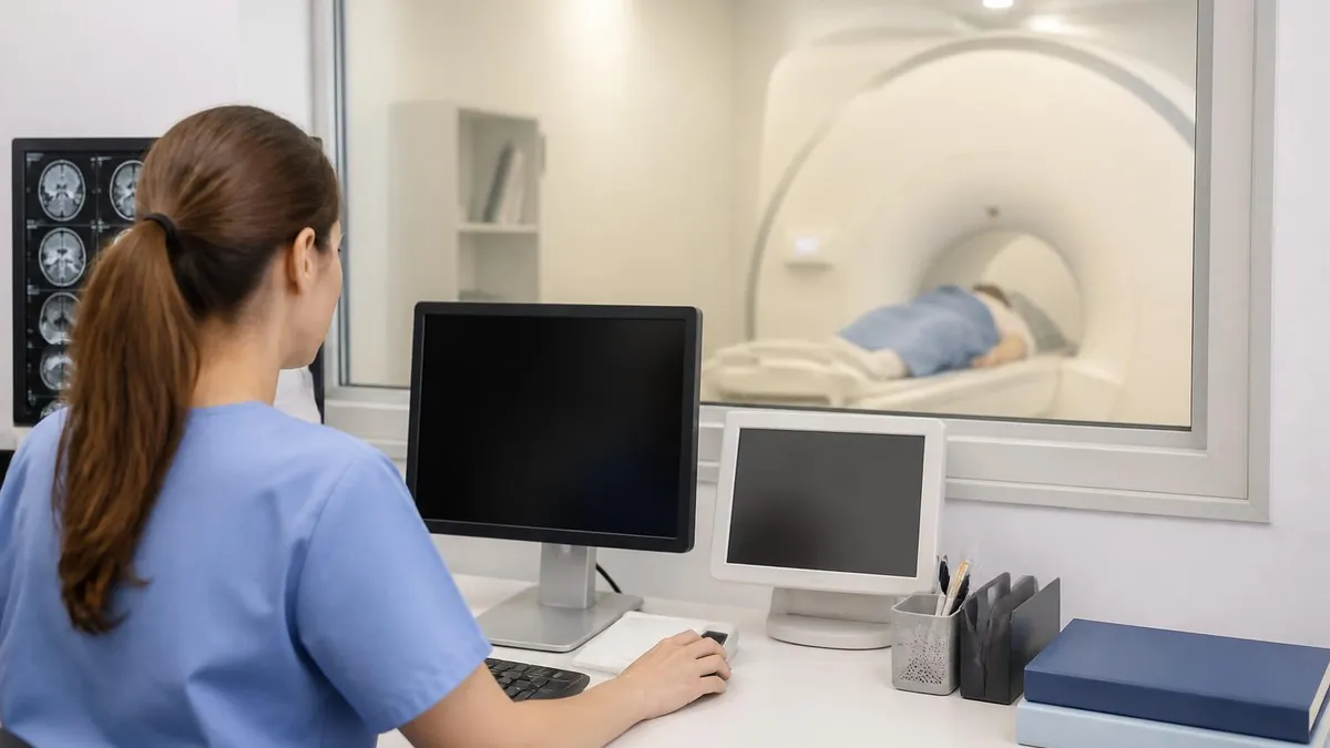
Communication happens through an intercom. The technologist will give you instructions—most importantly, to hold still during imaging sequences. Motion is the enemy of image quality. Even swallowing can blur certain sequences. When you need to stay absolutely still, it's usually only for a few minutes at a time.
If contrast is being given, you'll feel a brief cool sensation as the gadolinium is injected. Some people notice a slight metallic taste or mild warmth—both are temporary and harmless.
When the scan is done, the table slides out, the coil is removed, and you're free to go. There's no recovery time for a standard MRI (sedation is different). You can drive, eat, and resume normal activities immediately.
Understanding Your Brain MRI Results
After the scan, a radiologist—a physician who specializes in interpreting medical images—will review the images and write a report. This report goes to your ordering physician, who'll discuss the findings with you.
A few terms you might encounter:
- Normal brain MRI — all structures appear as expected; no abnormal signal, masses, or lesions detected
- T2/FLAIR hyperintensities — bright spots on T2 or FLAIR sequences; can indicate many things from small vessel disease to demyelination to prior injury
- Enhancing lesion — a lesion that takes up contrast, indicating active inflammation or a disrupted blood-brain barrier
- Mass effect — when a lesion is large enough to push surrounding structures out of position
- Hydrocephalus — enlarged ventricles, often from impaired cerebrospinal fluid drainage
- Cerebral atrophy — loss of brain volume, which can be age-related or associated with neurodegenerative disease
It's worth noting that incidental findings are common on brain MRIs. A small arachnoid cyst, minor white matter changes, or an asymmetric ventricle might appear on your report and mean absolutely nothing clinically. Your doctor will put findings in context based on your symptoms and history—the MRI report alone doesn't tell the whole story.
If you're studying for the MRI registry exam or working toward becoming an MRI technologist, understanding MRI safety protocols is non-negotiable. Contraindications, magnetic field zones, and implant screening procedures are heavily tested topics. The path starts well before the registry exam—if you're exploring this field professionally, looking into MRI tech school programs is the right first step.
Brain MRI vs CT Scan: When Is Each Used?
Patients often wonder why they're getting one versus the other. The short answer: CT is faster and better for bone and acute bleeding; MRI is slower but dramatically better for soft tissue detail.
In acute trauma or suspected hemorrhagic stroke, CT is usually done first—results in minutes, and it's excellent at detecting fresh blood. But when doctors need to evaluate brain tumors, MS, dementia, epilepsy, or any condition involving brain tissue itself, MRI wins on resolution and contrast. For a side-by-side breakdown of when each is preferred, see MRI vs CT scan.
The decision also involves practical considerations. MRI takes longer, is more expensive, and has contraindications (metal implants) that CT doesn't. Some patients can't lie still for 45 minutes. In those cases, CT may be the more practical choice even if MRI would theoretically provide more detail.
Functional MRI: A Different Kind of Brain Scan
Worth mentioning separately: functional MRI (fMRI) maps brain activity, not just structure. It detects changes in blood oxygenation that correspond to neural activity—the basic principle being that active brain regions demand more oxygen.
Clinical fMRI is used primarily for surgical planning. Before removing a brain tumor or performing epilepsy surgery, neurosurgeons need to know where critical functions like language, memory, and motor control are located in that specific patient's brain. fMRI can map these areas non-invasively so surgeons can plan their approach and minimize the risk of damaging eloquent cortex.
Research fMRI is a different beast—it's what you read about in studies investigating attention, emotion, addiction, and cognition. It's a powerful research tool, though interpreting fMRI data correctly requires serious statistical rigor.
Brain MRI for MRI Registry Candidates
If you're studying for the MRI registry exam—specifically the ARRT MRI examination—brain MRI physics and protocols are central to the exam. You'll need to understand sequence selection, artifact recognition, and clinical applications at a level beyond what this article covers.
Knowing which sequences detect acute stroke (DWI), which show MS lesions best (FLAIR), and why gadolinium enhances certain pathologies requires solid grounding in MRI physics and pathophysiology. It's not enough to memorize—you need to understand why.
Practice questions are the best way to test whether you actually understand the material or just recognize familiar patterns. The MRI registry exam is challenging, and test-taking strategy matters as much as content knowledge. Working through full-length practice tests under timed conditions helps you identify gaps before exam day.
If you're early in your career and exploring this field, understanding the full scope—from the physics of magnetic resonance imaging to clinical applications to safety protocols—gives you the context to learn more efficiently. The MRI tech salary reflects the specialized knowledge and responsibility the role demands.
Brain MRI is one of the most clinically impactful technologies in modern medicine. Whether you're a patient trying to understand your upcoming scan, a student preparing for the registry exam, or a curious reader who wants to understand how it works—it's worth understanding well. The more you know about what the scan does and doesn't show, the better equipped you are to ask the right questions and make sense of the results.
About the Author
Medical Laboratory Scientist & Clinical Certification Expert
Johns Hopkins UniversityDr. Sandra Kim holds a PhD in Clinical Laboratory Science from Johns Hopkins University and is certified as a Medical Technologist (MT) and Medical Laboratory Scientist (MLS) through ASCP. With 16 years of clinical laboratory experience spanning hematology, microbiology, and molecular diagnostics, she prepares candidates for ASCP board exams, MLT, MLS, and specialist certification tests.
Join the Discussion
Connect with other students preparing for this exam. Share tips, ask questions, and get advice from people who have been there.
View discussion (4 replies)