MRI vs CT Scan: Key Differences, Cost, Radiation & When Each Is Used 2026 June
Pass your MRI vs CT Scan: Key Differences, Cost, exam on the first attempt. Practice questions with detailed answer explanations, hints, and instant ✍🏼
MRI vs CT Scan: What's Actually Different?
Your doctor orders an imaging study and you're wondering — why an MRI instead of a CT scan, or the other way around? These two technologies look similar from the outside, but they work through completely different physics, deliver different risks, and reveal different things about your body.
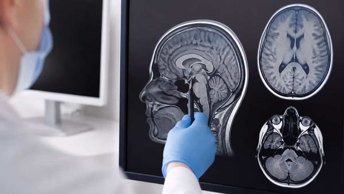
An MRI (Magnetic Resonance Imaging) uses powerful magnetic fields and radiofrequency waves to create detailed images of soft tissues. There is no ionizing radiation involved. The machine excites hydrogen atoms in your body's water molecules and measures how they realign — generating exquisitely detailed contrast between different tissue types.
A CT scan (Computed Tomography), sometimes called a CAT scan, fires X-rays from multiple angles around your body and uses a computer to reconstruct cross-sectional images. CT is significantly faster than MRI, handles patients with claustrophobia more readily, and produces superior images of dense structures like bone. It's the tool of choice in trauma bays and emergency departments where speed saves lives.
The choice between MRI and CT is never arbitrary. Radiologists select the modality that best answers the specific clinical question, balances diagnostic value against radiation risk, and fits the patient's physical situation. Here's how they differ across every dimension that matters.
The imaging field has evolved dramatically in recent years. MRI field strengths have increased to 3T and even 7T for research, improving resolution and enabling new applications. CT has moved to photon-counting detector technology, reducing radiation dose while dramatically increasing spatial resolution. AI-powered image reconstruction allows diagnostic-quality CT at doses 60–80% lower than conventional protocols. Both technologies continue advancing — yet their fundamental clinical niches remain stable, defined by physics that no software update can change.
Understanding these fundamentals makes you a better advocate for your own care. When a physician recommends an imaging study, they are weighing diagnostic yield, radiation risk, cost, availability, and your specific anatomy and medical history simultaneously. Knowing why they landed on MRI versus CT — or why they chose one over ultrasound or nuclear medicine — gives you the framework to ask informed questions and feel confident in the recommendation.
How Each Technology Works
Understanding the underlying physics explains why one scanner is recommended over another for specific conditions.
MRI physics: The MRI machine generates a powerful static magnetic field — typically 1.5 to 3 Tesla in clinical scanners. This field aligns hydrogen protons in your body's water molecules. The machine then pulses radiofrequency energy at those protons, knocking them out of alignment. When the pulse stops, they realign and release energy that detectors measure. Different tissues (fat, muscle, fluid, gray matter, white matter) have different relaxation times (T1 and T2), producing the contrast that makes MRI so diagnostically powerful. No radiation is used at any stage.
CT physics: A CT gantry rotates an X-ray tube around the patient, firing beams through the body at hundreds of angles. Detectors measure how much radiation is absorbed at each angle. A computer then performs back-projection reconstruction to build a three-dimensional dataset reformattable into any plane. Bone absorbs far more X-ray than air-filled lung or soft tissue, making CT exceptional for structural and density-based imaging. The tradeoff is ionizing radiation exposure to the patient.
Both modalities can use contrast agents to highlight blood vessels, tumors, or inflammation — but the agents differ significantly. MRI uses gadolinium-based agents (generally well-tolerated, rare allergic reactions, nephrotoxicity concern only in severe kidney disease). CT uses iodinated contrast (nephrotoxic at higher doses, more allergic reactions, hold metformin 48h after in renal patients).
MRI sequences are the key to its versatility. T1-weighted images show anatomy clearly — fat appears bright, fluid dark. T2-weighted images highlight fluid — making edema, herniated disc material, and tumor conspicuous. FLAIR (Fluid Attenuated Inversion Recovery) suppresses cerebrospinal fluid signal while keeping lesions bright, ideal for detecting MS plaques near ventricles. Gradient echo sequences detect hemorrhage and calcium. Diffusion-weighted imaging (DWI) maps water molecule movement — restricted diffusion in early stroke is positive within minutes of symptom onset, hours before CT or conventional MRI shows any change. This sequence-level specificity has no CT equivalent.
CT's key advantage is its quantitative nature. Tissue density is measured in Hounsfield units (HU) — a standardized scale where water is 0 HU, air is -1000 HU, and dense cortical bone is +1000 HU. This lets radiologists characterize materials precisely: blood (40–80 HU) is distinguishable from soft tissue tumor, fat (-80 to -120 HU) from muscle (35–55 HU), and calcification (>300 HU) from surrounding tissue.
The Hounsfield scale makes CT intrinsically quantitative in a way MRI, with its relative signal intensities, is not. This precision is exploited in lung cancer screening (measuring nodule density), cardiovascular imaging (coronary artery calcium scoring), and liver fat quantification — clinical applications where knowing exact tissue density drives clinical decisions.
Which Conditions Call for MRI?
MRI dominates whenever soft tissue contrast and detail are the diagnostic priority and time allows for a longer scan.
Brain and neurological conditions: MRI is the gold standard for brain tumor detection, multiple sclerosis lesion mapping, stroke evaluation (particularly diffusion-weighted imaging for early ischemic stroke), encephalitis, and dementia assessment. Superior soft tissue contrast allows visualization of subtle white matter changes, early infarcts, and small lesions that CT cannot resolve.
Spine imaging: Herniated discs, spinal cord compression, epidural abscesses, and spinal tumors are all evaluated with MRI. The ability to visualize the spinal cord itself — a soft tissue structure — is something CT cannot replicate without intrathecal contrast injection (myelography), a far more invasive procedure.
Joint and musculoskeletal assessment: Torn ligaments, meniscal tears, rotator cuff injuries, and cartilage damage are MRI territory. Sports medicine orthopedists rely on MRI for pre-surgical planning. A knee MRI can distinguish between a partial and complete ACL tear while simultaneously assessing the menisci, cartilage, and surrounding structures.
Prostate and pelvic imaging: Multiparametric prostate MRI has transformed prostate cancer detection, enabling targeted biopsy of suspicious lesions. Pelvic MRI is standard for uterine, ovarian, and rectal cancer staging. Cardiac MRI is increasingly used for myocardial viability assessment and cardiomyopathy characterization.
Cardiac MRI is increasingly used for myocardial viability assessment, cardiomyopathy characterization, and congenital heart disease evaluation — offering functional and structural detail that echocardiography and CT cannot always provide. For high-risk breast cancer patients, breast MRI supplements annual mammography screening, particularly when mammography and ultrasound findings are inconclusive or when tumor extent assessment is needed before surgery.
Functional MRI (fMRI) maps brain activity by detecting blood oxygenation changes. It is used preoperatively to identify eloquent cortex — areas controlling language, motor function, or vision — that surgeons must avoid when resecting brain tumors. This is MRI territory that CT cannot approach at all. Diffusion tensor imaging (DTI) traces white matter fiber tracts, useful in surgical planning and traumatic brain injury assessment. MRI spectroscopy provides metabolite information about tissue chemistry — helping distinguish radiation necrosis from tumor recurrence without biopsy.
MRI Key Concepts
What is the passing score for the MRI exam?
Most MRI exams require 70-75% to pass. Check the official exam guide for exact requirements.
How long is the MRI exam?
The MRI exam typically allows 2-3 hours. Time management is critical for success.
How should I prepare for the MRI exam?
Start with a diagnostic test, create a 4-8 week study plan, and take at least 3 full practice exams.
What topics does the MRI exam cover?
The MRI exam covers multiple domains. Review the official content outline for the complete list.
Which Conditions Call for CT?
CT wins whenever speed, bone detail, lung imaging, or emergency assessment is the priority.
Trauma: A polytrauma patient in the emergency department needs rapid, comprehensive injury assessment. CT provides a full-body survey — identifying intracranial hemorrhage, spinal fractures, pneumothorax, aortic injury, solid organ lacerations, and pelvic fractures — in a single fast study. MRI's 30–60 minute duration is incompatible with unstable trauma patients needing immediate surgery.
Chest and lung pathology: Pulmonary embolism, pneumonia, pleural effusion, lung cancer screening (low-dose CT), and pneumothorax are all CT-first indications. MRI performs poorly in the chest because the lungs contain little water (MRI's signal source) and cardiac/respiratory motion creates significant artifact.
Abdominal emergencies: Appendicitis, bowel obstruction, diverticulitis, kidney stones, and abdominal aortic aneurysm rupture are all evaluated with CT. A kidney stone invisible on plain X-ray appears immediately on non-contrast CT. CT abdomen/pelvis with contrast diagnoses appendicitis with sensitivity exceeding 95%.
Cancer staging: CT of the chest, abdomen, and pelvis with contrast remains the workhorse for initial cancer staging and treatment response — providing rapid whole-body survey with reasonable soft tissue detail and excellent vascular imaging.
CT angiography (CTA) has largely replaced conventional catheter angiography for evaluating the coronary arteries, aorta, pulmonary vasculature, and cerebral vessels. A CT coronary angiogram can rule out significant coronary artery disease in patients with atypical chest pain at far lower risk than invasive cardiac catheterization. CT colonography (virtual colonoscopy) provides full colon evaluation without sedation — though conventional colonoscopy remains preferred when therapeutic intervention (polyp removal) is anticipated simultaneously.
Low-dose CT lung cancer screening is now recommended annually for high-risk adults (ages 50–80, 20 pack-year smoking history, current or former smoker within 15 years). This represents CT at its most impactful — detecting early-stage lung cancer when it is still surgically resectable, dramatically improving survival rates compared to symptomatic detection. Positron emission tomography (PET) is almost always combined with CT (PET-CT) to fuse metabolic information with anatomical detail, the standard of care in oncology staging and treatment monitoring.
MRI Preparation Checklist
- ✓Remove all metal objects: jewelry, piercings, hairpins, hearing aids, watches
- ✓Inform the technologist of any metal implants, electronic devices, or prior surgeries
- ✓If contrast is ordered, blood draw to check kidney function (GFR) may be required
- ✓For abdominal/pelvic MRI, fast for 4–6 hours to reduce bowel motion artifact
- ✓Wear comfortable, loose clothing or change into a facility gown
- ✓Inform staff of claustrophobia — anxiolytic premedication or wide-bore MRI may be arranged
- ✓Verify all implants are MRI-conditional before scanning day
Claustrophobia and Patient Tolerance
The physical experience of MRI and CT differs substantially — for some patients, this determines which modality is feasible.
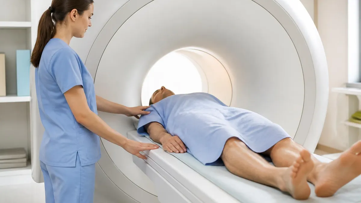
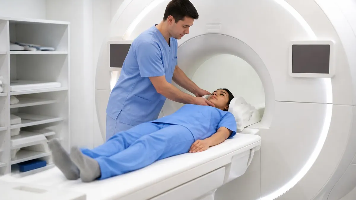
MRI claustrophobia: Standard closed-bore MRI scanners have a narrow tunnel (typically 60–70 cm bore diameter) that patients slide into completely. The machine is loud — gradient coils produce banging and clanging sounds reaching 110 decibels — and scans last 30–90 minutes. Solutions include anxiolytic premedication, wide-bore 3T systems (70 cm bore), open MRI systems (lower field strength, lower image quality), and in rare cases, general anesthesia for essential studies. Approximately 1–2% of patients cannot complete a standard closed-bore MRI due to claustrophobia.
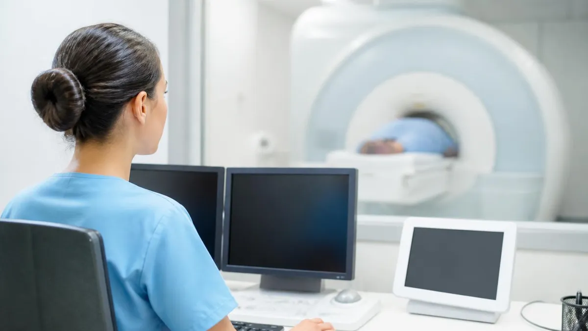
CT tolerance: CT is significantly more tolerable for claustrophobic patients. The CT gantry is a wide, open ring (typically 70–80 cm), patients pass through quickly, and the entire scan takes seconds to minutes. CT is also the only practical option for critically ill patients on ventilators, or patients with monitoring equipment that cannot enter an MRI suite due to magnetic field interference.
Metal implants and MRI safety: Ferromagnetic metal implants can be heated, displaced, or create imaging artifact in an MRI machine. This includes certain older pacemakers, cochlear implants, aneurysm clips, and metallic foreign bodies. Most modern orthopedic hardware (titanium rods, knee replacements) is MRI-conditional rather than MRI-incompatible, but implant verification is required before every MRI scan. CT has no metal contraindications — making it the default for patients with uncertain implant histories.
Pediatric patients present a special case for both modalities. Children struggle to remain still for 30–90 minute MRI scans, often requiring sedation or general anesthesia — which carry their own risks. CT's speed frequently eliminates sedation for children, a meaningful safety advantage. However, children are more radiosensitive than adults (their tissues divide more rapidly and they have more remaining life span for radiation effects to manifest), making the MRI preference for non-urgent pediatric imaging even stronger when clinically equivalent. Pediatric radiologists are expert at choosing the modality that minimizes both radiation risk and sedation risk for each clinical question.
Cost Comparison: Imaging Centers vs Hospital
Freestanding imaging centers charge 60–80% less than hospital-based radiology departments for identical studies. Brain MRI without contrast: $400–$800. Knee MRI: $500–$900. Head CT: $270–$500. Abdomen/pelvis CT: $400–$700. These cash-pay rates are widely available at independent imaging facilities and some health system price-transparency portals.
MRI Practice Test Questions
Prepare for the MRI - Magnetic Resonance Imaging exam with our free practice test modules. Each quiz covers key topics to help you pass on your first try.
MRI Knowledge
MRI Exam Questions covering Knowledge. Master MRI Test concepts for certification prep.
MRI Physics
Free MRI Practice Test featuring Physics. Improve your MRI Exam score with mock test prep.
MRI Anatomy and Pathology
MRI Test Prep for MRI Anatomy and Pathology. Practice MRI Quiz questions and boost your score.
MRI Anatomy and Positioning
MRI Questions and Answers on MRI Anatomy and Positioning. Free MRI practice for exam readiness.
MRI Contrast Agents
Free MRI Quiz on MRI Contrast Agents. MRI Exam prep questions with detailed explanations.
MRI Patient Care and Positioning
MRI Practice Questions for MRI Patient Care and Positioning. Build confidence for your MRI certification exam.
MRI Questions and Answers
MRI vs CT: Advantages and Disadvantages
- +MRI: No ionizing radiation — safe for children, pregnant women, repeated studies
- +MRI: Superior soft tissue contrast for brain, spine, joints, and pelvic organs
- +MRI: Multiple imaging sequences reveal tissue biology, not just anatomy
- +MRI: No radiation-induced cancer risk regardless of how many scans are needed
- +CT: Dramatically faster — complete scan in under 15 minutes, often seconds of scanning
- +CT: Superior bone and lung imaging — the best modality for fractures and chest pathology
- +CT: Works with metal implants — no ferromagnetic safety concerns
- +CT: Accessible 24/7 in most emergency departments without scheduling delays
- +CT: Better tolerated by claustrophobic patients due to open gantry design
- −MRI: Long scan times (30–90 min) — problematic for unstable or claustrophobic patients
- −MRI: Loud noise from gradient coils requires hearing protection
- −MRI: Metal implants may be contraindicated — thorough screening required before every scan
- −MRI: Higher cost at hospital-based facilities — $800–$3,500 for complex studies
- −MRI: Motion artifact from patient movement degrades image quality significantly
- −CT: Ionizing radiation — cumulative dose matters for children and frequent scanners
- −CT: Iodinated contrast nephrotoxicity risk — creatinine check required in renal patients
- −CT: Higher allergic reaction rate to iodinated contrast versus gadolinium
- −CT: Poor soft tissue contrast — misses subtle brain lesions, disc herniations, ligament tears
Choosing Between MRI and CT: What Patients Should Know
You rarely get to choose your imaging modality — that is your physician's call, based on what clinical question needs answering. But understanding the reasoning helps you participate meaningfully in your care and ask better questions.
If your doctor orders a CT when you expected an MRI (or vice versa), it is worth asking why. The answer will almost always be clinically sound — speed, the specific tissue being evaluated, implant safety, or the diagnostic question being asked. In some cases the choice is practical: MRI may not be available in the evening, or insurance authorization would delay your diagnosis unacceptably.
If you have significant claustrophobia or metal implants, tell your ordering physician before the scan is scheduled. There are often alternatives — wide-bore MRI, open MRI, CT — that can provide equivalent information without the barriers. If you are concerned about radiation and the study is elective, it is completely appropriate to ask whether MRI or ultrasound could answer the same question. For children especially, this conversation is worth having and radiologists expect it.
Radiation risk from CT should be understood in proportion. A single CT scan in an adult with an urgent medical question carries a small and well-defined risk that almost always is outweighed by the clinical benefit. The anxiety around radiation is often disproportionate to actual risk — particularly for adults over 40 whose remaining life span shortens the window for radiation-induced cancer to emerge. The principle of ALARA (As Low As Reasonably Achievable) means radiologists and technologists are actively minimizing your dose with every scan.
For patients who need repeated imaging over months or years — monitoring a known cancer, following MS lesions, tracking scoliosis — the conversation about radiation accumulation is important and proactive planning makes a real difference. Substituting MRI for CT wherever clinically equivalent reduces lifetime dose meaningfully. Ask your oncologist or neurologist whether MRI surveillance is feasible for your specific condition.
Both MRI and CT are safe, established, and highly valuable diagnostic tools. The differences between them are complementary rather than competitive — a hospital with both modalities and radiologists skilled in deploying each appropriately delivers better patient care than one restricted to either technology alone. When the imaging recommendation surprises you, ask. When radiation matters for your situation, say so. Informed patients get better care.
About the Author
Medical Laboratory Scientist & Clinical Certification Expert
Johns Hopkins UniversityDr. Sandra Kim holds a PhD in Clinical Laboratory Science from Johns Hopkins University and is certified as a Medical Technologist (MT) and Medical Laboratory Scientist (MLS) through ASCP. With 16 years of clinical laboratory experience spanning hematology, microbiology, and molecular diagnostics, she prepares candidates for ASCP board exams, MLT, MLS, and specialist certification tests.
Join the Discussion
Connect with other students preparing for this exam. Share tips, ask questions, and get advice from people who have been there.
View discussion (4 replies)