
Back MRI: A Complete Guide to Spine Imaging, Preparation, Results, and What to Expect
A back MRI guide covering how spine scans work, preparation, ❓ costs, what results mean, and how to read herniated discs, stenosis, and nerve findings.
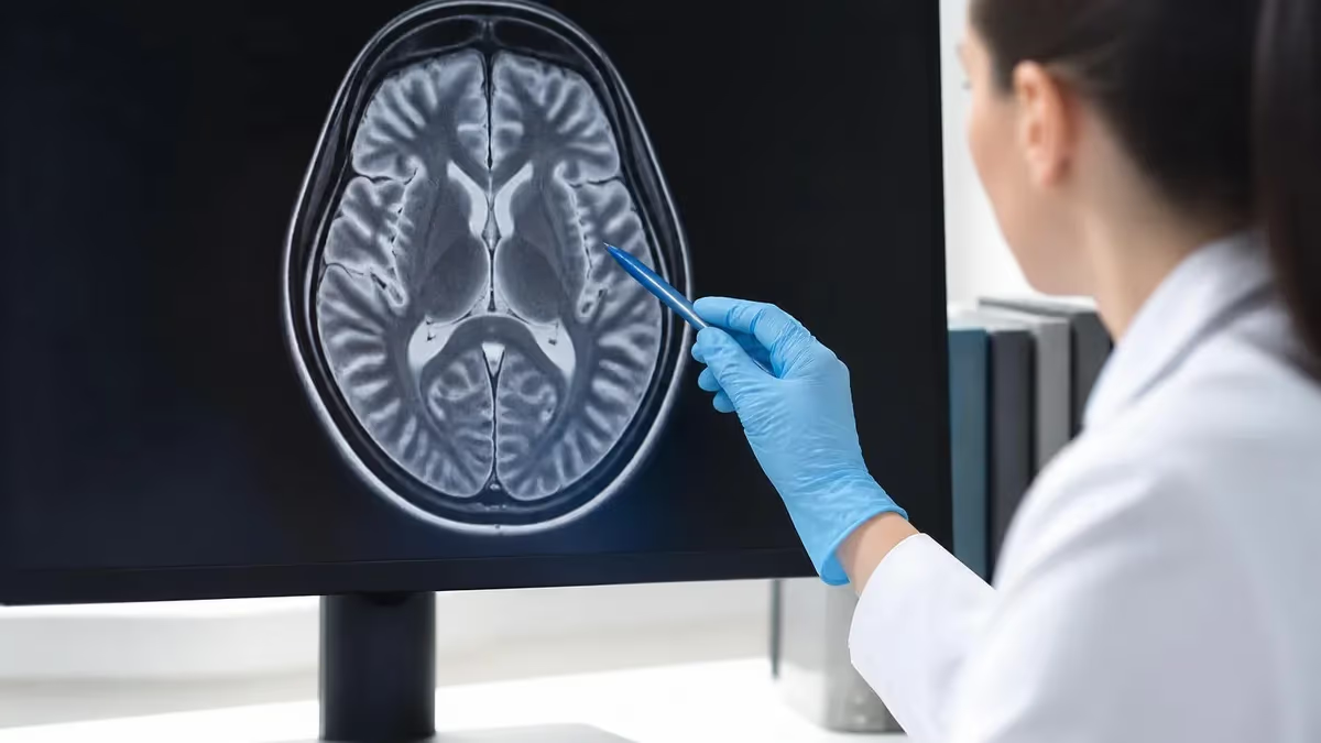
A back MRI is one of the most powerful diagnostic tools available for evaluating spinal pain, nerve compression, and soft-tissue injury that ordinary X-rays simply cannot reveal. Magnetic resonance imaging uses a strong magnetic field and radio waves rather than ionizing radiation, producing detailed cross-sectional pictures of vertebrae, discs, the spinal cord, and surrounding nerves. For patients living with chronic lower back pain, sciatica, or unexplained numbness, a back MRI often becomes the decisive test that explains symptoms and shapes a treatment plan.
Physicians order spinal MRI scans for many reasons: persistent pain lasting more than six weeks, leg weakness, suspected herniated discs, spinal stenosis, infection, tumors, or trauma. Because the technology distinguishes between fluid, fat, bone marrow, and nerve tissue with remarkable clarity, radiologists can pinpoint exactly which disc is bulging or which nerve root is being pinched. This level of detail makes MRI the gold standard when conservative treatments have failed and a clear anatomical answer is needed before surgery.
The lumbar spine—your lower back—is the most commonly imaged region because it bears the greatest mechanical load and is the frequent source of debilitating pain. However, MRI can target any spinal segment, including the thoracic and cervical areas. If your symptoms point toward your neck rather than your lower back, your doctor may instead request a focused study like an MRI of cervical spine, which uses the same physics but concentrates on the seven uppermost vertebrae and the nerves supplying the arms.
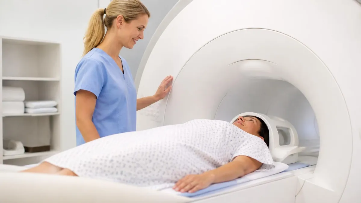
One of the biggest advantages of a back MRI is that it is completely non-invasive and painless. There are no needles unless contrast dye is required, no radiation exposure, and no recovery time afterward. You lie still on a padded table that slides into the scanner, and the machine does the rest. The trade-off is that you must remain motionless, and the scanner produces loud knocking and buzzing sounds that some patients find unsettling without earplugs or headphones.
Understanding what a back MRI involves—before, during, and after the appointment—helps reduce anxiety and ensures you get the most accurate results possible. Many patients arrive unsure whether they need contrast, how long the scan will take, or what the radiologist's report will actually mean. This guide walks through every stage of the process, from the moment your doctor writes the order to the day you sit down to review the findings and decide on next steps.
We will also cover practical concerns that matter to real patients: how much a spine MRI costs in the United States, whether insurance typically covers it, what to do if you are claustrophobic, and how to interpret common terms like "disc desiccation," "annular tear," and "foraminal narrowing" that appear in nearly every report. By the end, you will know exactly what to expect and how to be an informed participant in your own spinal care.
Back MRI by the Numbers
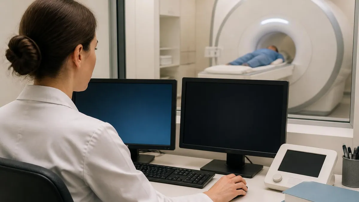
What Happens During a Back MRI
Check-In & Screening
Changing & Prep
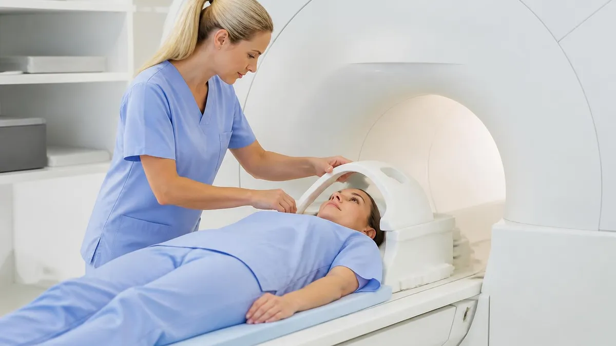
Positioning
Image Acquisition
Contrast Phase
Completion
Doctors do not order a back MRI lightly, because the test is costly and not every episode of back pain requires advanced imaging. Most acute low back pain resolves within four to six weeks with rest, physical therapy, and over-the-counter medication. Clinical guidelines therefore recommend imaging only when specific "red flag" symptoms appear or when pain persists despite conservative care. Knowing these triggers helps you understand why your physician believes a scan is genuinely necessary rather than simply reassuring.
The classic indications for a spine MRI include progressive leg weakness, numbness or tingling that follows a nerve pathway, and sciatica that radiates below the knee. These signs suggest a herniated disc or stenosis is compressing a nerve root. Bowel or bladder dysfunction combined with saddle numbness is a medical emergency called cauda equina syndrome, and it warrants an immediate, often same-day, MRI to prevent permanent neurological damage.
Other red flags push physicians toward urgent imaging as well. Unexplained weight loss, night pain, fever, or a history of cancer raises concern for tumor or infection within the vertebrae or spinal canal. Patients on immunosuppressants or intravenous drug users face higher infection risk, so a back MRI with contrast becomes the preferred study because gadolinium dramatically highlights abscesses, discitis, and osteomyelitis that plain images might miss entirely.
MRI is also routine before and after spinal surgery. Surgeons rely on detailed pre-operative images to plan the exact level and approach for a discectomy, laminectomy, or fusion. After surgery, contrast-enhanced MRI helps distinguish scar tissue—which lights up uniformly—from recurrent herniated disc material, which does not. This distinction is clinically vital because scar tissue is treated very differently from a true disc recurrence requiring another operation.
Trauma represents another major reason for spinal imaging. After a fall, car accident, or sports injury, MRI evaluates not only fractures but also ligament tears, spinal cord bruising, and bleeding that CT cannot fully characterize. While CT remains superior for visualizing bone detail, MRI excels at showing soft-tissue and neural injury, which is why the two studies are frequently complementary rather than interchangeable in trauma settings.
It is worth understanding that abnormal MRI findings are extremely common even in people without pain. Studies show that a large percentage of pain-free adults over forty have disc bulges or degeneration visible on imaging. This means a back MRI must always be interpreted alongside your symptoms and physical exam—an incidental finding is not automatically the cause of your pain, and good clinicians correlate the pictures with the patient sitting in front of them.
MRI Practice Test Questions
Prepare for the MRI - Magnetic Resonance Imaging exam with our free practice test modules. Each quiz covers key topics to help you pass on your first try.
MRI Knowledge
MRI Exam Questions covering Knowledge. Master MRI Test concepts for certification prep.
MRI Physics
Free MRI Practice Test featuring Physics. Improve your MRI Exam score with mock test prep.
MRI Anatomy and Pathology
MRI Test Prep for MRI Anatomy and Pathology. Practice MRI Quiz questions and boost your score.
MRI Anatomy and Positioning
MRI Questions and Answers on MRI Anatomy and Positioning. Free MRI practice for exam readiness.
MRI Contrast Agents
Free MRI Quiz on MRI Contrast Agents. MRI Exam prep questions with detailed explanations.
MRI Patient Care and Positioning
MRI Practice Questions for MRI Patient Care and Positioning. Build confidence for your MRI certification exam.
Lumbar, Thoracic, and Cervical Spine MRI Explained
The lumbar spine MRI covers the five lower vertebrae (L1–L5) plus the sacrum, the region responsible for most chronic back pain. Because this area bears your body's weight and powers bending and lifting, discs here degenerate and herniate most often. A lumbar study is the typical answer when patients describe sciatica, buttock pain, or numbness radiating down the leg.
Radiologists examine each disc level for bulging, herniation, and height loss, then assess the central canal and nerve-root exit channels for narrowing. Findings at L4–L5 and L5–S1 are especially common because these joints absorb the greatest mechanical stress. The report maps each level so your physician can correlate the images with the precise pattern of your symptoms.
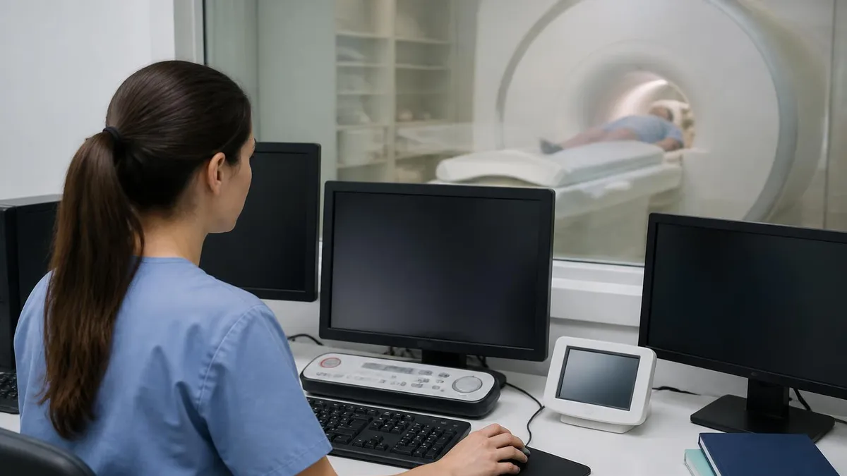
Back MRI: Advantages and Limitations
- +Uses no ionizing radiation, making it safe for repeated studies
- +Superior soft-tissue contrast reveals discs, nerves, and cord clearly
- +Detects herniation, stenosis, infection, and tumors others miss
- +Completely non-invasive and painless without contrast
- +Multi-plane images give surgeons precise pre-operative maps
- +Distinguishes scar tissue from recurrent disc after surgery
- −Expensive compared with X-ray and sometimes CT
- −Loud scanning noise and enclosed bore can trigger claustrophobia
- −Requires lying still for 30 to 60 minutes
- −Unsafe for some pacemakers and certain metal implants
- −Abnormal findings are common and may not explain pain
- −Contrast carries small risks for patients with kidney disease
Back MRI Preparation Checklist
- ✓Tell staff about any pacemaker, defibrillator, or implanted device.
- ✓Disclose all metal implants, screws, plates, or shrapnel.
- ✓Remove jewelry, watches, hairpins, and body piercings before the scan.
- ✓Leave credit cards and electronics outside the magnet room.
- ✓Inform the technologist if you might be pregnant.
- ✓Report kidney problems before any gadolinium contrast injection.
- ✓Wear comfortable, metal-free clothing or change into a gown.
- ✓Mention claustrophobia so sedation or an open MRI can be arranged.
- ✓Bring prior imaging or a CD for comparison if available.
- ✓Take prescribed anti-anxiety medication only with a driver present.
Metal and magnets do not mix
The MRI magnet is always on, even between scans. Ferromagnetic objects can become dangerous projectiles, and certain implants may heat or malfunction in the field. Always complete the screening form honestly and disclose every device, surgery, or metal fragment—your safety depends entirely on this conversation with the technologist.
Reading a back MRI report can feel overwhelming because radiologists use precise but unfamiliar terminology. Understanding the most common terms transforms an intimidating document into useful information you can discuss intelligently with your doctor. The report typically moves level by level down the spine, describing the discs, vertebrae, spinal canal, and nerve-root channels at each segment, then summarizing the most clinically significant findings in an "impression" at the end.
"Disc desiccation" simply means a disc has dried out and lost water content, a normal part of aging that appears dark on T2-weighted images. "Disc bulge" describes a disc that extends slightly beyond its normal margin around its whole circumference, while "herniation," "protrusion," or "extrusion" indicate that inner disc material has pushed out through a focal weak spot. The size and direction of a herniation matter far more than the label alone.
"Spinal stenosis" refers to narrowing of the central canal where the spinal cord or nerve bundle travels, while "foraminal stenosis" describes narrowing of the side channels where individual nerve roots exit. Either type can compress nerves and produce pain, numbness, or weakness. The report usually grades these as mild, moderate, or severe, and severe narrowing that matches your symptoms is often what prompts referral to a spine surgeon for evaluation.
An "annular tear" or "annular fissure" is a crack in the tough outer ring of a disc. These can be painful sources of discogenic back pain even without a frank herniation. "Modic changes" describe inflammatory or degenerative changes in the vertebral bone marrow next to a damaged disc and are increasingly recognized as a marker of mechanical back pain in some patients, guiding more targeted treatment decisions.
You may also see "spondylolisthesis," meaning one vertebra has slipped forward relative to the one below it, and "facet arthropathy," describing arthritis of the small joints that link the vertebrae. Both contribute to stenosis and instability. The radiologist measures the degree of slippage and notes whether it appears stable or potentially progressive, information that strongly influences whether bracing, therapy, or surgery is recommended for your particular case.
The single most important section is the "impression," where the radiologist distills everything into a few prioritized statements. This is where the findings most likely to explain your symptoms appear. Remember that imaging always requires clinical correlation: a moderate disc bulge on your scan only matters if it lines up with the nerve distribution of your actual pain. Bring your report to your appointment and ask your physician to walk through each finding with you.
Loss of bladder or bowel control, numbness in the groin or inner thighs, or rapidly worsening leg weakness can signal cauda equina syndrome—a surgical emergency. Do not wait for a routine appointment. Go to the emergency room immediately, as delayed treatment can cause permanent nerve damage and paralysis.
The cost of a back MRI in the United States varies dramatically depending on where you have it performed, whether contrast is used, and the type of insurance you carry. Prices commonly range from about $400 at a freestanding outpatient imaging center to well over $3,500 at a hospital-based radiology department. This wide gap means that simply choosing an independent facility instead of a hospital can save hundreds or even thousands of dollars for the identical study and image quality.
Most insurance plans cover medically necessary spine MRI, but many require prior authorization. Your physician's office must submit documentation showing that conservative treatments were tried or that red-flag symptoms are present. If authorization is denied, the office can appeal with additional clinical notes. Patients with high-deductible plans should ask for the negotiated cash price up front, since paying directly is sometimes cheaper than billing insurance before the deductible is met.
Safety remains the central consideration for any MRI. Because the magnet is permanently energized, patients with certain cardiac pacemakers, cochlear implants, aneurysm clips, or metallic foreign bodies in the eye may be unable to undergo the scan. Many modern implants are now labeled "MRI conditional," meaning they are safe under specific parameters. Knowing whether your hardware is compatible is essential, and you can learn more about MRI safety materials and which metals pose risks.
Claustrophobia is one of the most common obstacles to completing a back MRI. The traditional closed-bore scanner surrounds your body in a narrow tube, which can provoke significant anxiety. Solutions include oral anti-anxiety medication prescribed beforehand, wide-bore scanners with more room, and open MRI units that sacrifice some image quality for comfort. Telling the scheduling staff about your claustrophobia in advance ensures the right accommodation is arranged before you arrive.
If your scan requires contrast, a gadolinium-based agent is injected through an IV to highlight inflammation, infection, tumors, and surgical scar tissue. Gadolinium is generally very safe, but patients with severe kidney impairment require careful screening because their bodies clear the agent more slowly. A simple blood test measuring kidney function is often checked before contrast is given to anyone with known renal disease or risk factors.
After your back MRI, the images are reviewed by a board-certified radiologist who produces a written report, usually within 24 to 48 hours. Your referring physician then interprets those findings in the context of your symptoms and examination. The scan itself is only one piece of the puzzle; the real value emerges when your doctor combines the imaging, your history, and your physical exam to build a treatment plan tailored specifically to you.
Getting the most out of your back MRI starts well before you reach the scanner. Arrive at least fifteen minutes early to complete paperwork without rushing, and use the restroom beforehand since you will need to lie still for up to an hour. Wear loose, comfortable clothing free of zippers, snaps, and metal so you may be able to skip changing into a gown. Bringing a list of your symptoms helps the technologist confirm the correct region is being imaged.
Staying perfectly still is the single most important thing you can do during the scan. Motion blurs the images and can force the technologist to repeat sequences, lengthening your time in the bore. Before each sequence begins, the technologist will tell you how long it lasts—anywhere from one to seven minutes. Use those windows to settle into a comfortable position, breathe steadily, and avoid shifting your hips or legs until the sequence is complete.
If noise bothers you, request earplugs and headphones; many centers offer music to help you relax. Closing your eyes before entering the bore and keeping them closed throughout is a simple trick that helps claustrophobic patients tolerate the enclosed space. Some facilities provide a mirror angled so you can see out of the tube, and most place a squeeze-ball alarm in your hand so you can signal the technologist at any moment if you become uncomfortable.
When your results arrive, resist the urge to self-diagnose from internet searches alone. The terminology is genuinely complex, and findings that sound alarming are frequently age-related and harmless. Instead, schedule a follow-up specifically to review the report. Write down your questions in advance—ask which finding explains your pain, whether the issue is stable, and what the realistic treatment options are, from physical therapy to injections to surgery.
Keep a copy of your images and report for your own records, ideally on a CD or through a patient portal. If you seek a second opinion or change physicians, having the actual images rather than just the written report saves time and avoids unnecessary repeat scans. Comparison with prior studies is also enormously valuable, because radiologists can determine whether a finding is new, stable, or progressing over months or years.
Finally, remember that a back MRI is a diagnostic tool, not a treatment in itself. The vast majority of patients with disc bulges, mild stenosis, and degenerative changes improve with conservative care such as targeted physical therapy, core strengthening, weight management, and activity modification. Surgery is reserved for specific situations like progressive weakness, intractable nerve pain, or instability. Use your scan results as a roadmap for an informed conversation, and partner with your physician to choose the least invasive path that addresses your goals.
Being an engaged, prepared patient consistently leads to better experiences and clearer answers. Understanding why the test was ordered, how to prepare, what the machine does, and what the report means puts you in control of your spinal health rather than feeling like a passive bystander. The more you know going in, the more confident and reassured you will feel when you finally see your own spine on the screen.
MRI Questions and Answers

MRI of Cervical Spine: Complete Guide to Imaging, Anatomy, Pathology, and Patient Preparation
MRI Medical Abbreviation: What MRI Stands For and Why It Matters

Knee MRI Images: A Complete Guide to Reading, Understanding, and Interpreting Knee Scans
ICD 10 Codes for MRI: Complete Guide to Abnormal MRI Coding, Documentation, and Reimbursement
Is Nickel Titanium MRI Compatible? A Complete Guide to MRI Safety Materials
About the Author

Medical Laboratory Scientist & Clinical Certification Expert
Johns Hopkins UniversityDr. Sandra Kim holds a PhD in Clinical Laboratory Science from Johns Hopkins University and is certified as a Medical Technologist (MT) and Medical Laboratory Scientist (MLS) through ASCP. With 16 years of clinical laboratory experience spanning hematology, microbiology, and molecular diagnostics, she prepares candidates for ASCP board exams, MLT, MLS, and specialist certification tests.
Join the Discussion
Connect with other students preparing for this exam. Share tips, ask questions, and get advice from people who have been there.
View discussion (8 replies)