Is Nickel Titanium MRI Compatible? A Complete Guide to MRI Safety Materials
Is nickel titanium MRI compatible? Learn which implant materials are MRI safe, ✍🏼 conditional, or unsafe, with practical screening tips for techs.
If you work in magnetic resonance imaging or are about to be scanned with a stent or orthodontic wire in your body, the question is nickel titanium MRI compatible comes up almost daily in screening rooms across the country. Nickel titanium, more commonly known by the trade name Nitinol, is one of the most widely used biomedical alloys in modern medicine and shows up in stents, guidewires, archwires, vena cava filters, and orthopedic staples.
For the vast majority of these devices the answer is yes, nickel titanium is considered MRI Conditional, but the full story involves heating, artifact, field strength, and device-specific testing that every technologist should understand.
The reason nickel titanium behaves so well inside a magnetic resonance scanner comes down to its crystalline structure. Nitinol is a near-equiatomic intermetallic compound of nickel and titanium that forms an austenitic or martensitic phase depending on temperature. Despite the high nickel content, the alloy is only weakly paramagnetic, meaning its magnetic susceptibility sits close to that of human tissue. That low susceptibility produces minimal translational and rotational force inside a 1.5 Tesla or 3 Tesla magnet, which is the single biggest safety concern for any implanted metal.
However, MRI safety is never a binary yes-or-no decision based on material alone. The American Society for Testing and Materials, through ASTM F2503, classifies every device as MR Safe, MR Conditional, or MR Unsafe, and almost all nitinol implants fall into the conditional category. That means they can be scanned, but only under documented conditions involving static field strength, gradient slew rate, specific absorption rate, and scan duration. Skipping any of those conditions can lead to burns, image artifact, or in rare cases displacement.
For technologists preparing for board exams or for radiology residents reviewing safety protocols, understanding the science behind nitinol compatibility is essential. The Joint Commission considers MRI screening errors a sentinel event, and the FDA receives hundreds of MRI-related adverse event reports each year. Most are preventable with proper material identification, manufacturer card review, and adherence to the patient's specific implant labeling rather than assumptions based on alloy class alone.
This guide walks through everything you need to know about nickel titanium in the MRI environment, from the underlying metallurgy to clinical screening workflows. We will cover heating risk, artifact size, common nitinol devices, conditional scan parameters, and the practical decisions technologists make when a patient cannot produce an implant card. If you want a refresher on basic terminology first, our guide to the MRI medical abbreviation explains the language used throughout this article.
By the time you finish reading, you will be able to explain why a nitinol coronary stent can safely enter a 3T scanner six weeks after placement, why a nitinol aneurysm clip may still require extra screening, and how to document the conditional approval in your worklist. The goal is to move from memorized rules toward genuine understanding, which is what board examiners, lead technologists, and patient safety officers want to see in practice.
This information reflects current ACR Manual on MR Safety recommendations and standard industry practice as of 2026. Always defer to your facility's policy, the implant manufacturer's instructions for use, and the supervising radiologist's judgment when conflicts arise. Material science is consistent, but device approvals change as new evidence emerges, and a current MRI safety screener will always outperform a textbook memory.
Nickel Titanium in MRI by the Numbers
Material Classifications in MRI
The device poses no known hazards in any MRI environment. These are nonconductive, nonmetallic, and nonmagnetic items such as plastic or silicone components. The label uses a green square with the letters MR.
The device has been demonstrated safe under specified conditions. Nearly all nitinol implants fall here, with conditions defining field strength, SAR limits, gradient performance, and minimum healing time. Labeled with a yellow triangle.
The device poses hazards in all MRI environments and must never be brought near the scanner. Ferromagnetic items such as older aneurysm clips or carbon steel tools fall here. Labeled with a red circle and slash.
Many implants placed before 2005 lack ASTM F2503 labeling. These require manufacturer lookup, original operative report review, or fluoroscopic identification before a scan can proceed under conditional approval.
To understand why nickel titanium is generally MRI conditional rather than fully MR safe, it helps to look at what happens at the atomic level when nitinol enters a strong magnetic field. Pure nickel is ferromagnetic at room temperature with a Curie point near 354 degrees Celsius, but when alloyed in roughly equal proportions with titanium and processed into the austenitic phase, the resulting compound loses almost all of that ferromagnetism. The electronic configuration shifts, and the bulk material becomes weakly paramagnetic instead.
Paramagnetic materials are attracted to magnetic fields, but the force is several orders of magnitude weaker than ferromagnetic attraction. Inside a 1.5T scanner the displacement force on a typical nitinol stent is measured in fractions of a Newton, well below the gravitational force already acting on the device. That is why a nitinol vena cava filter does not tug, twist, or migrate during a routine abdominal MRI, even though it contains a metal commonly associated with magnetic behavior.
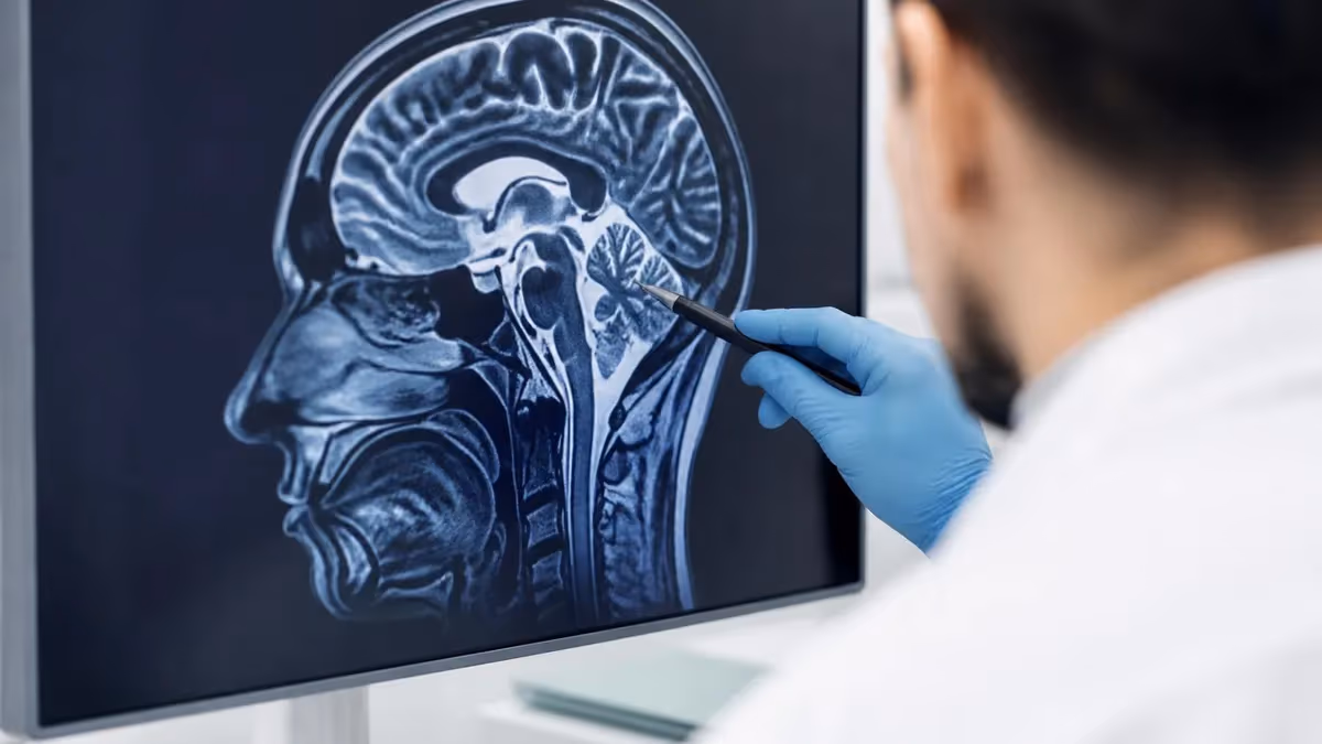
Artifact, on the other hand, is a separate concern. Even weakly paramagnetic materials distort the local magnetic field enough to create signal voids on gradient echo sequences. The artifact halo around a nitinol stent is typically two to five millimeters wide, which is acceptable for most diagnostic questions but can obscure pathology immediately adjacent to the device. This is why radiologists sometimes order spin echo or short echo time sequences when imaging close to nitinol hardware.
Specific absorption rate, or SAR, is the third pillar of conditional approval. Long, electrically conductive structures act as antennas inside the radiofrequency field, concentrating energy at their tips and potentially heating surrounding tissue. Nitinol guidewires used during interventional procedures have been measured heating up to several degrees in worst-case configurations, which is why diagnostic MRI is generally avoided while such temporary devices are in place.
Implanted nitinol devices behave very differently because they are short, encapsulated, and not connected to external leads. Testing under ASTM F2182 typically shows temperature rises below two degrees Celsius at standard whole-body SAR levels of 2 watts per kilogram. That is biologically negligible, particularly in vascularized tissue where blood flow rapidly carries away any modest heat buildup before it can cause cellular damage.
The reason manufacturers still require conditional labeling rather than full MR safe approval is regulatory rather than physical. ASTM F2503 reserves the green MR Safe designation for items that are nonmetallic, nonconductive, and nonmagnetic — three criteria nitinol cannot meet because it is metallic and conducts electricity. Even with negligible heating and force, the alloy must therefore carry the conditional label, which is a labeling rule, not a warning of clinical danger.
For a deeper look at how scanner technology has evolved to accommodate modern implant materials, including dedicated low-SAR sequences and conditional imaging protocols, our overview of the history of MRI traces the developments that made today's safety standards possible.
MRI Practice Test Questions
Prepare for the MRI - Magnetic Resonance Imaging exam with our free practice test modules. Each quiz covers key topics to help you pass on your first try.
MRI Knowledge
MRI Exam Questions covering Knowledge. Master MRI Test concepts for certification prep.
MRI Physics
Free MRI Practice Test featuring Physics. Improve your MRI Exam score with mock test prep.
MRI Anatomy and Pathology
MRI Test Prep for MRI Anatomy and Pathology. Practice MRI Quiz questions and boost your score.
MRI Anatomy and Positioning
MRI Questions and Answers on MRI Anatomy and Positioning. Free MRI practice for exam readiness.
MRI Contrast Agents
Free MRI Quiz on MRI Contrast Agents. MRI Exam prep questions with detailed explanations.
MRI Patient Care and Positioning
MRI Practice Questions for MRI Patient Care and Positioning. Build confidence for your MRI certification exam.
MRI Safety Materials: How Nitinol Compares
At 1.5 Tesla, nearly every modern nitinol device carries an MR Conditional label permitting routine scanning. Translational and rotational forces are minimal because the alloy is only weakly paramagnetic, and tested heating remains well under two degrees Celsius at whole-body SAR up to 2 W/kg. Artifact halos average around two to three millimeters, which preserves diagnostic quality for most regions of interest including vascular and abdominal imaging.
Technologists working at 1.5T can scan nitinol stents, filters, and orthodontic wires confidently after verifying the device card and respecting any post-placement waiting period. The default whole-body SAR mode is almost always acceptable, and only when imaging is performed directly over a long nitinol implant should the radiologist consider switching to first-level controlled mode or reducing flip angle to limit cumulative energy deposition.
Nickel Titanium in the MRI Environment: Pros and Cons
- +Weakly paramagnetic with minimal displacement force at 1.5T and 3T
- +Low artifact halo allows diagnostic imaging of adjacent anatomy
- +Heating remains under 2 degrees Celsius at standard SAR levels
- +Widely used in stents, filters, and orthodontics with extensive safety data
- +Most modern devices carry clear ASTM F2503 conditional labeling
- +Compatible with both spin echo and many gradient echo sequences
- +Endothelialization within weeks further reduces any residual concerns
- −Cannot be classified MR Safe due to metallic conductivity requirement
- −Long nitinol guidewires can heat significantly during interventional MRI
- −Artifact obscures pathology directly within a few millimeters of the device
- −7T compatibility is rarely tested and generally not approved
- −Legacy implants placed before 2005 may lack documented conditions
- −Requires manufacturer card review and SAR-mode verification before scanning
- −Patients with unidentified nitinol hardware delay scanning until verified
MRI Screening Checklist for Nickel Titanium Implants
- ✓Verify the implant manufacturer, model number, and lot from the patient's device card.
- ✓Confirm ASTM F2503 labeling status: MR Safe, MR Conditional, or MR Unsafe.
- ✓Document the maximum static field strength approved, typically 1.5T or 3T.
- ✓Check the whole-body SAR limit and B1+rms ceiling listed in the IFU.
- ✓Confirm the minimum waiting period after device placement, often six weeks.
- ✓Identify the maximum gradient slew rate the device tolerates.
- ✓Note any scan duration caps required to manage cumulative heating.
- ✓Review the operative report if no device card is available.
- ✓Communicate conditions to the technologist and supervising radiologist.
- ✓Document final approval and chosen scan mode in the worklist and report.
Conditional does not mean dangerous
The MR Conditional label on a nitinol implant is a regulatory description, not a warning. It tells you the device has been tested and approved under specific scan parameters that almost every clinical 1.5T and 3T scanner already meets by default. The real risk is failing to verify the conditions, not the alloy itself.
Nickel titanium appears in more medical devices than most patients realize, and many of those devices arrive at the MRI suite without much advance warning. Coronary and peripheral arterial stents account for the largest single category, with millions placed worldwide each year. The vast majority are self-expanding nitinol mesh designs because the alloy's shape memory property allows the stent to be compressed inside a delivery catheter and then return to its programmed expanded shape once deployed at body temperature.
Inferior vena cava filters represent another common nitinol implant. These small, umbrella-shaped devices sit inside the IVC to trap clots before they reach the lungs. Modern filters such as the Cook Celect, Bard Denali, and Cordis OptEase are all nitinol-based and carry 1.5T and 3T conditional labeling. They are routinely imaged during abdominal MRI when patients need follow-up for clot burden or filter migration concerns.
Orthodontics is the third major source. Nickel titanium archwires are used during the initial alignment phase of treatment because their superelasticity applies gentle, consistent force over a wide range of tooth movement. Patients undergoing brain or head MRI with active orthodontic appliances generally do not need to remove the wires, though significant artifact in the oral cavity is unavoidable. The wires themselves are safe at 1.5T and 3T.
Orthopedic surgery has adopted nitinol for memory staples used in foot, ankle, and small bone fusions. The staples are inserted in their martensitic phase at cool temperatures and then warmed by the patient's body to spring into a compressive position. Postoperative MRI is routinely performed when needed, with the staples behaving similarly to other small implants in terms of minimal force and modest artifact within a few millimeters.
Interventional radiology relies heavily on nitinol guidewires for navigation through tortuous vessels. These wires are not permanent implants but are critical to mention because intraprocedural MRI guidance is a growing field. When a nitinol wire extends along a long, relatively straight vascular path, resonant RF coupling can heat the tip dramatically, which is why conventional diagnostic MRI is contraindicated while such temporary devices are in the body.
Less common but worth knowing are nitinol aneurysm clips, occlusion devices for atrial septal defects, esophageal stents, biliary stents, and orthopedic spinal rods that use nitinol rods or wires as part of a hybrid construct. Each of these has its own conditional approval, and none should be assumed safe based on alloy alone. Always check the specific manufacturer card or operative report.
Patients sometimes confuse nitinol with other titanium-based materials, asking whether their hip replacement counts as nitinol. Commercially pure titanium and Ti-6Al-4V used in orthopedics are different alloys from nickel titanium. They also carry MR Conditional labels at 1.5T and 3T, but the conditions and artifact patterns differ. Educating patients about this distinction reduces confusion during the screening interview.
Material class alone is not a substitute for device-specific conditional labeling. Two stents from different manufacturers, both made of nitinol, may have very different SAR limits, B1+rms ceilings, or post-placement waiting periods. Never approve a scan based on the alloy name without reviewing the actual implant card or manufacturer instructions for use.
Heating and artifact are the two practical concerns technologists weigh whenever a nitinol device enters the scan suite, and understanding the magnitude of each helps you make confident clinical decisions. Heating is governed primarily by SAR, device geometry, position relative to the body coil, and scan duration. Short, encapsulated implants such as stents and filters dissipate heat efficiently through surrounding vasculature, while long conductive structures can act as resonant antennas and concentrate energy at their tips.
For typical permanent nitinol implants, peer-reviewed bench testing under ASTM F2182 consistently shows temperature rises of less than two degrees Celsius even after fifteen minutes of continuous scanning at the FDA upper limit of 4 watts per kilogram. In vivo this is even safer because blood flow continually removes heat from the implant surface. Clinically significant burns from nitinol implants are essentially unheard of when manufacturer conditions are respected.
Artifact behavior depends on pulse sequence selection. Gradient echo sequences are the most susceptible to nitinol-related distortion, producing dark signal voids that extend several millimeters beyond the actual device edge. Spin echo and turbo spin echo sequences are far more forgiving because the 180-degree refocusing pulses compensate for static field inhomogeneity. Short echo times and high readout bandwidth further reduce artifact size and improve diagnostic quality in nearby tissue.
Clinical decision-making becomes most difficult when a patient cannot identify their implant or when the device card is unavailable. In these scenarios, the ACR Manual on MR Safety recommends treating the patient as potentially MR Unsafe until proper documentation is obtained. This might involve calling the implanting facility, requesting old operative reports, or in some cases performing a low-dose fluoroscopic shot to identify the device by shape.
Some facilities maintain in-house databases of common implants linked to ASTM F2503 conditional labeling, which dramatically speeds up screening. Online resources such as MRIsafety.com and the manufacturer's own digital catalogs are also indispensable. Many radiology departments now build these references directly into their RIS workflow so the technologist can look up a device by model number without leaving the worklist.
When a nitinol device is approved as conditional but the requested protocol exceeds the labeled limits, options include adjusting the protocol, switching to a lower field scanner, or imaging the region of interest using an alternative modality. Our overview of MRI alternatives explains when CT, ultrasound, or other modalities provide equivalent diagnostic information without the safety constraints of MRI.
The takeaway for technologists, residents, and registry candidates is that nitinol is a material you will encounter constantly, and the science strongly supports its safety in well-documented conditional scanning. Confidence comes from knowing where the data lives, who labels devices, what parameters matter, and how to escalate when documentation is incomplete. Master those workflows and you will rarely cancel a scan unnecessarily for a nickel titanium implant.
Translating all of this into reliable day-to-day practice requires a few habits that experienced MRI technologists develop over time. The first is treating the screening interview as a conversation rather than a checklist. When a patient mentions any kind of stent, filter, wire, or staple, ask follow-up questions about when and where it was placed, who placed it, and whether they were given a card. Patients often forget they have implants, and gentle prompts uncover important history.
The second habit is verifying through more than one source whenever possible. A device card is excellent, but cross-referencing the card number against the manufacturer's online registry confirms that no recalls or labeling changes have occurred since placement. This step takes thirty seconds and can prevent the very rare situation where a device has been reclassified after a postmarket safety review. Document the date you confirmed the labeling along with the source.
The third habit involves scan parameter discipline. When a nitinol device has a B1+rms limit, set the system to first-level controlled operating mode if needed and verify the calculated B1+rms before each sequence. Modern scanners display this value in the protocol editor. If a sequence exceeds the limit, reducing flip angle or adding TR usually brings it back within range without sacrificing image quality.
Communication with the supervising radiologist is the fourth habit. Even when a conditional scan is clearly approved on paper, a quick verbal confirmation with the on-call radiologist documents shared responsibility and gives the radiologist a chance to suggest sequence modifications that improve diagnostic value near the implant. This is particularly useful for stented vessels where dedicated post-contrast sequences with low artifact can be substituted.
The fifth habit is documentation. Every conditional scan should generate a worklist note recording the device model, the SAR mode selected, the maximum B1+rms used, and the time at which conditions were verified. This protects the patient, the technologist, and the facility if any question arises later about whether the scan was performed safely. Good documentation is the single best legal protection against rare adverse events.
Finally, ongoing education is essential. ASTM standards, FDA guidance, and ACR guidelines evolve every few years. Attending annual MRI safety refreshers, reviewing the latest ACR Manual on MR Safety, and following society publications keeps your practice current. Material science does not change quickly, but conditional labeling rules and recommended workflows certainly do, and yesterday's safe practice may not match today's expectations.
For technologists preparing for the ARRT MRI registry or ARMRIT certification, expect at least two to four questions on implant materials, including specific scenarios about nitinol. Knowing the material classifications, the meaning of conditional labeling, the typical heating range, and the role of ASTM F2503 will let you answer those questions quickly and move on to harder problems involving pulse sequences and contrast pharmacology.
MRI Questions and Answers

MRI Medical Abbreviation: What MRI Stands For and Why It Matters

The History of MRI: From Discovery to Modern Medicine

Knee MRI Images: A Complete Guide to Reading, Understanding, and Interpreting Knee Scans

Noise of MRI Machine: Why MRI Scanners Are So Loud and What to Expect
MRI Alternatives: When CT, Ultrasound, X-Ray, and PET Make More Sense Than an MRI
About the Author
Medical Laboratory Scientist & Clinical Certification Expert
Johns Hopkins UniversityDr. Sandra Kim holds a PhD in Clinical Laboratory Science from Johns Hopkins University and is certified as a Medical Technologist (MT) and Medical Laboratory Scientist (MLS) through ASCP. With 16 years of clinical laboratory experience spanning hematology, microbiology, and molecular diagnostics, she prepares candidates for ASCP board exams, MLT, MLS, and specialist certification tests.
Join the Discussion
Connect with other students preparing for this exam. Share tips, ask questions, and get advice from people who have been there.
View discussion (6 replies)