
Abnormal MRI ICD 10: A Complete Guide to Coding, Documentation, and Clinical Interpretation
Abnormal MRI ICD 10 guide: 🆕 how to code abnormal findings (R90, R93), document results, avoid denials, and interpret results for accurate reimbursement.
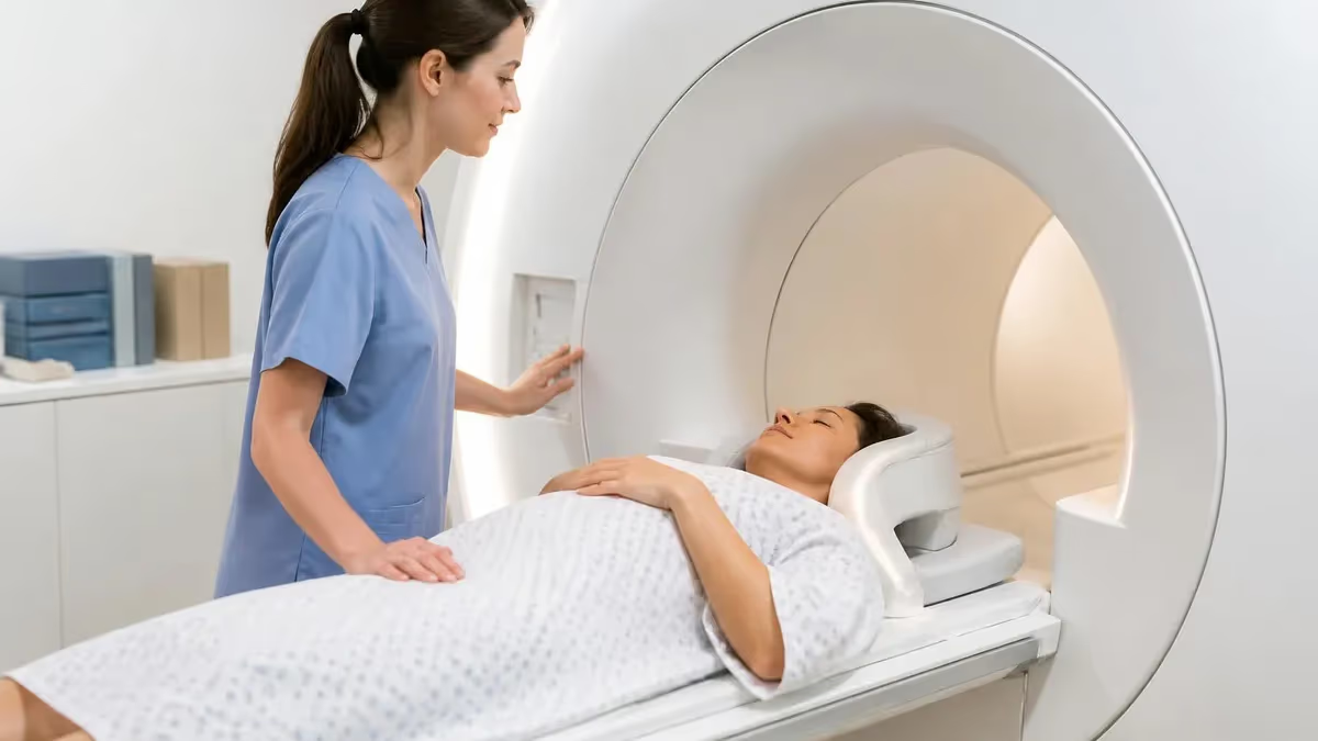
Understanding abnormal mri icd 10 coding is essential for radiologists, coders, technologists, and billing specialists who translate complex imaging findings into the standardized language insurers require. When an MRI reveals something outside normal anatomy, the report cannot simply say "abnormal" — it must be matched to a precise ICD-10-CM code that justifies the study, supports medical necessity, and protects reimbursement. Getting this right is the difference between a clean claim and a costly denial that delays patient care.
ICD-10-CM, the diagnostic code set used across the United States since October 2015, contains more than 70,000 codes. A small but critical subset describes abnormal findings on diagnostic imaging that have no established diagnosis yet. The most frequently used category for abnormal MRI results is R90, covering abnormal findings on diagnostic imaging of the central nervous system, while R93 captures abnormal findings from imaging of other body structures. These "R" codes belong to Chapter 18, which houses symptoms, signs, and abnormal clinical and laboratory findings.
Why does a separate code exist for an abnormal but unspecified finding? Because imaging frequently identifies an abnormality before a definitive diagnosis is confirmed. A radiologist may see an unexpected mass, lesion, or signal change that requires follow-up, biopsy, or correlation. Until a specific disease is established, the abnormal-findings code accurately reflects the clinical reality and supports any recommended next steps without overstating what is actually known.
For the people who handle these reports daily, precision matters at every stage. A technologist documents the indication, a radiologist dictates the impression, and a coder selects the code that maps to that impression. If any link in this chain is vague, the claim may be questioned. Payers increasingly scrutinize imaging because MRI is expensive, and an abnormal-findings code paired with weak documentation is a common reason for audits and recoupment requests.
This guide walks through the full workflow: which ICD-10 codes apply to abnormal MRI results, how to document findings so they survive payer review, how coders avoid the traps of unspecified codes, and how clinical interpretation connects to the billing record. Whether you are studying for a registry exam or working a busy coding queue, mastering this material builds confidence and reduces rework across the imaging department.
We will also connect the coding side to the clinical side, because the strongest coders understand what radiologists actually see on a scan. Knowing the difference between an incidental finding and a clinically significant abnormality — and how each is phrased in a report — helps you choose codes that are both accurate and defensible. By the end, you will have a practical framework you can apply to real reports tomorrow.
Abnormal MRI ICD 10 by the Numbers
Key ICD-10 Codes for Abnormal MRI Findings
Abnormal findings on diagnostic imaging of the central nervous system, including R90.0 intracranial space-occupying lesion and R90.89 other abnormal findings on brain and head imaging.
Abnormal findings on diagnostic imaging of other body sites, such as R93.5 abdominal regions, R93.6 limbs, and R93.7 other musculoskeletal structures identified on MRI.
Abnormal and inconclusive findings on diagnostic imaging of breast, used when breast MRI reveals a lesion or density requiring further workup before a definitive diagnosis.
Abnormal results of function studies, occasionally relevant when functional MRI or specialized sequences return abnormal results that do not yet map to a specific disease.
Z codes describe encounters for screening or follow-up. When MRI is done without symptoms, the reason for the exam may require a Z code rather than an abnormal-findings code.
Translating an abnormal MRI into the correct ICD-10 code begins with the radiologist's impression, the single most important section of any report for coding purposes. Coders are trained to assign codes from confirmed, documented findings — not from their own interpretation of images. If the impression states "3 mm enhancing lesion in the left frontal lobe, etiology uncertain," the coder cannot leap to a tumor diagnosis. Instead, an abnormal-findings code such as R90.0 may apply until a definitive diagnosis is established through biopsy or follow-up.
The first rule of imaging coding is to code the established diagnosis when one exists. If the MRI confirms a herniated disc, multiple sclerosis plaques, or a meniscal tear, you code that specific condition, not an abnormal-findings code. The R90 and R93 families are reserved for situations where the imaging shows something genuinely abnormal but the underlying disease has not yet been named. This hierarchy prevents the overuse of vague codes that payers flag.
Second, understand the role of signs and symptoms. When MRI is ordered for headache, low back pain, or numbness, those symptom codes (such as R51 for headache or M54 for back pain) often justify the study even before results return. After the scan, the coding may shift to a confirmed diagnosis or to an abnormal-findings code, depending on what the radiologist documents. Knowing when to use the symptom versus the result is a core coding judgment.
Third, incidental findings deserve careful handling. MRI frequently reveals unrelated abnormalities — a small renal cyst seen on a lumbar spine study, for instance. Whether and how to code these depends on facility policy and whether the finding affects management. Many incidental findings are documented but not separately coded if they have no clinical impact, while others trigger a recommendation for follow-up that supports an additional code.
Fourth, laterality and specificity matter enormously in ICD-10. The code set demands left versus right, acute versus chronic, and anatomic precision whenever the documentation supports it. A coder who selects an unspecified code when a more specific one was available risks denials and audit flags. This is why strong communication between coders and radiologists — through queries when documentation is ambiguous — is a hallmark of a well-run imaging operation.
Finally, sequencing rules govern which code comes first on a claim. The principal diagnosis reflects the condition chiefly responsible for the encounter, and secondary codes capture comorbidities or incidental findings. For outpatient imaging, the reason the exam was ordered usually drives sequencing, with abnormal-findings codes added to document what the scan actually revealed. Mastering this sequence keeps claims clean and reduces back-and-forth with payers, which is exactly the efficiency a busy department needs.
MRI Practice Test Questions
Prepare for the MRI - Magnetic Resonance Imaging exam with our free practice test modules. Each quiz covers key topics to help you pass on your first try.
MRI Knowledge
MRI Exam Questions covering Knowledge. Master MRI Test concepts for certification prep.
MRI Physics
Free MRI Practice Test featuring Physics. Improve your MRI Exam score with mock test prep.
MRI Anatomy and Pathology
MRI Test Prep for MRI Anatomy and Pathology. Practice MRI Quiz questions and boost your score.
MRI Anatomy and Positioning
MRI Questions and Answers on MRI Anatomy and Positioning. Free MRI practice for exam readiness.
MRI Contrast Agents
Free MRI Quiz on MRI Contrast Agents. MRI Exam prep questions with detailed explanations.
MRI Patient Care and Positioning
MRI Practice Questions for MRI Patient Care and Positioning. Build confidence for your MRI certification exam.
Documentation and Medical Necessity for Abnormal MRI ICD 10
Medical necessity starts before the patient enters the scanner. The ordering provider must document a clear clinical indication — the symptom, sign, or condition prompting the MRI. A vague order like "rule out pathology" rarely satisfies payers. Strong orders specify the symptom, duration, prior treatments tried, and the clinical question the imaging should answer, giving coders defensible justification.
This indication often becomes the initial ICD-10 code used for prior authorization. Payers compare the documented indication against their coverage policies, so aligning the order with covered diagnoses upfront prevents downstream denials. When the scanned region and the indication match, the claim has a far smoother path through adjudication and payment.
Using Abnormal-Findings (R) Codes: Strengths and Limitations
- +Accurately reflects imaging that shows abnormality without a confirmed diagnosis
- +Supports medical necessity for recommended follow-up studies
- +Prevents overstating a diagnosis that has not been confirmed
- +Keeps the record honest during diagnostic uncertainty
- +Maps cleanly to radiologist impressions that note uncertainty
- +Useful when biopsy or correlation is still pending
- −Considered nonspecific and may trigger payer scrutiny
- −Can lead to denials if documentation is thin
- −Should never replace a confirmed, codeable diagnosis
- −Overuse signals incomplete documentation in audits
- −May require additional symptom codes to justify the exam
- −Does not capture laterality or disease-specific detail
Pros and Cons at a Glance
| Pros | Cons |
|---|---|
| Accurately reflects imaging that shows abnormality without a confirmed diagnosis | Considered nonspecific and may trigger payer scrutiny |
| Supports medical necessity for recommended follow-up studies | Can lead to denials if documentation is thin |
| Prevents overstating a diagnosis that has not been confirmed | Should never replace a confirmed, codeable diagnosis |
| Keeps the record honest during diagnostic uncertainty | Overuse signals incomplete documentation in audits |
| Maps cleanly to radiologist impressions that note uncertainty | May require additional symptom codes to justify the exam |
| Useful when biopsy or correlation is still pending | Does not capture laterality or disease-specific detail |
Abnormal MRI ICD 10 Coding Accuracy Checklist
- ✓Confirm the radiologist's impression before assigning any code.
- ✓Code the established diagnosis whenever one is documented.
- ✓Use R90 or R93 only when no specific diagnosis is confirmed.
- ✓Match the diagnosis code to the CPT procedure billed.
- ✓Capture laterality, size, and location when documented.
- ✓Add symptom codes that justify why the MRI was ordered.
- ✓Query the radiologist when documentation is ambiguous.
- ✓Handle incidental findings per facility policy.
- ✓Sequence the principal diagnosis correctly on the claim.
- ✓Verify the indication aligns with payer medical policy.
- ✓Document any recommendation for follow-up imaging.
- ✓Avoid unspecified codes when a specific code is available.
Code from the impression, not your interpretation
Coders must assign diagnosis codes only from what the radiologist explicitly documents in the report — never from their own reading of the images. When the impression confirms a diagnosis, code it specifically. When it notes an abnormality without a confirmed cause, use the appropriate R90 or R93 abnormal-findings code and query the physician if anything is unclear.
Reimbursement for MRI hinges on whether the diagnosis codes support medical necessity, and abnormal-findings codes occupy a delicate position in that calculus. Because R90 and R93 are nonspecific by design, payers may pay an initial diagnostic MRI under these codes while questioning repeat studies that rely on the same vague justification. Understanding how payers view these codes helps billing teams anticipate which claims will sail through and which will land in a manual review queue.
Denials related to imaging frequently trace back to one of three problems: the indication did not match the payer's covered diagnoses, the documentation failed to support the level of service, or the diagnosis code did not justify the specific CPT procedure billed. Abnormal-findings codes can be perfectly appropriate, but when paired with weak documentation they invite scrutiny. The remedy is rarely a different code — it is stronger, more specific documentation from the ordering provider and the radiologist.
Prior authorization is the front line of MRI reimbursement. Most commercial payers and many Medicare Advantage plans require pre-approval for advanced imaging through a radiology benefit manager. The indication submitted during authorization must align with the codes that later appear on the claim. A mismatch between the authorized diagnosis and the billed diagnosis is a common and avoidable reason for denial, even when the study itself was clinically warranted and properly performed.
When a denial does arrive, the appeal process leans heavily on the medical record. A strong appeal attaches the order, the full radiology report, and any clinical notes that establish why the MRI was necessary and what the abnormal finding means for the patient's care. Demonstrating that the abnormal finding prompted a recommended next step — follow-up imaging, specialist referral, or biopsy — reinforces that the study delivered actionable clinical value.
Tracking denial patterns is one of the most valuable things a revenue team can do. If a particular payer routinely denies lumbar MRI coded with an abnormal-findings code, that pattern signals a documentation gap to fix at the source. Feedback loops between billing, coding, and the ordering providers turn recurring denials into one-time lessons, steadily improving the clean-claim rate and shortening the revenue cycle for the entire imaging operation.
Finally, compliance considerations run underneath everything. Upcoding to a more reimbursable but unsupported diagnosis is fraud, and downcoding to a vague abnormal-findings code when a specific diagnosis was documented leaves legitimate revenue on the table. The ethical and financial sweet spot is the same: code exactly what the documentation supports, no more and no less. That discipline protects the organization, the patient, and the integrity of the entire billing record.
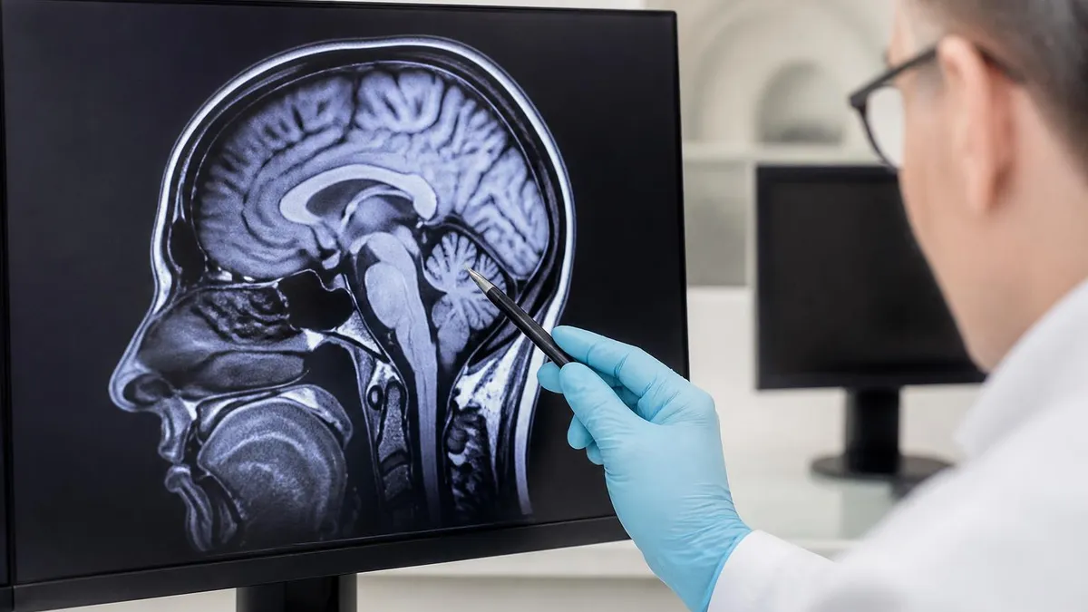
One of the most preventable MRI denials happens when the diagnosis used for prior authorization does not match the diagnosis billed on the final claim. Always reconcile the authorized indication with the radiologist's documented findings before the claim drops, and update authorizations when the clinical picture changes.
Coders who understand the clinical side of imaging make sharper, more defensible decisions, so a working grasp of MRI interpretation is genuinely valuable even for non-clinical staff. MRI produces images based on how hydrogen protons in tissue respond to a strong magnetic field and radiofrequency pulses. Different sequences — T1-weighted, T2-weighted, FLAIR, diffusion-weighted — highlight different tissue properties, and an abnormality often appears as a change in signal intensity compared to surrounding normal tissue on one or more of these sequences.
On a T1-weighted image, fat appears bright and fluid appears dark, making it excellent for anatomy. On a T2-weighted image, fluid appears bright, which is why edema, inflammation, and many lesions stand out so clearly. When a radiologist describes "T2 hyperintensity" or "restricted diffusion," they are noting a signal abnormality that may represent anything from a benign cyst to acute ischemia. The clinical context determines whether that finding is significant or incidental, and the report's impression captures that judgment.
Contrast-enhanced MRI adds another dimension. Gadolinium-based contrast agents highlight areas of increased vascularity or a breakdown in the blood-brain barrier, helping distinguish active disease from old, stable changes. An enhancing lesion behaves differently from a non-enhancing one, and radiologists weigh enhancement patterns heavily when forming an impression. For coders, the presence of enhancement and the radiologist's interpretation of it can be the deciding factor between an abnormal-findings code and a confirmed diagnosis.
Anatomy literacy helps coders too. Knowing that the corpus callosum sits centrally in the brain, that the meniscus cushions the knee, or that intervertebral discs separate the vertebrae lets a coder follow the report's logic and spot when laterality or location should be coded.
You do not need to be a radiologist, but recognizing the structures named in a report makes it far easier to select the most specific code the documentation supports and to know when something is missing. If you want to see how this plays out in practice, reviewing real knee MRI images alongside their reports is a powerful way to connect findings to codes.
The concept of an incidental finding is worth revisiting here from the clinical angle. A brain MRI ordered for headache may reveal a small, asymptomatic meningioma. Clinically, the radiologist documents it and may recommend interval follow-up. For coding, this incidental finding may warrant its own code if it affects management, but the headache that prompted the study still drives the medical-necessity narrative. Understanding both threads keeps the record accurate.
Ultimately, the clinical and coding worlds are two views of the same patient. The radiologist describes what the magnet revealed; the coder translates that description into the language of claims. When both parties share vocabulary and respect each other's constraints, abnormal findings are documented precisely, coded accurately, and reimbursed appropriately. That shared understanding is the foundation of a high-functioning imaging service and the reason cross-training pays dividends across the department.
If you want the full picture, the abnormal mri of brain guide walks through it step by step.
For a deeper look at this topic, see our guide to abnormal mri of the brain.
Putting all of this into daily practice comes down to a few repeatable habits that keep abnormal MRI coding accurate and audit-ready. Start every coding task by reading the impression first, then working backward through the findings to confirm the story holds together. If the impression names a definitive diagnosis, code it specifically and move on. If it describes an abnormality without a confirmed cause, reach for the appropriate R90 or R93 code and check whether a symptom code is needed to explain why the study was ordered in the first place.
Build a personal reference of the abnormal-findings codes you use most often. For brain and CNS imaging, R90.0 for intracranial space-occupying lesion and R90.89 for other abnormal findings come up constantly. For musculoskeletal and abdominal MRI, the R93 subcodes by region are your workhorses. Knowing these cold means you spend less time searching the code book and more time confirming that the documentation truly supports your selection, which is where accuracy is actually won.
Never hesitate to query the radiologist when something is ambiguous. A well-crafted query is not a criticism — it is a professional request for clarification that protects everyone. Ask specific, non-leading questions: "Is the left frontal lesion benign, malignant, or undetermined based on this study?" The answer either confirms a specific code or validates the abnormal-findings code, and the documented response becomes part of the defensible record if the claim is ever reviewed.
Reconcile the order, the authorization, and the claim before anything drops. These three documents should tell one consistent clinical story — same anatomy, same indication, same logic. When they diverge, pause and investigate rather than pushing the claim through, because a five-minute check now prevents a multi-week denial and appeal later. Many departments build this reconciliation into a pre-bill edit so the system flags mismatches automatically before a human ever sees the claim.
Stay current with annual ICD-10-CM updates, which take effect each October 1. Codes are added, revised, and retired every year, and an outdated code is an instant denial. Subscribe to your professional organization's coding updates, and review the changes that touch radiology specifically. The same discipline applies to payer policies, which shift frequently and quietly; a covered indication this quarter may require new documentation next quarter.
Finally, treat denials as data, not defeats. Every denied imaging claim carries a lesson about documentation, code selection, or payer policy. Log the reason, identify the pattern, and feed that insight back to ordering providers and radiologists. Over time, this closed loop dramatically improves your clean-claim rate. Combined with steady study — practice questions, anatomy review, and physics fundamentals — these habits turn abnormal MRI coding from a source of stress into a reliable, confident routine you can sustain across a long career.
Sample MRI - Magnetic Resonance Imaging Practice Questions
Try these questions from our free MRI - Magnetic Resonance Imaging practice tests. The correct answer and an explanation follow each question.
Wraparound (aliasing) artifact in MRI is caused by:
- A. Structures outside the FOV contributing signal that is folded back into the image
- B. Gradient non-linearity at the edges of the bore
- C. T2* dephasing from metallic objects
- D. Phase errors from pulsatile flow
Answer: A. Structures outside the FOV contributing signal that is folded back into the image
When anatomy extends beyond the selected FOV in the phase-encode direction, it is aliased and appears superimposed on the opposite side of the image.
Fast spin echo sequences, in contrast to spin echo, have
- A. T2 weighting decreased but susceptibility rose
- B. Diminished susceptibility and T2 weighting
- C. T2 weighting increased but susceptibility decreased
- D. elevated susceptibility and elevated T2 weighting
Answer: C. T2 weighting increased but susceptibility decreased
Fast Spin Echo (FSE) sequences utilize multiple 180-degree refocusing pulses, which effectively reduce the impact of magnetic susceptibility artifacts by repeatedly rephasing spins. This leads to decreased susceptibility artifacts compared to conventional spin echo. Additionally, FSE sequences can achieve stronger T2 weighting due to the extended echo train and multiple echoes contributing to the signal, often resulting in increased T2 weighting.
Which type of MRI coil surrounds the entire body and is used to transmit RF energy uniformly?
- A. Surface coil
- B. Phased-array coil
- C. Body (volume) coil
- D. Gradient coil
Answer: C. Body (volume) coil
The body (volume) coil is built into the bore and provides uniform RF transmission across the entire patient.
Which MRI sequence is most sensitive for detecting acute subarachnoid hemorrhage?
- A. T2-weighted FSE
- B. FLAIR (Fluid Attenuated Inversion Recovery)
- C. Gradient Echo (GRE)
- D. Diffusion-Weighted Imaging (DWI)
Answer: B. FLAIR (Fluid Attenuated Inversion Recovery)
FLAIR suppresses CSF signal, making subarachnoid blood appear hyperintense against a dark CSF background.
Take the full MRI - Magnetic Resonance Imaging practice test
MRI Questions and Answers
ICD 10 Codes for MRI: Complete Guide to Abnormal MRI Coding, Documentation, and Reimbursement

MRI of Cervical Spine: Complete Guide to Imaging, Anatomy, Pathology, and Patient Preparation
MRI Medical Abbreviation: What MRI Stands For and Why It Matters

Knee MRI Images: A Complete Guide to Reading, Understanding, and Interpreting Knee Scans
Is Nickel Titanium MRI Compatible? A Complete Guide to MRI Safety Materials
About the Author

Medical Laboratory Scientist & Clinical Certification Expert
Johns Hopkins UniversityDr. Sandra Kim holds a PhD in Clinical Laboratory Science from Johns Hopkins University and is certified as a Medical Technologist (MT) and Medical Laboratory Scientist (MLS) through ASCP. With 16 years of clinical laboratory experience spanning hematology, microbiology, and molecular diagnostics, she prepares candidates for ASCP board exams, MLT, MLS, and specialist certification tests.
Join the Discussion
Connect with other students preparing for this exam. Share tips, ask questions, and get advice from people who have been there.
View discussion (8 replies)