What Can an MRI Detect? A Complete Guide to MRI Findings
What can a MRI detect? Learn the conditions, diseases, and abnormalities MRI scans reveal across the brain, spine, joints, organs, and soft tissues.
If you have ever wondered what can a MRI detect, the short answer is remarkable: magnetic resonance imaging is one of the most versatile diagnostic tools in modern medicine, capable of revealing soft tissue abnormalities, tumors, vascular conditions, joint injuries, neurological diseases, and dozens of other findings that x-rays and ultrasound simply cannot show with the same clarity. Because MRI uses powerful magnets and radio waves rather than ionizing radiation, it can be repeated safely and used to monitor disease progression over months and years.
The strength of MRI lies in its ability to differentiate between tissue types. By manipulating timing parameters such as TR, TE, and TI, radiologists can highlight water, fat, blood, or specific pathology. A T2-weighted sequence makes inflammation and edema glow bright white, while a T1-weighted post-contrast scan lights up enhancing tumors and active multiple sclerosis plaques. This biochemical sensitivity is why MRI has become the gold standard for brain, spinal cord, musculoskeletal, and pelvic imaging.
Patients are often referred for an MRI when a physical exam, lab work, or another imaging study produces an unclear result. For example, a CT scan might show a suspicious area in the liver, but only MRI can characterize whether it is a benign hemangioma, focal nodular hyperplasia, or hepatocellular carcinoma. Likewise, a sports physician may suspect a meniscal tear in the knee, but MRI confirms the diagnosis and grades the severity before surgery is even considered.
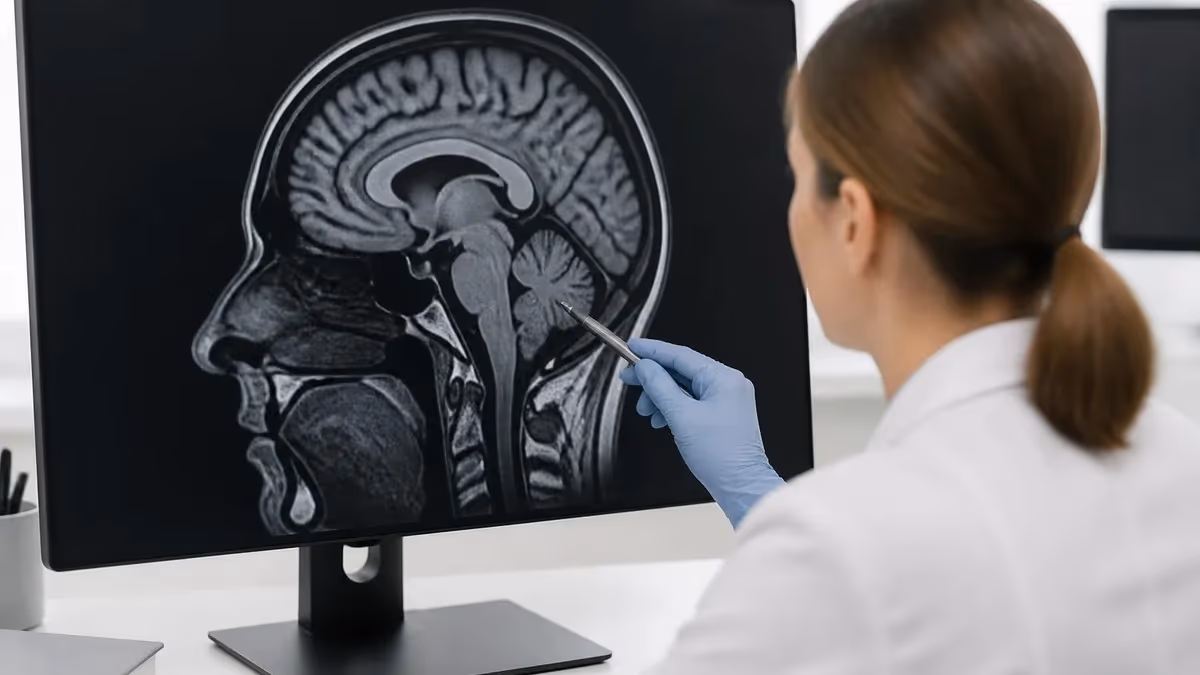
Neurological applications are perhaps the most well-known. MRI can detect strokes within minutes of symptom onset using diffusion-weighted imaging, identify demyelinating lesions in multiple sclerosis, locate brain tumors as small as a few millimeters, and characterize hemorrhages by their age. It also visualizes the spinal cord, nerve roots, and intervertebral discs, making it essential for patients with back pain, sciatica, or suspected cord compression.
Beyond the nervous system, MRI excels at evaluating joints, muscles, tendons, ligaments, and cartilage. Orthopedic surgeons rely on MRI to plan repairs of rotator cuff tears, ACL ruptures, labral injuries, and stress fractures invisible on x-ray. In cardiology, cardiac MRI assesses myocardial viability after a heart attack, quantifies ejection fraction with extraordinary precision, and detects conditions like myocarditis, amyloidosis, and arrhythmogenic cardiomyopathy.
This guide walks through everything an MRI can reveal, organized by body region and clinical scenario. You will learn how contrast agents change what is visible, why certain sequences are chosen for certain questions, and what limitations still exist. For a deeper look at how contrast enhances diagnostic accuracy, see our overview of MRI With and Without Contrast: How It Works, What to Expect before your appointment.
Whether you are a patient preparing for a scan, a student studying for the registry, or a clinician brushing up on MRI capabilities, understanding the breadth of detectable pathology will help you interpret findings and ask better questions. MRI is not infallible, but its diagnostic yield is unmatched across most subspecialties, and new techniques like MR spectroscopy, functional MRI, and 7-Tesla imaging continue to push the boundaries of what we can see inside the human body without a single incision.
What MRI Detects by the Numbers
Brain and Neurological Conditions MRI Can Detect
MRI detects acute ischemic strokes within minutes using diffusion-weighted imaging, far earlier than CT. It also identifies chronic infarcts, transient ischemic attacks, and small vessel disease that contribute to vascular dementia.
From benign meningiomas to aggressive glioblastomas, MRI characterizes mass lesions by location, enhancement pattern, and surrounding edema. Contrast-enhanced T1 sequences reveal tumors as small as two to three millimeters in diameter.
MRI is the cornerstone of MS diagnosis, showing characteristic demyelinating plaques in periventricular, juxtacortical, infratentorial, and spinal cord locations. Active lesions enhance with gadolinium, helping clinicians monitor disease activity.
MR angiography detects unruptured aneurysms, arteriovenous malformations, and vascular stenosis without injecting iodinated contrast. Time-of-flight sequences map the circle of Willis with submillimeter resolution for surgical planning.
Volumetric MRI quantifies hippocampal atrophy in Alzheimer disease, frontotemporal volume loss, and Parkinson-related changes in the substantia nigra. Pattern recognition helps distinguish neurodegenerative subtypes early.
Spinal MRI is one of the most frequently ordered studies in radiology, and for good reason. The spine houses the spinal cord, nerve roots, vertebral bodies, intervertebral discs, facet joints, ligaments, and paraspinal muscles, all of which can be sources of pain or neurological deficit. Unlike x-rays, which only show bony alignment, MRI visualizes every soft tissue structure in exquisite detail, making it indispensable for evaluating back pain, sciatica, radiculopathy, myelopathy, and trauma.
Disc herniations are among the most common findings on lumbar and cervical MRI. The scan reveals whether a disc bulge is broad-based, focally protruding, extruded, or sequestered, and shows precisely which nerve root is compressed. This level of detail directly guides treatment, helping surgeons decide between conservative management, epidural injection, microdiscectomy, or fusion. Sagittal T2 sequences highlight disc dehydration, while axial cuts demonstrate the exact relationship to the thecal sac.
Spinal stenosis, both central and foraminal, is another major target of MRI. Patients with neurogenic claudication often have severe canal narrowing from hypertrophied ligamentum flavum, facet arthropathy, and disc material. MRI quantifies the cross-sectional area of the canal and identifies the most stenotic level, which correlates with symptom localization. Without MRI, surgical planning for laminectomy or decompression would rely on guesswork.
Tumors of the spine fall into three categories: extradural, intradural extramedullary, and intramedullary. MRI distinguishes between them based on location and enhancement. Metastatic disease from breast, lung, prostate, or kidney often involves vertebral bodies and the epidural space. Schwannomas and meningiomas typically sit intradurally outside the cord, while ependymomas and astrocytomas grow within the cord itself. Early detection is critical to prevent permanent neurological injury.
Infections like discitis, osteomyelitis, and epidural abscess are medical emergencies that MRI identifies before they cause cord compression or sepsis. Characteristic findings include disc space hyperintensity on T2, endplate destruction, and rim-enhancing fluid collections after contrast. In immunocompromised patients, MRI also reveals tuberculous spondylitis and fungal infections that mimic malignancy on other modalities.
Trauma evaluation has been revolutionized by spinal MRI. While CT remains the first-line study for fractures, MRI is essential for assessing cord contusion, ligamentous injury, epidural hematoma, and traumatic disc herniation. The STIR sequence is particularly sensitive to bone marrow edema, identifying occult fractures that CT might miss in osteoporotic patients. For acronyms and abbreviations you will encounter in spine reports, check our explainer on MRI Medical Abbreviation: What MRI Stands For and Why It Matters.
Finally, MRI is the only modality that reliably evaluates the spinal cord itself. Conditions like transverse myelitis, syringomyelia, cord infarction, and demyelination from neuromyelitis optica all have signature appearances on cord imaging. Pediatric applications include tethered cord, spinal dysraphism, and syringohydromyelia, often diagnosed before surgical correction. The breadth of spinal pathology MRI can detect makes it irreplaceable in both elective workups and emergent presentations.
What an MRI Can Detect in Joints and Soft Tissue
The knee is the most commonly imaged joint in musculoskeletal MRI. Scans routinely detect medial and lateral meniscal tears, classifying them by morphology as horizontal, oblique, radial, longitudinal, or complex. MRI accuracy approaches ninety percent for meniscal pathology and exceeds ninety-five percent for complete ACL ruptures, which appear as discontinuity of fibers with surrounding edema.
Beyond ligaments and menisci, knee MRI evaluates articular cartilage using fat-suppressed proton density sequences. It identifies chondral defects, osteochondral lesions, and early osteoarthritis. Bone marrow edema, occult fractures, Baker cysts, patellar tendinopathy, and plica syndrome all have distinctive appearances. The scan also rules out tumors and infection when clinical suspicion is high.
MRI vs Other Imaging: Strengths and Trade-offs
- +Exceptional soft tissue contrast unmatched by CT or ultrasound
- +No ionizing radiation, making it safe for repeated scans and pregnancy after first trimester
- +Multi-planar imaging in sagittal, coronal, axial, and oblique planes without repositioning
- +Functional sequences like DWI, MRS, and fMRI add physiological information
- +Detects pathology before structural damage is visible on x-ray
- +MR angiography images blood vessels without iodinated contrast
- +Excellent for cartilage, ligaments, tendons, brain, and spinal cord
- −Long scan times of thirty to ninety minutes can be difficult for claustrophobic patients
- −Contraindicated for many pacemakers, cochlear implants, and ferromagnetic foreign bodies
- −Significantly more expensive than CT or ultrasound, often $500 to $3,000 per study
- −Motion artifacts degrade images, requiring breath-holds or sedation in children
- −Poor visualization of cortical bone and pulmonary parenchyma
- −Gadolinium contrast carries small risks of NSF in severe renal disease
- −Limited availability in rural areas and longer scheduling waits than CT
Pre-MRI Checklist: What to Tell Your Technologist
- ✓List every implanted device including pacemakers, defibrillators, neurostimulators, and cochlear implants
- ✓Disclose any prior surgery involving metal clips, coils, stents, or orthopedic hardware
- ✓Mention occupational metal exposure such as welding or grinding that could leave eye fragments
- ✓Report pregnancy status or possibility of pregnancy before any scan
- ✓Bring documentation of MRI conditional devices including model numbers when possible
- ✓Notify the staff of severe claustrophobia so sedation or open MRI can be arranged
- ✓Disclose kidney disease or low GFR before gadolinium contrast administration
- ✓Remove all jewelry, watches, hearing aids, hairpins, and metallic clothing
- ✓List allergies especially to gadolinium-based contrast agents
- ✓Confirm the body part and clinical question with the technologist to ensure correct protocol
MRI Detects What Other Scans Cannot
Up to forty percent of patients with normal CT or x-ray findings have clinically significant pathology revealed on subsequent MRI. This is especially true for ligamentous injuries, early cancers, demyelinating disease, and bone marrow disorders. When clinical suspicion remains high after a negative initial scan, MRI is often the definitive next step.
Cardiac MRI has emerged as the reference standard for evaluating heart muscle function, viability, and structure. Unlike echocardiography, which can be limited by poor acoustic windows, cardiac MRI provides reproducible measurements of ejection fraction, ventricular volumes, and wall motion. It detects myocardial infarction by late gadolinium enhancement, which highlights scarred, non-viable tissue. Cardiologists use this information to decide whether revascularization will improve function or whether the area is permanently damaged.
Beyond ischemic disease, cardiac MRI diagnoses myocarditis using the updated Lake Louise criteria, which combine T2 mapping for edema, T1 mapping for fibrosis, and extracellular volume calculations. It identifies hypertrophic cardiomyopathy, arrhythmogenic right ventricular cardiomyopathy, cardiac amyloidosis, sarcoidosis, and iron overload from hemochromatosis or thalassemia. Each condition has signature patterns of enhancement and tissue characterization that guide treatment.
Vascular MRI, including magnetic resonance angiography and venography, evaluates arteries and veins throughout the body without nephrotoxic iodinated contrast. MRA detects carotid stenosis, renal artery stenosis in hypertensive patients, peripheral arterial disease, and aortic aneurysms or dissections. Time-resolved sequences can capture arterial and venous phases in a single acquisition, while phase-contrast techniques quantify flow velocity and direction.
Abdominal MRI is increasingly used as a problem-solving tool. Liver MRI characterizes focal lesions with extraordinary specificity, distinguishing hemangiomas, focal nodular hyperplasia, adenomas, and hepatocellular carcinoma based on enhancement patterns through arterial, portal venous, and delayed phases. Hepatobiliary contrast agents like gadoxetate sodium add functional information about hepatocyte uptake, useful for detecting metastases and characterizing equivocal nodules in cirrhotic patients.
Pancreatic and biliary MRI, performed as MRCP, visualizes the bile ducts and pancreatic duct without endoscopy. It detects choledocholithiasis, primary sclerosing cholangitis, cholangiocarcinoma, intraductal papillary mucinous neoplasms, and chronic pancreatitis. The non-invasive nature of MRCP has dramatically reduced the need for diagnostic ERCP, reserving the latter for therapeutic interventions only.
Pelvic MRI offers unmatched detail for gynecologic, prostate, and rectal conditions. In women, it stages endometrial and cervical cancers, characterizes ovarian masses, maps endometriosis, and evaluates uterine fibroids before myomectomy or embolization. In men, multiparametric prostate MRI with diffusion-weighted imaging has transformed prostate cancer detection, replacing systematic biopsies with targeted lesion-directed sampling. For rectal cancer, MRI determines T-stage, mesorectal fascia involvement, and nodal status with surgical accuracy.
Renal and adrenal MRI complement CT by characterizing indeterminate lesions. Chemical shift imaging distinguishes lipid-rich adrenal adenomas from non-adenomas in seconds, while in-phase and out-of-phase sequences identify steatosis in the liver and kidneys. For pheochromocytoma, MRI shows the classic light bulb bright T2 signal. The breadth of abdominal and pelvic pathology MRI detects has made it indispensable in oncology, gastroenterology, and women's health.
Patients with non-MRI-conditional pacemakers, ferromagnetic aneurysm clips, cochlear implants, or metallic foreign bodies in the eye can suffer serious injury or death inside the scanner. Always complete the full safety screening and bring device cards. When in doubt, the MRI must be deferred until safety is confirmed by manufacturer documentation.
Despite its versatility, MRI has real limitations that every patient and clinician should understand. Cortical bone produces almost no signal on conventional sequences because it lacks mobile protons, so MRI is poor at detecting hairline cortical fractures, subtle calcifications, and dental pathology. CT remains the first-line study for acute trauma, pulmonary nodules, kidney stones, and acute hemorrhage in the first few hours. MRI complements rather than replaces these other modalities.
Motion is the enemy of MRI image quality. A patient who cannot hold still for ninety seconds at a time will produce blurry, non-diagnostic images. This affects pediatric patients, individuals with dementia, claustrophobic adults, and anyone in significant pain. Solutions include sedation, anesthesia, faster sequences, motion-correction algorithms, and open-bore scanners. Even so, certain body parts like the small bowel and beating heart require specialized techniques like cine imaging and breath-holds.
Resolution limits also matter. While 3-Tesla scanners can detect lesions as small as two to three millimeters in the brain, peripheral structures like cranial nerves, fine vascular branches, and microscopic metastases may still be invisible. Functional changes often precede structural ones, which is why a normal MRI does not exclude all disease. Conditions like early Alzheimer disease, mild traumatic brain injury, and small fiber neuropathy may show subtle or no findings on routine imaging.
Contrast considerations add another layer of complexity. Gadolinium agents cross the disrupted blood-brain barrier and enhance tumors, infections, and active demyelination, but they should be used cautiously in patients with severe renal impairment due to nephrogenic systemic fibrosis risk. Newer macrocyclic agents have minimal risk, but every contrast study still requires renal function assessment and informed consent. Patients should also be aware that trace gadolinium can deposit in the brain after multiple contrast studies, though clinical significance remains under investigation.
Cost and access remain practical barriers. An MRI study in the United States typically costs between five hundred and three thousand dollars before insurance, compared to a few hundred dollars for CT or ultrasound. Wait times for outpatient MRI can stretch weeks in rural areas, delaying diagnosis of cancers and progressive neurologic conditions. Health systems are addressing this with extended hours, mobile MRI units, and abbreviated protocols that reduce scan time to under fifteen minutes for screening indications.
Patient-specific factors can also limit MRI quality. Metallic hardware like spinal fusion rods, joint replacements, and dental implants creates susceptibility artifact that distorts nearby anatomy. Body habitus affects coil placement and signal-to-noise ratio in larger patients. Even modern wide-bore systems have weight and circumference limits. For tips on imaging through orthodontic appliances, our guide on MRI With Braces: Safety, Image Quality, and What to Expect explains practical workarounds.
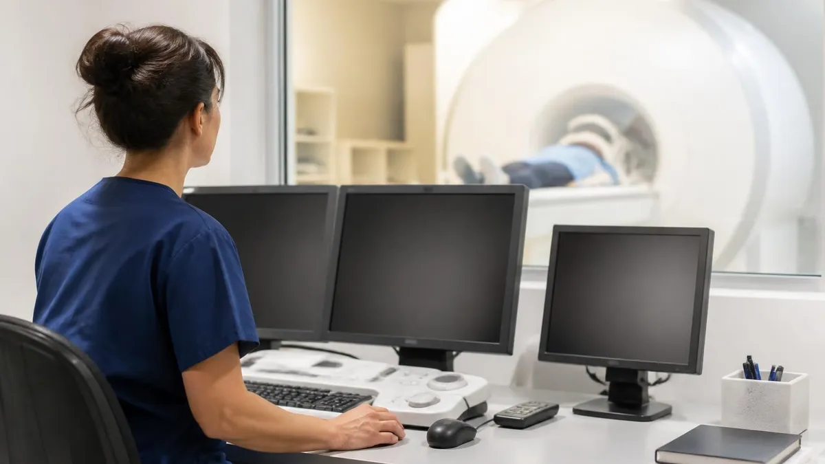
Finally, MRI interpretation is operator-dependent. A scan is only as good as the radiologist reading it and the protocol used to acquire it. Subspecialty expertise in neuroradiology, musculoskeletal imaging, abdominal imaging, and cardiac MRI dramatically improves diagnostic accuracy. When findings are equivocal or unexpected, a second opinion from a fellowship-trained radiologist often changes the diagnosis and management plan. Understanding both what MRI can and cannot do helps set realistic expectations for every clinical encounter.
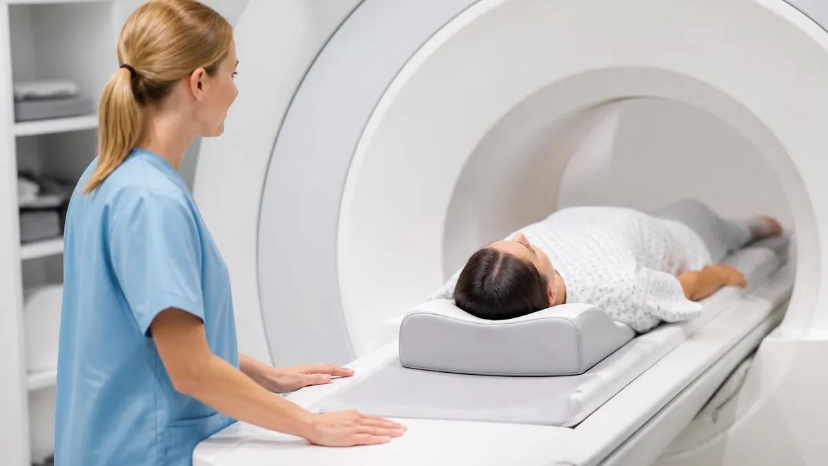
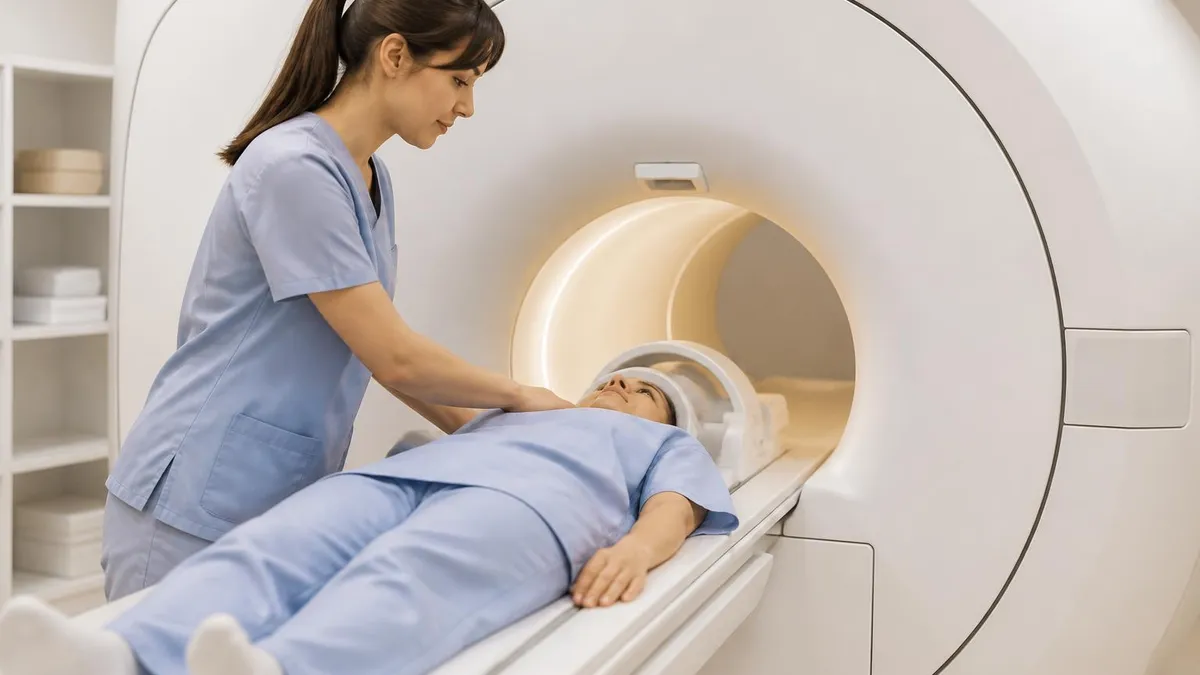
Preparing for an MRI is straightforward but matters more than most patients realize. Arrive thirty minutes early to complete safety screening, bring a list of all implants and surgeries, and wear loose comfortable clothing free of metal. If you are claustrophobic, ask your ordering provider about oral anxiolytics or an open MRI option ahead of time. Eating and drinking are usually fine unless you are scheduled for an abdominal scan, MRCP, or general anesthesia, in which case fasting instructions will be provided.
During the scan, you will lie on a padded table that slides into the bore of the magnet. The technologist will give you a squeeze ball for emergencies and headphones to muffle the loud knocking sounds produced by gradient coils. Stay as still as possible during each sequence, which typically lasts two to six minutes. Breath-holds may be required for chest, cardiac, or abdominal exams. If contrast is part of your protocol, expect a small IV in your arm and a cool sensation during injection.
After the scan, you can resume normal activities immediately unless you received sedation. Drink plenty of water to help clear contrast through the kidneys. Results are usually available within twenty-four to seventy-two hours, depending on the facility. Your ordering provider will discuss findings and next steps. For routine outpatient studies, requesting a copy of both the report and the image disc allows for future comparison and second opinions if needed.
Understanding common MRI terminology empowers patients to participate in their own care. Phrases like incidental finding, T2 hyperintensity, enhancement, restricted diffusion, and Modic changes appear frequently in reports. Most incidental findings are benign and require no action, but some warrant follow-up or referral. Always read the impression section carefully and ask your provider to translate medical jargon into actionable next steps. A good clinician will spend time explaining what was found and what it means for you.
For students preparing for the ARRT MRI registry, the breadth of pathology covered in this article maps directly to the exam content outline. Expect questions on signal characteristics of common lesions, sequence selection for specific clinical questions, contraindications, safety zones, and artifact recognition. Memorizing tissue contrast on T1, T2, FLAIR, STIR, and post-contrast sequences forms the foundation of pathology recognition. Practice questions across multiple difficulty levels solidify this knowledge.
Clinicians ordering MRI should provide clear clinical history on the requisition. Generic indications like back pain or headache yield generic protocols. Specifying the clinical question, suspected pathology, prior imaging, and relevant lab values allows the radiologist to tailor sequences for maximum diagnostic yield. Adding contrast appropriately, requesting MR angiography when vascular pathology is suspected, and selecting the correct field of view all impact outcomes. A well-considered order is the first step toward an accurate diagnosis.
Looking forward, MRI continues to evolve rapidly. Artificial intelligence is reducing scan times by sixty percent or more using deep learning reconstruction. New sequences like APT-CEST imaging detect tumor metabolism without contrast. Seven-Tesla scanners are entering clinical use for ultra-high-resolution brain imaging. The next decade will likely see MRI screening for cancers, faster pediatric protocols, and expanded use in emergency departments. The list of what an MRI can detect grows every year, reinforcing its central role in modern medicine.
MRI Questions and Answers
About the Author
Medical Laboratory Scientist & Clinical Certification Expert
Johns Hopkins UniversityDr. Sandra Kim holds a PhD in Clinical Laboratory Science from Johns Hopkins University and is certified as a Medical Technologist (MT) and Medical Laboratory Scientist (MLS) through ASCP. With 16 years of clinical laboratory experience spanning hematology, microbiology, and molecular diagnostics, she prepares candidates for ASCP board exams, MLT, MLS, and specialist certification tests.