Will a Brain MRI Show a Stroke? What MRI Reveals About Brain Injury, Bleeding, and Recovery
Will a brain MRI show a stroke? Yes — 📝 and within minutes. Learn what MRI shows, which sequences detect bleeds, and how scans guide treatment.
Will a brain MRI show a stroke? In almost every clinical situation, yes — and often within minutes of the event. Magnetic resonance imaging is currently the most sensitive widely available test for detecting both ischemic strokes (caused by blocked arteries) and hemorrhagic strokes (caused by bleeding). Specifically, the diffusion-weighted imaging (DWI) sequence can reveal injured brain tissue as early as 3 to 30 minutes after symptom onset, long before changes appear on a standard CT scan. This early detection window is one of the main reasons emergency departments increasingly rely on MRI for stroke workups.
An MRI does far more than confirm that a stroke has happened. It tells the neurologist exactly where the damage sits, how large the affected territory is, which vascular distribution is involved, and whether the tissue is salvageable or already infarcted. By comparing diffusion images with perfusion images, radiologists can identify the "ischemic penumbra" — the at-risk but still living tissue surrounding the dead core. That distinction directly influences whether a patient receives thrombolytics, undergoes mechanical thrombectomy, or is managed medically with antiplatelets and risk-factor control.
For patients and families, an MRI report after a suspected stroke can read like another language. Terms such as restricted diffusion, FLAIR hyperintensity, T2 shine-through, susceptibility blooming, microbleeds, and chronic gliosis all describe specific findings on specific pulse sequences. Each tells a piece of the story: when the stroke happened, what type it was, whether old strokes are present, and how aggressive the underlying vascular disease appears to be.
MRI is also uniquely valuable for stroke mimics. Conditions like complex migraines, focal seizures, hypoglycemia, multiple sclerosis flares, brain tumor mris, and abscesses can present with sudden neurologic symptoms identical to stroke. CT often cannot reliably distinguish these. MRI, especially when combined with contrast and advanced sequences, frequently provides the answer that changes treatment. This is why neurologists often say that imaging is not just confirmation — it is part of the diagnosis itself.
Beyond the acute setting, MRI continues to serve stroke patients during recovery. Follow-up scans measure how much tissue ultimately survived, identify silent strokes that occurred between visits, and detect small vessel disease, atrophy, or microbleeds that influence long-term medication choices. For physicians studying for boards or technologists preparing for the registry, understanding stroke imaging is foundational — and reviewing the basics of how an MRI test works helps tie together physics, anatomy, and pathology.
This guide walks through everything an MRI can show about a stroke: which sequences detect which findings, how timing affects mri for migraines see, what the typical workflow looks like in the emergency department, and the limitations every reader should understand. Whether you are a patient who just had a scan, a caregiver trying to interpret a report, or a clinician sharpening your skills, you will leave with a clear picture of why MRI has become the imaging gold standard for stroke.
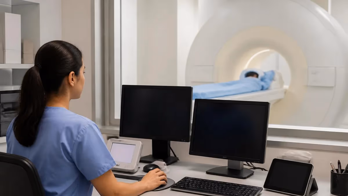
We will also address common patient concerns — claustrophobia, contrast safety, scan length, and what to expect when the scanner is loud and the table feels narrow. Stroke imaging is fast in most modern hospitals, but the experience can still be intimidating. Understanding the process helps reduce anxiety and improves the quality of the images radiologists rely on for diagnosis.
Brain MRI and Stroke by the Numbers
Stroke Types a Brain MRI Can Detect
Caused by a clot blocking blood flow. DWI shows bright restricted diffusion within minutes, with corresponding dark spots on the ADC map confirming true cytotoxic edema rather than artifact.
Bleeding inside the brain from a ruptured vessel. Susceptibility-weighted imaging (SWI) and gradient echo (GRE) sequences show dark blooming where blood products distort the magnetic field.
Tiny deep strokes from small vessel disease, often in basal ganglia, thalamus, or pons. MRI detects lesions as small as 3 mm — far below CT resolution — making it ideal for these subtle infarcts.
Brief stroke-like episodes that resolve. DWI catches about 30-50% of true TIAs as small punctate restrictions, helping risk-stratify patients who appear neurologically normal at evaluation.
Strokes at the borderzones between major arterial territories, often from hypotension or carotid stenosis. MRI shows a characteristic chain-of-lakes pattern not always visible on CT imaging.
The reason MRI outperforms CT in stroke imaging comes down to physics — and the sequences neuroradiologists order in a dedicated stroke protocol are specifically designed to interrogate brain tissue at different molecular timescales. Diffusion-weighted imaging exploits the random Brownian motion of water molecules. In healthy tissue, water moves freely. When a stroke causes cells to swell and die (cytotoxic edema), water becomes trapped inside, restricting its motion. DWI lights up these areas brilliantly, often before any structural change appears.
Apparent diffusion coefficient (ADC) maps are calculated from DWI data and serve as the essential confirmation step. True acute ischemia shows bright DWI with dark ADC. If both DWI and ADC appear bright, the finding is called T2 shine-through and is not a stroke — it could be old gliosis, MS plaque, or vasogenic edema. Reading these two sequences together is foundational for any clinician interpreting an emergency MRI, and it is one of the highest-yield concepts on radiology boards.
FLAIR (fluid-attenuated inversion recovery) suppresses cerebrospinal fluid, making subtle parenchymal abnormalities easier to see. In mri for stroke FLAIR is critical for determining whether an infarct is hyperacute or already several hours old. The "DWI-FLAIR mismatch" — bright on DWI but invisible on FLAIR — suggests the stroke is less than 4.5 hours old, which is the thrombolytic treatment window. This single mismatch can decide whether a patient receives tPA.
Susceptibility-weighted imaging (SWI) and gradient echo (GRE) sequences detect hemorrhage with exquisite sensitivity. Iron in blood products distorts the local magnetic field, creating a dramatic dark "blooming" artifact. SWI can also reveal cerebral microbleeds — tiny prior bleeds that signal amyloid angiopathy or hypertensive vasculopathy, both of which dramatically change anticoagulation decisions. CT misses most microbleeds entirely.
MR angiography (MRA) shows the cerebral vessels themselves. Time-of-flight MRA requires no contrast and can identify large vessel occlusions in the carotid, middle cerebral, basilar, or vertebral arteries. For patients being considered for mechanical thrombectomy, MRA confirms the target vessel. Contrast-enhanced MRA provides higher resolution but requires gadolinium, which is generally avoided in patients with severe renal dysfunction.
Perfusion-weighted imaging (PWI) measures cerebral blood flow and volume. By comparing the DWI "core" (already dead tissue) with the PWI "penumbra" (tissue at risk but still alive), physicians estimate how much brain can still be saved. A large mismatch favors aggressive reperfusion therapy. This concept revolutionized stroke care after the DAWN and DEFUSE-3 trials extended thrombectomy windows from 6 to 24 hours in selected patients.
Together these sequences make MRI a multi-dimensional stroke detector. Understanding what each one shows — and what it cannot show — separates competent stroke imaging from expert interpretation. If you are new to MRI terminology, reviewing the meaning of MRI and related acronyms can help anchor the alphabet soup of T1, T2, DWI, FLAIR, GRE, SWI, MRA, and PWI before you dive deeper.
MRI Practice Test Questions
Prepare for the MRI - Magnetic Resonance Imaging exam with our free practice test modules. Each quiz covers key topics to help you pass on your first try.
MRI Knowledge
MRI Exam Questions covering Knowledge. Master MRI Test concepts for certification prep.
MRI Physics
Free MRI Practice Test featuring Physics. Improve your MRI Exam score with mock test prep.
MRI Anatomy and Pathology
MRI Test Prep for MRI Anatomy and Pathology. Practice MRI Quiz questions and boost your score.
MRI Anatomy and Positioning
MRI Questions and Answers on MRI Anatomy and Positioning. Free MRI practice for exam readiness.
MRI Contrast Agents
Free MRI Quiz on MRI Contrast Agents. MRI Exam prep questions with detailed explanations.
MRI Patient Care and Positioning
MRI Practice Questions for MRI Patient Care and Positioning. Build confidence for your MRI certification exam.
What an MRI Shows Across Stroke Timing
In the first 24 hours after a stroke, MRI is far more sensitive than CT. Diffusion-weighted imaging (DWI) becomes positive within minutes, showing bright restricted diffusion in the affected tissue while the corresponding ADC map appears dark. FLAIR is often normal during the first 4 to 6 hours, which is actually clinically useful: the DWI-positive, FLAIR-negative mismatch suggests the stroke is recent enough for thrombolytic therapy or thrombectomy.
Hemorrhagic strokes show characteristic patterns based on blood breakdown products. Hyperacute hemorrhage (under 12 hours) appears isointense to slightly hyperintense on T2 because oxyhemoglobin is not yet paramagnetic. Gradient echo and SWI sequences are the most sensitive for catching even small acute bleeds, which is why every modern stroke protocol includes them as standard imaging.
MRI vs. CT for Detecting Stroke
- +Detects ischemic stroke within minutes via DWI, hours before CT changes appear
- +Identifies lacunar and brainstem strokes that CT routinely misses entirely
- +Reveals cerebral microbleeds invisible to CT, guiding anticoagulation choices
- +Distinguishes acute, subacute, and chronic strokes on the same exam easily
- +Quantifies penumbra with perfusion imaging to guide thrombectomy decisions
- +No ionizing radiation, making it safer for repeat imaging and younger patients
- −Longer scan time (6-15 minutes) than CT (under 5 minutes) in most centers
- −Less available at night and in rural emergency departments than CT scanners
- −Cannot be used safely with many pacemakers, certain implants, or metal foreign bodies
- −Contraindicated in unstable patients who cannot lie flat or hold still
- −Gadolinium contrast carries small risks in patients with severe renal dysfunction
- −More expensive than CT, with higher reimbursement and scheduling challenges
Reading a Stroke MRI: Step-by-Step Checklist
- ✓Confirm patient name, scan date, and clinical history of acute neurologic deficit
- ✓Start with DWI: identify any areas of restricted diffusion (bright signal)
- ✓Cross-check ADC map: true acute infarct is bright on DWI and dark on ADC
- ✓Review FLAIR for parenchymal hyperintensity matching the DWI lesion
- ✓Assess DWI-FLAIR mismatch to estimate stroke age and treatment window
- ✓Examine SWI or GRE for blooming artifact suggesting acute or chronic blood products
- ✓Check MRA for large vessel occlusion in carotid, MCA, basilar, or vertebral arteries
- ✓Look at perfusion maps for core/penumbra mismatch if thrombectomy is considered
- ✓Map the lesion to a specific vascular territory (MCA, ACA, PCA, watershed, lacunar)
- ✓Compare with prior imaging when available to separate old strokes from new ones
Bright DWI + Negative FLAIR = Stroke Under 4.5 Hours Old
When a stroke is bright on diffusion-weighted imaging but invisible on FLAIR, the lesion is statistically very likely to be less than 4.5 hours old. This mismatch was validated in the WAKE-UP trial and now guides thrombolytic decisions in patients with unknown symptom onset, including those who wake up with deficits. Knowing this single rule can change a patient's outcome.
Despite its strengths, MRI is not infallible — and understanding its limitations is essential for anyone reading or ordering these scans. Roughly 5% of acute ischemic strokes are initially DWI-negative, particularly very small brainstem and posterior fossa lesions, or strokes imaged in the first 30 minutes. Repeat imaging at 24 hours often reveals the lesion. Clinicians must never let a negative MRI override a clear clinical syndrome, especially when symptoms persist or evolve over time.
Stroke mimics are a major source of diagnostic confusion. Hypoglycemia can cause unilateral weakness with restricted diffusion that resolves once glucose is corrected. Status epilepticus produces cortical DWI changes from prolonged neuronal firing rather than ischemia. Hemiplegic migraine, posterior reversible encephalopathy syndrome (PRES), and certain encephalitides can all generate findings that look stroke-like. Careful clinical correlation, repeat imaging, and sometimes MR spectroscopy help separate these from true infarcts.
Motion artifact remains a real challenge. Stroke patients often cannot hold still — they may be confused, dysphasic, vomiting, or seizing. Severe motion blurs DWI and can either obscure a real lesion or create artifact that mimics one. Newer scanners with multi-shot DWI, propeller acquisitions, and motion correction algorithms have improved robustness, but technologists still need to optimize sedation, padding, and coil positioning to get diagnostic images in agitated patients.
Metallic implants are an absolute or relative contraindication depending on type. Older pacemakers, cochlear implants, certain aneurysm clips, retained metal in the eye, and some neurostimulators preclude MRI entirely. MR-conditional implants can usually be scanned under specific protocols. Every patient must complete a thorough safety screening, and any ambiguity should trigger a call to the manufacturer or a review of the implant's MRI safety documentation before scanning.
Gadolinium contrast adds value in some stroke cases — particularly for vasculitis, tumor-mimics, and certain angiographic protocols — but is not required for the standard stroke workup. Patients with severe kidney disease (eGFR under 30) face a small risk of nephrogenic systemic fibrosis, though modern macrocyclic agents have dramatically reduced this concern. Always confirm renal function and pregnancy status before administering contrast in any setting, particularly in the emergency department.
Posterior fossa strokes deserve special attention. The brainstem and cerebellum are small, highly eloquent, and notoriously difficult to image because of CSF pulsation and bone artifact from the skull base. A small pontine infarct that produces life-threatening symptoms may show only a 4-mm DWI spot. Reviewing thin-slice axial and sagittal sequences carefully, and correlating with the clinical exam, prevents these critical strokes from being missed during initial interpretation.
Finally, MRI cannot show what it was not designed to show. Hyperacute hemorrhage under one hour can be subtle because oxyhemoglobin is not yet paramagnetic. Subarachnoid hemorrhage is often better detected by CT in the first 6 hours despite advances in FLAIR-based detection. Knowing when to combine MRI with CT, lumbar puncture, or CT angiography is the mark of an experienced stroke clinician working in modern emergency care.
Every minute of large vessel ischemic stroke destroys approximately 1.9 million neurons. MRI should never delay tPA administration in eligible patients when CT is faster and adequate to rule out hemorrhage. Door-to-needle goals remain under 60 minutes, and door-to-groin puncture for thrombectomy under 90 minutes. Always follow your hospital's stroke protocol and defer to the on-call neurologist for imaging modality selection.
After the acute stroke is diagnosed and treated, MRI continues to play a central role in recovery and long-term care. A follow-up scan at 24 to 72 hours measures the final infarct volume, which correlates with disability outcomes and informs rehabilitation planning. If thrombolytics or thrombectomy were given, this scan also screens for hemorrhagic transformation — a feared complication in which the reperfused infarct bleeds, sometimes catastrophically requiring surgical intervention or reversal agents.
Many patients have silent strokes — small infarcts that produce no symptoms but appear on MRI. These are common in older adults and in patients with atrial fibrillation, untreated hypertension, or diabetes. Their presence dramatically raises the risk of future symptomatic stroke and dementia. When neurologists see silent strokes on routine imaging, they often intensify blood pressure control, add or strengthen antiplatelet therapy, and screen aggressively for occult atrial fibrillation with extended cardiac monitoring.
White matter hyperintensities on FLAIR, often called leukoaraiosis or small vessel disease, accompany many strokes. While not strokes themselves, they reflect chronic ischemic injury to small penetrating arteries and predict both stroke recurrence and cognitive decline. Quantifying their burden (Fazekas score) has become standard reporting practice in many centers. A high burden often shifts management toward aggressive vascular risk factor modification rather than additional procedures.
Cerebral microbleeds detected on SWI are now recognized as critical biomarkers. Lobar microbleeds suggest cerebral amyloid angiopathy, which carries a high risk of hemorrhage if patients are anticoagulated. Deep microbleeds usually indicate hypertensive vasculopathy. The number, location, and distribution of microbleeds directly influence whether atrial fibrillation patients receive warfarin, a DOAC, or no anticoagulation at all — decisions that can mean the difference between preventing and causing a stroke.
For technologists and radiologists who want to deepen their understanding of brain MRI patterns beyond stroke, exploring related anatomy is invaluable. Reviewing the approach to systematic image reading used in knee MRI reinforces the same disciplined pattern-recognition habits that apply to neurologic imaging. The same principles of comparing sequences, identifying signal intensity, and recognizing pathology translate across body regions and clinical contexts seen daily.
Patient experience matters enormously in stroke imaging. Many stroke survivors are anxious, claustrophobic, or unable to communicate normally. Family presence, clear explanations, eye contact, headphones with calming music, and a brief practice run through the bore can dramatically improve cooperation and image quality. Some centers now use prism glasses, open-bore scanners, or low-dose anxiolytics for severely claustrophobic patients without compromising the diagnostic sequences needed for clinical decision-making.
Finally, MRI is increasingly used in stroke prevention research. Functional MRI, diffusion tensor imaging, and arterial spin labeling are revealing how the brain reorganizes after stroke and how rehabilitation reshapes neural networks. These tools remain mostly research-grade today but are quickly entering clinical practice in academic medical centers. The next decade will likely bring AI-assisted stroke MRI interpretation that flags lesions, quantifies penumbra, and predicts outcomes faster than any human reader could.
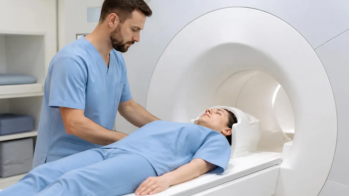
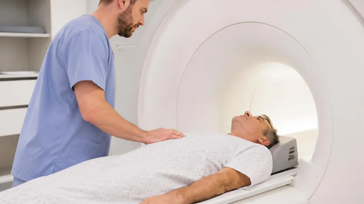
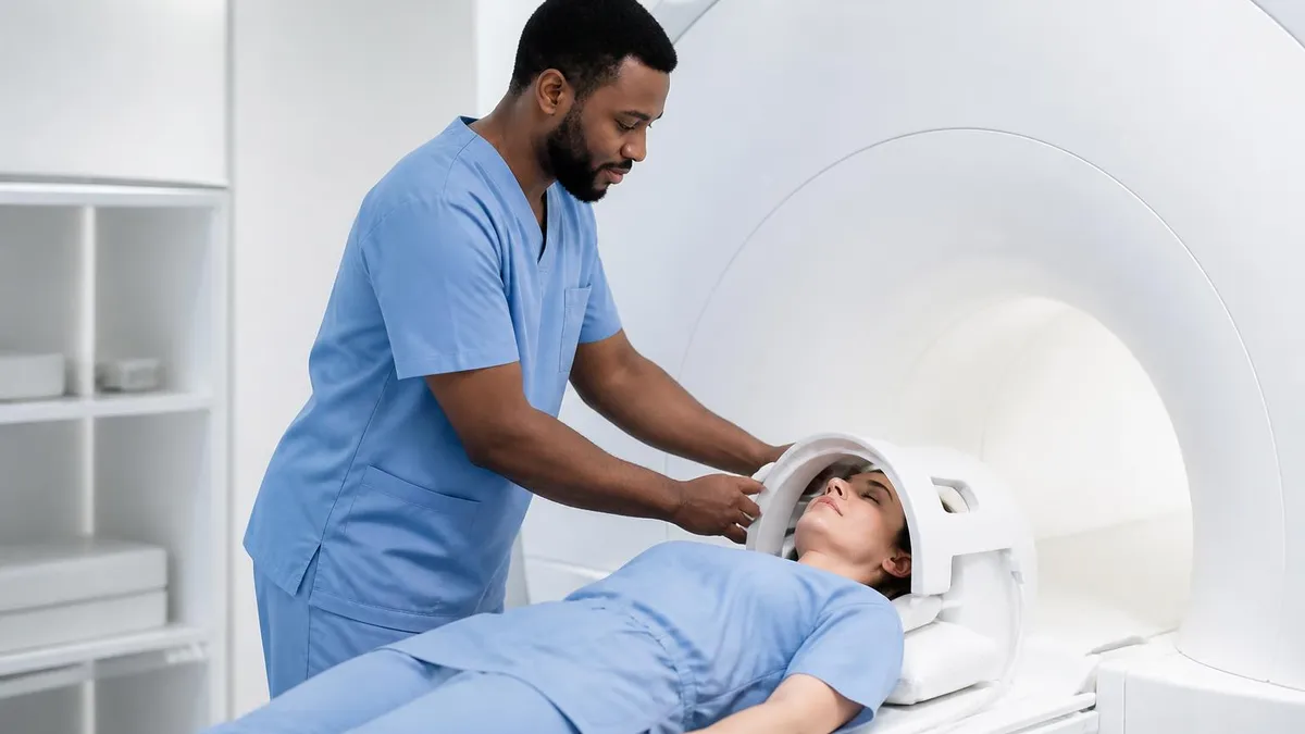
If you or a loved one has been scheduled for a brain MRI to evaluate possible stroke, a few practical preparation tips can make the experience smoother. Wear comfortable clothing without metal — no zippers, underwire bras, or metallic appliques. Remove all jewelry, watches, hearing aids, and dentures before the scan. Inform staff about any implants, surgeries, or metal exposure history, including welding or shrapnel. A thorough safety screening is non-negotiable, and honesty about prior procedures protects you from real risk.
The scan itself usually takes 6 to 15 minutes for a stroke protocol, though some advanced studies with perfusion and angiography may run 25 to 30 minutes. You will lie on your back, head inside a coil shaped like a helmet. Loud knocking, beeping, and humming sounds are normal — they come from the gradient coils rapidly switching during image acquisition. Earplugs or headphones reduce the noise significantly, and most centers now offer music through the headset during the entire scan.
If you are claustrophobic, tell the technologist before you arrive. Many scanners are now wider-bore designs with significantly more head room. Some hospitals offer open MRI systems, though these typically produce lower-quality images and may not be appropriate for acute stroke evaluation. A small dose of oral lorazepam, taken about an hour before the scan, helps many patients tolerate the bore without affecting image quality. Family members can often stay in the room with you to provide reassurance.
Movement is the enemy of good MRI images. Even small shifts during the diffusion sequence can blur an entire stroke into unreadability. Practice lying still, breathe normally, and focus on a single point of imagined detail in your mind. The technologist will provide instructions through the intercom and check on you between sequences. If you need to stop, squeeze the safety bulb placed in your hand — the staff will come immediately to help you out of the scanner.
After the scan, you can usually resume normal activity immediately unless you received sedation or contrast. Gadolinium contrast occasionally causes mild nausea, headache, or a metallic taste, all of which typically resolve within a few hours. Drinking plenty of water helps clear the contrast through your kidneys. If you develop a rash, breathing difficulty, or persistent symptoms, contact your physician right away for evaluation and management of a possible adverse reaction.
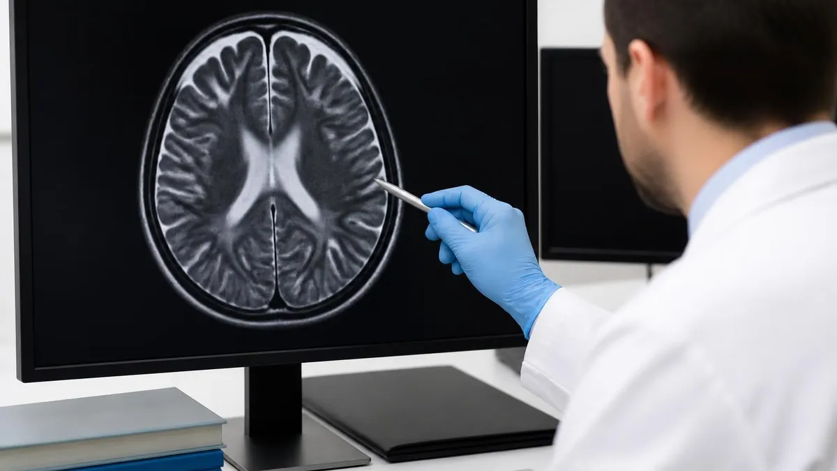
Reports are usually available to the ordering physician within hours, and abnormal findings are often communicated immediately by the radiologist via a critical results phone call. Patients can typically access the report through their hospital portal within a day. Reading your own MRI report can be daunting — radiologists write for other physicians, not patients. Ask your neurologist or primary care provider to translate findings like "restricted diffusion," "FLAIR hyperintensity," "microbleeds," or "chronic small vessel ischemic changes" in everyday language.
For anyone preparing for radiology boards, MRI registries, or neuroscience exams, mastering brain stroke imaging is one of the highest-yield investments you can make. Stroke questions appear on virtually every neuroimaging exam, and the same DWI, FLAIR, SWI, and ADC patterns recur constantly. Use practice questions that show real images, force you to make sequence-specific calls, and build a mental checklist you can apply under exam-time pressure when seconds count and clarity matters most.
MRI Questions and Answers
What Is an MRI Test? How Magnetic Resonance Imaging Scans Diagnose Disease in 2026

MRI Medical Abbreviation: What MRI Stands For and Why It Matters

The History of MRI: From Discovery to Modern Medicine

Knee MRI Images: A Complete Guide to Reading, Understanding, and Interpreting Knee Scans

Noise of MRI Machine: Why MRI Scanners Are So Loud and What to Expect
About the Author
Medical Laboratory Scientist & Clinical Certification Expert
Johns Hopkins UniversityDr. Sandra Kim holds a PhD in Clinical Laboratory Science from Johns Hopkins University and is certified as a Medical Technologist (MT) and Medical Laboratory Scientist (MLS) through ASCP. With 16 years of clinical laboratory experience spanning hematology, microbiology, and molecular diagnostics, she prepares candidates for ASCP board exams, MLT, MLS, and specialist certification tests.
Join the Discussion
Connect with other students preparing for this exam. Share tips, ask questions, and get advice from people who have been there.
View discussion (6 replies)