Ischaemic Stroke MRI: Complete Guide to Imaging, Sequences, and Diagnosis
🎓 Ischaemic stroke MRI guide: DWI, ADC, FLAIR, perfusion sequences, timing windows, and what radiologists look for in acute stroke imaging.
An ischaemic stroke MRI is the single most powerful imaging tool available for diagnosing acute brain infarction, mapping salvageable tissue, and guiding time-critical treatment decisions in the emergency department. While CT remains the front-line study at many hospitals because of its speed and accessibility, MRI has become the reference standard for confirming small, early, or posterior-circulation strokes that CT routinely misses. Understanding how each sequence contributes to the diagnosis is essential for technologists, residents, and registry candidates alike.
The clinical urgency behind stroke imaging cannot be overstated. Roughly 1.9 million neurons die every minute that a large-vessel occlusion goes untreated, which is why the phrase "time is brain" dominates every stroke protocol. MRI helps clinicians answer three critical questions within minutes of the patient entering the scanner: is this an infarct, how old is it, and is there a penumbra of tissue that could still be rescued with thrombolysis or thrombectomy?
Diffusion-weighted imaging (DWI) is the cornerstone of the stroke protocol because cytotoxic oedema restricts the movement of water molecules within minutes of arterial occlusion. A bright DWI lesion paired with a dark apparent diffusion coefficient (ADC) map is the imaging signature of acute infarction, and this pattern can be reliably detected as early as thirty minutes after symptom onset. No other modality offers comparable sensitivity in the hyperacute window.
Beyond DWI, a complete stroke MRI integrates FLAIR, gradient-echo or susceptibility-weighted imaging, MR angiography of the neck and circle of Willis, and increasingly a perfusion sequence. Each adds a unique piece of the puzzle, from confirming when a stroke started in a wake-up patient to detecting microbleeds that would change anticoagulation decisions. For a refresher on the underlying terminology, our MRI medical abbreviation guide breaks down every acronym you will encounter in a stroke report.
This article walks through the complete imaging workflow used in modern comprehensive stroke centres, from the moment the patient is moved onto the table to the final radiology report that drives intervention. It covers indications, contraindications, sequence selection, timing windows, common pitfalls, and the specific findings that distinguish embolic from lacunar from watershed infarcts. Whether you are studying for the ARRT advanced MRI exam or interpreting your first call shift, the concepts here form the practical backbone of acute neuroimaging.
The reader should expect a balance of physics, anatomy, and clinical reasoning. Stroke MRI is a discipline where understanding a single sequence in isolation is rarely useful. The diagnostic power emerges from pattern recognition across the protocol, recognising how DWI, ADC, FLAIR, and angiographic data combine to localise the lesion, estimate its age, and predict its trajectory over the next forty-eight hours.
Finally, this guide reflects current 2026 practice in US comprehensive stroke centres, including updated extended-window selection criteria, the role of MRI in mechanical thrombectomy decisions out to twenty-four hours, and the growing use of automated post-processing software that quantifies core and penumbra volumes in seconds. Bookmark this page and use it as a reference whenever a stroke alert pages overhead.
Ischaemic Stroke MRI by the Numbers
The Acute Stroke MRI Workflow
Stroke Alert Activation
Safety Screening
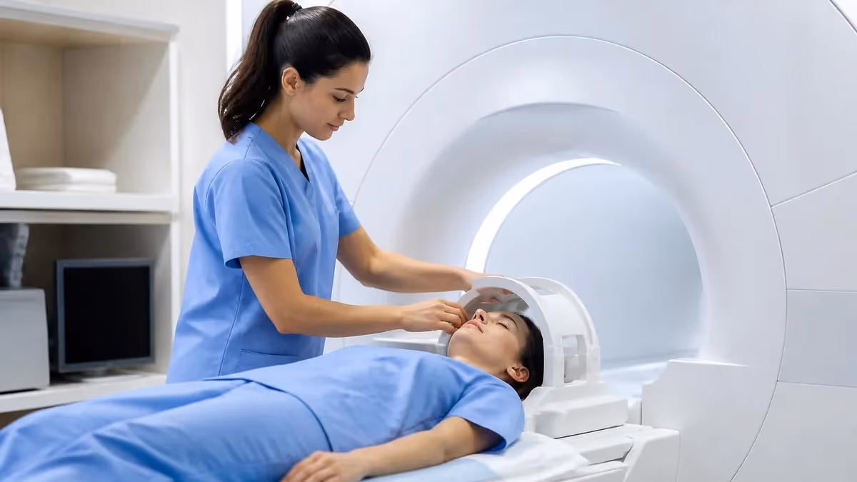
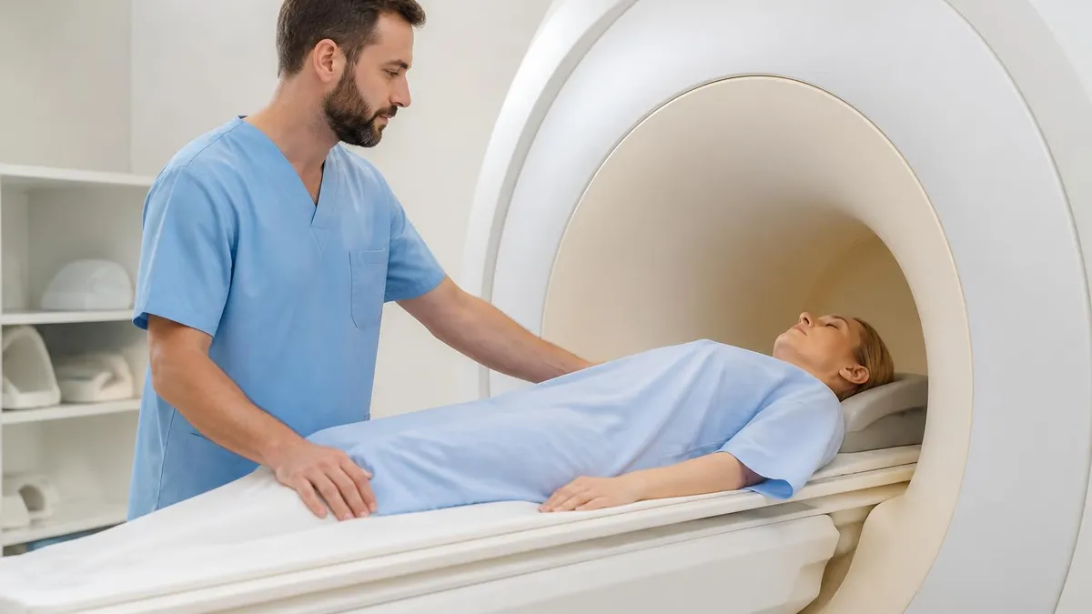
Acquisition
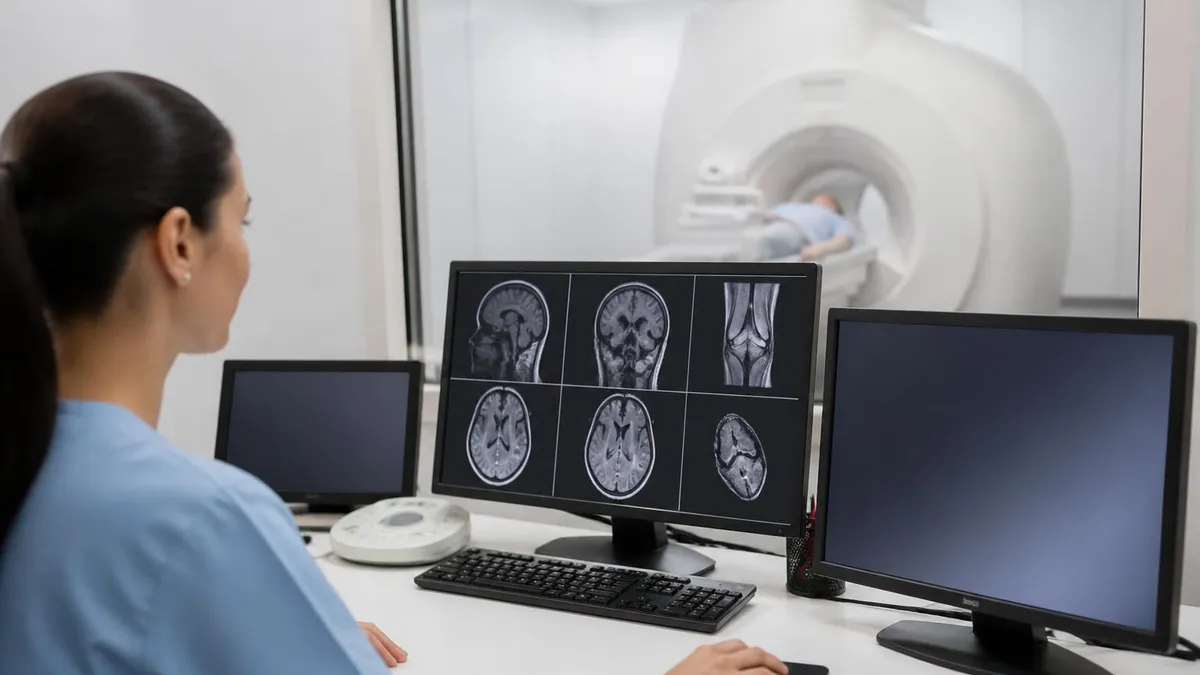
Real-Time Interpretation
Treatment Decision
Follow-Up Imaging
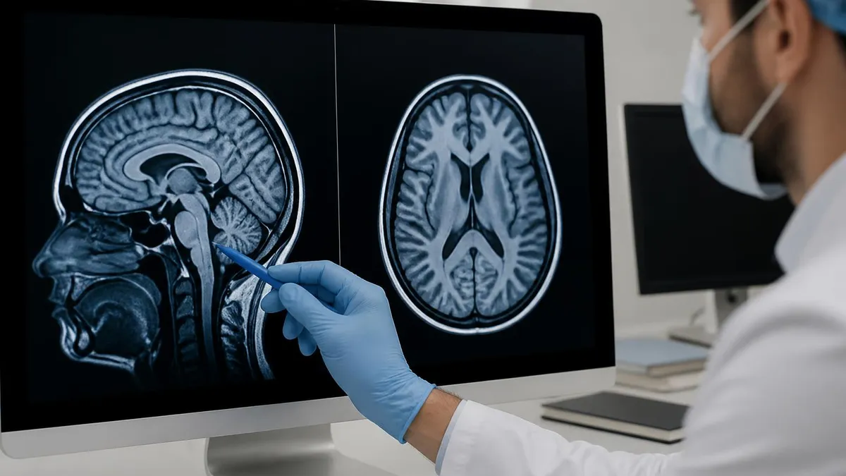
Diffusion-weighted imaging works by applying strong, paired motion-probing gradients that dephase and rephase water molecules. In healthy brain, water diffuses freely and signal is suppressed. In acutely infarcted tissue, failure of the sodium-potassium ATPase pump causes water to shift from the extracellular to the intracellular compartment, where its motion is restricted. That restricted water retains signal, producing the characteristic bright lesion that is the imaging hallmark of an acute infarct.
The apparent diffusion coefficient map is calculated mathematically from DWI images acquired at different b-values, most commonly b=0 and b=1000. The ADC map removes T2 shine-through artefact and gives a quantitative measure of diffusion. True restricted diffusion appears bright on DWI and dark on ADC, while T2 shine-through lesions appear bright on both. Mastering this distinction is the single most important interpretive skill in stroke imaging.
DWI sensitivity peaks between three and seven days after onset, then signal slowly normalises through a process called pseudonormalisation. ADC values typically drop within minutes, reach their nadir at one to four days, and pseudonormalise at seven to ten days. After that, ADC may even rise above baseline as the tissue evolves toward encephalomalacia. Knowing this temporal curve allows the radiologist to estimate stroke age from imaging alone, which becomes critical in wake-up strokes or patients who cannot give a history.
The DWI-FLAIR mismatch concept exploits this temporal evolution. In the first four and a half hours, an infarct is typically visible on DWI but not yet on FLAIR, because cytotoxic oedema precedes the vasogenic oedema that drives FLAIR signal. A DWI-positive, FLAIR-negative lesion strongly suggests the stroke is within the thrombolysis window, even in patients who awoke with symptoms and cannot specify an onset time. This finding has expanded treatment eligibility for thousands of wake-up stroke patients.
Sensitivity for acute infarction approaches ninety-five percent with DWI, far exceeding non-contrast CT which detects only thirty to forty percent of acute strokes in the first six hours. The advantage is most dramatic in the posterior fossa, where beam hardening from the skull base obscures CT detail and brainstem or cerebellar infarcts are routinely missed until they become catastrophic. MRI sees these lesions cleanly. For historical context on how diffusion imaging transformed neurology, our history of MRI article traces the technology from Lauterbur's first images to modern stroke protocols.
Despite its sensitivity, DWI is not infallible. Up to seven percent of acute strokes are DWI-negative on initial imaging, especially small brainstem lesions, very early lacunes, and infarcts in the watershed zones. When clinical suspicion is high and DWI is negative, the protocol should include perfusion imaging or a repeat scan in twenty-four hours, because missed posterior-circulation strokes carry particularly grim outcomes if untreated.
Finally, DWI is not specific to stroke. Abscesses, hypercellular tumours like lymphoma and medulloblastoma, hyperviscous cysts, status epilepticus, and severe demyelination can all restrict diffusion. Pattern recognition, vascular territory mapping, and correlation with FLAIR and post-contrast imaging usually resolve the differential, but a high-quality reading always considers mimics before committing to a stroke diagnosis.
MRI Practice Test Questions
Prepare for the MRI - Magnetic Resonance Imaging exam with our free practice test modules. Each quiz covers key topics to help you pass on your first try.
MRI Knowledge
MRI Exam Questions covering Knowledge. Master MRI Test concepts for certification prep.
MRI Physics
Free MRI Practice Test featuring Physics. Improve your MRI Exam score with mock test prep.
MRI Anatomy and Pathology
MRI Test Prep for MRI Anatomy and Pathology. Practice MRI Quiz questions and boost your score.
MRI Anatomy and Positioning
MRI Questions and Answers on MRI Anatomy and Positioning. Free MRI practice for exam readiness.
MRI Contrast Agents
Free MRI Quiz on MRI Contrast Agents. MRI Exam prep questions with detailed explanations.
MRI Patient Care and Positioning
MRI Practice Questions for MRI Patient Care and Positioning. Build confidence for your MRI certification exam.
Key Sequences in an Ischaemic Stroke MRI
Diffusion-weighted imaging is acquired as an echo-planar sequence with b-values of 0 and 1000 s/mm², typically completing in under a minute. The bright lesion on DWI represents restricted water motion, the imaging signature of cytotoxic oedema seen within thirty minutes of arterial occlusion. ADC maps confirm the restriction is genuine and not T2 shine-through, which is essential to avoid false positives in chronic lesions.
Quantitative ADC thresholds around 620 × 10⁻⁶ mm²/s are used by automated software to estimate the irreversibly damaged core. Volumes under seventy millilitres typically predict good thrombectomy outcomes, while larger cores carry higher haemorrhage risk. Always inspect the ADC map directly rather than relying on the colour overlay, as motion or susceptibility artefact can produce misleading regions.
MRI vs CT for Acute Stroke: Trade-Offs
- +Detects acute infarct within thirty minutes of onset, far earlier than CT
- +Far superior visualisation of posterior fossa and brainstem strokes
- +Quantifies core and penumbra without iodinated contrast in many protocols
- +DWI-FLAIR mismatch identifies wake-up strokes eligible for thrombolysis
- +No ionising radiation, safer for young patients and repeat imaging
- +Simultaneous evaluation of parenchyma, vessels, and perfusion in one study
- −Longer acquisition time than non-contrast CT, even with abbreviated protocols
- −Limited availability overnight or at smaller community hospitals
- −Strict contraindications for pacemakers, metallic implants, and claustrophobia
- −Motion artefact is common in agitated, aphasic, or hemiparetic patients
- −Higher cost and more demanding staffing requirements per study
- −Susceptibility artefact at the skull base can obscure small infarcts
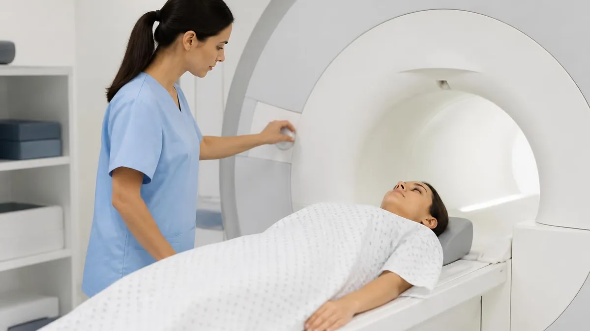
Pre-Scan Safety and Quality Checklist for Stroke MRI
- ✓Confirm absence of MR-unsafe implants using a written and verbal screening form
- ✓Document last known well time and current neurological deficit on the requisition
- ✓Check renal function if gadolinium contrast may be required for perfusion or vessel wall imaging
- ✓Ensure IV access in the arm closer to the scanner door for rapid contrast administration
- ✓Pre-program the abbreviated stroke protocol with DWI, FLAIR, GRE, MRA, and optional perfusion
- ✓Position the patient with foam padding to minimise motion in hemiparetic limbs
- ✓Use the smallest head coil that maintains coverage for the best signal-to-noise ratio
- ✓Verify that motion-correction and parallel-imaging accelerations are activated
- ✓Have post-processing software ready for ADC, MTT, CBF, CBV, and Tmax maps
- ✓Notify the neuroradiologist before the first sequence completes for real-time review
The Six-Minute Stroke Protocol
A modern abbreviated stroke MRI runs DWI, FLAIR, GRE, and three-dimensional time-of-flight MRA in approximately six minutes when parallel imaging and simultaneous multislice techniques are used. Adding perfusion extends the study by another three to four minutes. This compressed protocol delivers ninety-five percent of the diagnostic information of a full stroke MRI and is now the standard at comprehensive stroke centres worldwide.
Even an experienced neuroradiologist can be tricked by an ischaemic stroke MRI, and most diagnostic errors fall into a small number of recurring categories. The most common is mistaking T2 shine-through for true restricted diffusion. A subacute or chronic lesion with high T2 signal will appear bright on DWI simply because DWI carries underlying T2 weighting. The ADC map resolves the ambiguity: true restriction is dark on ADC, while shine-through is bright. Never call a stroke on DWI alone without confirming on ADC.
A second pitfall is failing to recognise the stroke mimics. Cerebral abscesses restrict diffusion because viscous pus impedes water motion, and they often present with focal deficit. Hypercellular tumours, especially lymphoma and medulloblastoma, restrict diffusion through high nuclear-to-cytoplasmic ratio packing. Status epilepticus can produce cortical and thalamic diffusion restriction that resolves within days. Postictal Todd's paralysis is a frequent stroke mimic in the ED, and the imaging can be misleadingly similar.
Susceptibility artefact at the skull base, frontal sinuses, and surgical sites can obscure small infarcts and create spurious areas of signal dropout. In patients with deep brain stimulators, dental hardware, or aneurysm clips, even MR-conditional devices generate distortion that limits sensitivity. Always inspect non-EPI sequences when EPI quality is degraded, and consider repeat imaging when the clinical syndrome is convincing but the EPI study is non-diagnostic.
Wake-up strokes pose unique challenges. The patient cannot specify symptom onset, so traditional time-based eligibility rules do not apply. The DWI-FLAIR mismatch becomes the operative criterion: if the infarct is visible on DWI but not yet on FLAIR, the lesion is presumed to be under four and a half hours old and the patient may receive thrombolytics. Quantitative FLAIR signal ratios are increasingly used to standardise this judgement and reduce interobserver variability.
Lacunar infarcts in the basal ganglia, thalamus, and pons are easy to miss because of their small size and the dense vascular anatomy in those regions. A focused approach is to scroll DWI slowly and compare side-to-side symmetry of the deep grey nuclei. Microinfarcts under three millimetres still produce clinically devastating syndromes when they involve eloquent territory like the corticospinal tract or thalamic sensory nuclei, so careful inspection is non-negotiable.
Watershed infarcts in the anterior or posterior border zones suggest hypoperfusion rather than embolism and demand a different workup. They are often bilateral, linear, and located between major vascular territories. Recognising the watershed pattern points the clinician toward investigation of carotid stenosis, hypotension during sleep or surgery, or systemic hypoperfusion, and changes the secondary prevention plan from anticoagulation toward revascularisation.
Finally, haemorrhagic transformation can occur within hours of an ischaemic stroke, especially after reperfusion. Always inspect GRE or SWI carefully on the post-treatment scan and quantify any blooming as petechial, confluent, or frank haematoma. The European Cooperative Acute Stroke Study classification of haemorrhagic transformation guides management and helps predict clinical worsening before it becomes apparent at the bedside.
A bright DWI lesion alone is never sufficient to diagnose acute stroke. T2 shine-through from chronic infarcts, gliosis, or white matter disease can mimic restricted diffusion. The diagnosis requires a bright DWI lesion paired with a dark ADC map. Failure to confirm on ADC is the single most common cause of false-positive stroke calls and unnecessary thrombolysis exposure.
A well-structured ischaemic stroke MRI report does more than describe findings; it translates imaging into actionable clinical guidance. The opening sentence should state whether an acute infarct is present, its vascular territory, and its approximate age based on DWI, ADC, and FLAIR signal. Stroke neurologists make immediate decisions from this first line, so clarity matters more than literary style.
Territory localisation should follow standard anatomic conventions: middle cerebral artery superior or inferior division, anterior cerebral artery, posterior cerebral artery, lenticulostriate, anterior or posterior choroidal, thalamoperforator, pontine perforator, or cerebellar (PICA, AICA, SCA). Identifying the vascular territory tells the clinician which artery to interrogate on MRA and guides the search for proximal occlusion that would be amenable to mechanical thrombectomy.
Core volume should be quantified whenever extended-window thrombectomy is being considered. Automated software now provides ADC-defined core in millilitres within seconds of acquisition, and most comprehensive stroke centres include this number in the report. Volumes under seventy millilitres generally support intervention, while volumes above one hundred millilitres predict futile reperfusion and increased haemorrhage risk under current selection criteria.
Vessel status from MRA must be described in terms of occlusion location and length. A proximal M1 occlusion, basilar occlusion, or internal carotid terminus occlusion is a thrombectomy target. Distal M3 or M4 occlusions and small perforator infarcts are not. The report should explicitly state whether a large-vessel occlusion is present, since this is the single most important determinant of treatment escalation.
Perfusion findings deserve their own paragraph when included. Report the Tmax greater than six second volume as the at-risk tissue, the ADC-defined core, and the mismatch ratio. A mismatch ratio above 1.8 and an absolute mismatch above fifteen millilitres support thrombectomy in the six to twenty-four hour window. Increasingly, automated software embeds these numbers directly into the PACS report template.
Comparison with prior imaging is essential, particularly in patients with known cerebrovascular disease. Distinguishing a new acute infarct from chronic encephalomalacia changes management entirely. For broader context on how MRI integrates with other imaging modalities in patient workups, see our complete knee MRI images guide for an analogous deep dive into reading and interpreting structured MRI studies.
Finally, the impression should list haemorrhage status, vessel status, core and penumbra volumes if available, and a clear recommendation. Phrases like "findings support consideration of mechanical thrombectomy" or "no large-vessel occlusion; medical management appropriate" remove ambiguity and accelerate decision-making in the time-critical stroke pathway. Reports that bury the recommendation in the body or hedge excessively slow down treatment and worsen patient outcomes.
For technologists running the stroke protocol, a few practical habits separate smooth studies from chaotic ones. Pre-program the abbreviated protocol as a single keystroke selection in the scanner console so that no time is wasted choosing sequences during an active stroke alert. Verify the protocol weekly to catch software updates that may have reset parameters or coil selections back to defaults.
Patient communication during stroke imaging is often impaired by aphasia, neglect, or reduced level of consciousness. Use clear, slow instructions, demonstrate breath-holds with gestures, and have a family member or interpreter at the bedside when possible. Reassure the patient at the start of every sequence and watch the camera for movement so you can pause and reposition before motion ruins a critical DWI run.
Use foam padding generously to immobilise the affected limb and reduce involuntary movement. Positioning the head with the canthomeatal line perpendicular to the table improves anatomic consistency across follow-up scans and makes the radiologist's job easier. A small wedge under the knees relieves back strain and reduces shifting during the eight to ten minute study.
For perfusion imaging, double-check the contrast injector settings before the patient enters the room. A power injector running at four to five millilitres per second with a saline chaser delivers the tight bolus needed for accurate Tmax and mean transit time maps. A sluggish injection ruins the perfusion study even when every other sequence is perfect, and there is no opportunity to repeat in an acute stroke.
For residents and fellows, build a personal reading routine that always inspects the same sequences in the same order: DWI first, then ADC, then FLAIR, then GRE, then MRA, then perfusion. This habit ensures you never miss a critical finding because you scrolled in the wrong direction or skipped a sequence under time pressure. Pattern recognition develops through this disciplined repetition more than through any textbook.
Track your own cases. Keep a brief log of stroke MRIs you read, including territory, core volume, vessel status, and clinical outcome. Reviewing these cases monthly accelerates pattern recognition far more than passive reading, and it builds the mental library you will draw on at three in the morning when a stroke alert comes in and decisions cannot wait. Most residents are stunned at how quickly they improve once they start logging cases systematically.
Finally, never read in isolation. The strongest stroke programs run multidisciplinary case reviews where neuroradiology, neurology, and neurosurgery sit together and dissect treatment decisions. Learning why a thrombectomy succeeded or failed, why a thrombolysis bled, or why a wake-up stroke was correctly selected for treatment builds clinical judgement that no algorithm or scoring system can replicate. Make the time for these conferences whenever they are offered.
MRI Questions and Answers

MRI Medical Abbreviation: What MRI Stands For and Why It Matters

The History of MRI: From Discovery to Modern Medicine

Knee MRI Images: A Complete Guide to Reading, Understanding, and Interpreting Knee Scans

Noise of MRI Machine: Why MRI Scanners Are So Loud and What to Expect
Full Body MRI Cost: What You'll Pay in 2026 and Why
About the Author
Medical Laboratory Scientist & Clinical Certification Expert
Johns Hopkins UniversityDr. Sandra Kim holds a PhD in Clinical Laboratory Science from Johns Hopkins University and is certified as a Medical Technologist (MT) and Medical Laboratory Scientist (MLS) through ASCP. With 16 years of clinical laboratory experience spanning hematology, microbiology, and molecular diagnostics, she prepares candidates for ASCP board exams, MLT, MLS, and specialist certification tests.
Join the Discussion
Connect with other students preparing for this exam. Share tips, ask questions, and get advice from people who have been there.
View discussion (6 replies)