Can You See a Brain Bleed on an MRI? What an MRI Can Detect 2026 July
Can you see a brain bleed on an MRI? Learn what MRI detects, 🧠 how blood appears on each sequence, and when CT is faster for acute bleeds.
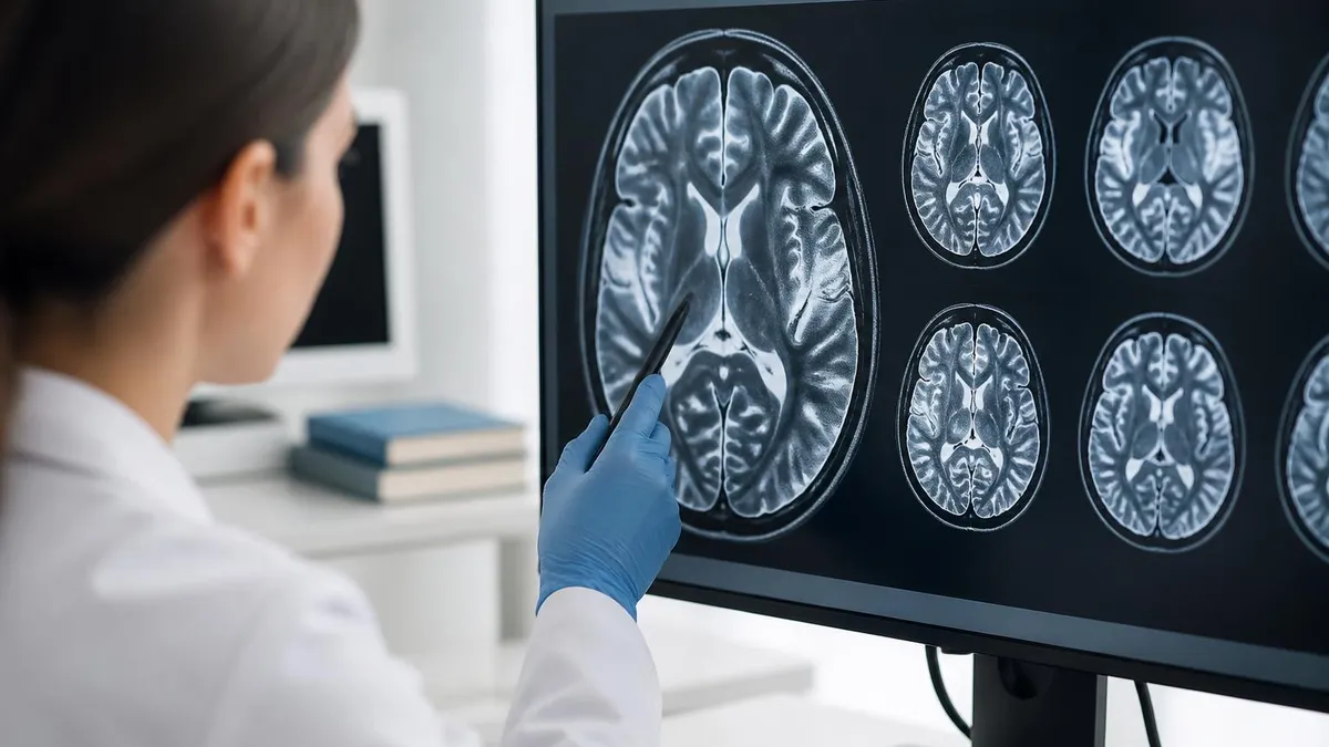
Can you see a brain bleed on an MRI? Yes — and in many situations MRI sees intracranial hemorrhage with remarkable clarity, including tiny microbleeds that a CT scan would miss entirely. Magnetic resonance imaging uses powerful magnetic fields and radiofrequency pulses to map the behavior of hydrogen protons inside tissue, and blood products produce very specific, time-dependent signals on different MRI sequences. That sensitivity makes MRI one of the most powerful diagnostic tools in modern neuroimaging, especially for subacute and chronic bleeds.
That said, MRI is rarely the first scan ordered in the emergency room when a stroke is suspected. CT remains the workhorse for acute hemorrhage because it is faster, more widely available, and excellent at spotting fresh blood within minutes of symptom onset. MRI shines later — when clinicians need to date a bleed, find microhemorrhages from amyloid angiopathy, evaluate a hemorrhagic tumor, or distinguish old blood from new injury. Understanding what an MRI can detect helps patients and clinicians choose the right test.
The reason MRI is so sensitive to blood comes down to chemistry. As red blood cells break down outside a vessel, hemoglobin passes through several iron-containing stages: oxyhemoglobin, deoxyhemoglobin, methemoglobin, and finally hemosiderin. Each form has different magnetic properties, and each one alters T1 and T2 relaxation in a predictable way. Radiologists exploit this by combining multiple sequences — T1, T2, FLAIR, diffusion-weighted imaging, and gradient echo or susceptibility-weighted imaging — to date a bleed within hours or days.
This article walks through exactly what MRI can detect beyond a brain bleed: strokes, tumors, demyelinating disease, infections, traumatic injury, vascular malformations, and small-vessel disease. We will explain how each pathology looks on the screen, why certain sequences are ordered, and what the limitations are. Whether you are a patient preparing for a scan, a student preparing for boards, or a clinician brushing up on MRI principles, the goal is to give you a clear, practical picture of how the technology works in real diagnostic settings.
We will also tackle the most common questions readers ask: how soon after a bleed will MRI show it, can MRI miss a hemorrhage, what does a chronic bleed look like, and when should a CT be ordered instead. Each answer is grounded in published radiology practice and the standard imaging protocols used in U.S. hospitals. Practical examples are included so you can connect the physics to what actually appears on a report.
If you are studying for the ARRT MRI registry, the ASRT advanced certification, or any radiologic technology board exam, the same principles covered here appear repeatedly on test questions. You can test yourself with our FREE MRI Knowledge Questions and Answers after reading. Knowing how blood ages on MRI is high-yield material — both for clinical practice and for the registry.
By the end of this guide, you will know not just whether a bleed can be seen on MRI, but how the appearance changes over hours, days, weeks, and years, what other pathologies share the differential, and when imaging teams pivot between MRI and CT. Let us start with the numbers that matter most.
MRI Brain Bleed Detection by the Numbers
Stages of a Brain Bleed on MRI
Hyperacute (0–24 hours)
Acute (1–3 days)
Early Subacute (3–7 days)
Late Subacute (1–4 weeks)
Chronic (Months to years)
To answer how MRI sees a brain bleed in practical terms, it helps to understand what the machine is actually measuring. MRI does not image blood directly — it images the magnetic environment that hemoglobin breakdown products create. When red blood cells leak into brain tissue, the iron inside hemoglobin shifts through several oxidation states, each with a different number of unpaired electrons. Those electrons distort the local magnetic field, and that distortion is what generates contrast on the images you see.
In the first few hours after a bleed, the iron is still bound to oxygen as oxyhemoglobin. Oxyhemoglobin is diamagnetic — it does not perturb the field much — so the bleed looks fairly bland on standard sequences. This is one reason an early hyperacute bleed can be tricky on MRI and why CT is often ordered first in the emergency department. CT instead detects the density of the protein-rich clot itself, which is bright on the scan from the moment the bleed forms.
Once oxygen is consumed, the iron converts to deoxyhemoglobin, which is paramagnetic. Now the bleed becomes strikingly dark on T2-weighted images and on gradient-echo or susceptibility-weighted sequences. This is the stage where MRI starts to outperform CT for sensitivity. A small bleed that looks subtle on CT can be unmistakable on a gradient-echo image because of the so-called blooming artifact — the dark signal looks larger than the actual lesion.
From there, hemoglobin continues to evolve into methemoglobin, both inside and outside intact red cells. Intracellular methemoglobin produces T1 brightness with T2 darkness; extracellular methemoglobin produces brightness on both. Eventually, macrophages clean up the debris and store the iron as hemosiderin, which leaves a permanent dark rim. Radiologists use the combination of T1, T2, FLAIR, and susceptibility sequences to estimate how old the bleed is.
This dating ability is one of MRI's biggest advantages. If a patient presents with confusion and a CT shows blood, MRI can tell the team whether the bleed is fresh or weeks old. That distinction changes management: an acute bleed may require blood-pressure control, reversal of anticoagulation, or surgical evacuation, while a chronic bleed may simply need follow-up. Read more about mri medical abbreviation for a foundational overview of the modality.
Sensitivity also depends on the magnet. A 3-Tesla scanner picks up subtle susceptibility differences better than a 1.5-Tesla unit, which is why many stroke and dementia clinics now use 3T MRI for routine evaluation. Coil choice, slice thickness, and echo time all matter as well. A poorly optimized protocol can miss small microbleeds even on a strong magnet, which is why radiologic technologists and MRI physicists are trained extensively on sequence selection.
Finally, patient cooperation affects everything. Brain bleeds can cause confusion, headache, vomiting, or restlessness. If the patient moves during the long acquisitions, motion artifact can mimic or obscure pathology. That is why technologists often use fast sequences and prioritize gradient-echo or SWI early in the scan — to capture the most clinically critical information before the patient becomes too uncomfortable to hold still.
MRI Practice Test Questions
Prepare for the MRI - Magnetic Resonance Imaging exam with our free practice test modules. Each quiz covers key topics to help you pass on your first try.
MRI Knowledge
MRI Exam Questions covering Knowledge. Master MRI Test concepts for certification prep.
MRI Physics
Free MRI Practice Test featuring Physics. Improve your MRI Exam score with mock test prep.
MRI Anatomy and Pathology
MRI Test Prep for MRI Anatomy and Pathology. Practice MRI Quiz questions and boost your score.
MRI Anatomy and Positioning
MRI Questions and Answers on MRI Anatomy and Positioning. Free MRI practice for exam readiness.
MRI Contrast Agents
Free MRI Quiz on MRI Contrast Agents. MRI Exam prep questions with detailed explanations.
MRI Patient Care and Positioning
MRI Practice Questions for MRI Patient Care and Positioning. Build confidence for your MRI certification exam.
Key MRI Sequences for Detecting a Brain Bleed
T1 and T2 are the two foundational MRI sequences and provide most of the anatomic context for a brain study. On T1, fat is bright and water is dark, which gives crisp gray-white matter contrast. T2 reverses that pattern, making CSF and edema appear bright and emphasizing pathology. Together they let radiologists see structure, mass effect, and surrounding swelling.
For a brain bleed, T1 and T2 also help establish age. Subacute bleeds with methemoglobin are classically bright on T1, while chronic hemosiderin appears very dark on T2. A radiologist who sees a bright T1 lesion with mass effect in the basal ganglia, for example, immediately considers a subacute hypertensive hemorrhage and orders correlation with susceptibility sequences for confirmation.
MRI vs CT for Detecting a Brain Bleed
- +Detects microbleeds smaller than 2 mm on SWI
- +Dates hemorrhage by hemoglobin breakdown stage
- +No ionizing radiation, safer for repeat imaging
- +Superior sensitivity for subacute and chronic bleeds
- +Identifies underlying tumors, AVMs, and cavernomas
- +Multiparametric — diffusion, perfusion, and spectroscopy add information
- −Slower acquisition than CT — minutes versus seconds
- −Less available after hours in many U.S. hospitals
- −Patient must be still and screened for metal
- −Contraindicated for some pacemakers and ferromagnetic implants
- −Hyperacute bleeds can look subtle on standard sequences
- −Higher cost and longer scheduling delays
What an MRI Can Detect Beyond a Brain Bleed
- ✓Acute and chronic ischemic strokes using diffusion-weighted imaging
- ✓Brain tumors, including primary gliomas and metastatic lesions
- ✓Multiple sclerosis plaques in white matter and the spinal cord
- ✓Cerebral aneurysms and arteriovenous malformations on MRA
- ✓Cavernous malformations and developmental venous anomalies
- ✓Encephalitis, meningitis, and brain abscesses with contrast
- ✓Traumatic axonal injury and contusions after head trauma
- ✓Hydrocephalus, cysts, and congenital malformations
- ✓Cerebral amyloid angiopathy through microbleed patterns
- ✓Neurodegenerative changes in Alzheimer's and Parkinson's disease
- ✓Pituitary microadenomas and sellar masses
- ✓Cranial nerve pathology, including acoustic neuromas
Susceptibility-weighted imaging is up to six times more sensitive than CT for cerebral microbleeds
Studies in stroke and dementia clinics show that SWI detects roughly 5–6 times more microbleeds than gradient-echo imaging and far more than CT. This sensitivity is critical for diagnosing cerebral amyloid angiopathy, predicting bleeding risk on anticoagulants, and guiding thrombolysis decisions in acute stroke. If your radiology report mentions microhemorrhages, that information almost always came from SWI.
MRI and CT are not rivals — they are complementary tools, and a smart imaging workup uses each one for what it does best. CT is the first-line claustrophobia mri someone arrives in the emergency department with sudden-onset headache, focal weakness, or altered mental status. A non-contrast head CT takes about thirty seconds, fits virtually every patient including those with pacemakers, and immediately identifies acute hemorrhage as bright signal against the darker brain. For triage decisions about thrombolysis or surgery, CT is hard to beat.
MRI enters the picture once the patient is stable and the team needs more information. If the CT is positive for blood, MRI can characterize the bleed — is there an underlying tumor, an aneurysm, a cavernous malformation, or simply hypertensive damage? If the CT is negative but symptoms persist, MRI can find the small ischemic stroke, the early encephalitis, or the subtle subarachnoid bleed that CT missed. Each scan answers different questions, and experienced clinicians choose deliberately.
Cost and access also matter in U.S. practice. CT scanners are everywhere; MRI is concentrated in larger hospitals and outpatient imaging centers. A community emergency department may not have MRI available overnight. Insurance authorization can delay outpatient MRI by days or weeks, while CT is almost always covered when ordered urgently. These realities shape the imaging pathway as much as physics does, and patients should ask their clinicians about both options.
For specific situations, MRI has clear advantages. Suspected acute ischemic stroke is the prime example. Diffusion-weighted imaging detects cytotoxic edema within minutes of an arterial occlusion — long before CT shows any change. That sensitivity has reshaped stroke care over the past two decades. Combined with MR angiography and perfusion imaging, MRI can identify salvageable brain tissue and guide thrombectomy decisions even hours after symptom onset.
Posterior fossa imaging is another MRI strength. The brainstem and cerebellum are partially obscured by beam-hardening artifact on CT. MRI avoids this entirely and shows the brainstem in crisp detail. Patients with vertigo, double vision, or cranial nerve symptoms benefit from MRI because subtle posterior circulation strokes can be invisible on CT yet obvious on diffusion-weighted MRI.
Pediatric and pregnant patients also benefit from MRI's lack of ionizing radiation. While CT delivers a meaningful radiation dose to the developing brain or fetus, MRI uses only magnetic fields and radio waves. For repeated follow-up imaging — for example in a child with a brain tumor or a pregnant woman with new neurologic symptoms — MRI is usually preferred. Gadolinium contrast is used cautiously, but the underlying scan itself is considered very safe.
Ultimately, the question is not whether MRI is better than CT, but which scan answers the clinical question fastest and most accurately. For acute hemorrhage in a crashing patient, CT wins. For dating a bleed, finding microbleeds, characterizing a mass, or evaluating subtle stroke, MRI is unmatched. For more on when other modalities make sense, see our guide to when CT, ultrasound, X-ray, and PET make more sense than an MRI.
If you or a loved one have sudden weakness, slurred speech, or severe headache, do not wait for an MRI. CT is faster, more available, and excellent for ruling out acute hemorrhage so that clot-busting treatment can begin. MRI is added later to clarify the diagnosis. Every minute counts — call emergency services immediately when stroke symptoms appear.
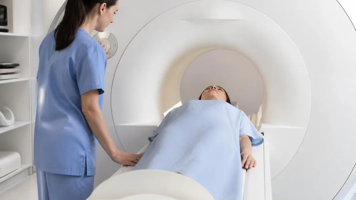
Even with all of its strengths, MRI has limitations that patients and clinicians need to understand. The scanner is a large bore surrounded by a powerful magnet, which means metal implants, cochlear devices, certain pacemakers, and retained shrapnel can be contraindications. Every patient must be carefully screened with a safety questionnaire before entering the room. A small but real number of devices are MR-conditional only at specific field strengths, and the technologist's job is to verify each item before the scan begins.
Claustrophobia is another common barrier. The bore feels narrow, the scan can last 30 to 60 minutes, and the gradient coils produce loud knocking sounds throughout. Up to 10 percent of patients struggle to complete a brain MRI without sedation. Wide-bore and open-MRI systems help, as do mild anxiolytics and good patient coaching. For an inside look at why the machines are so loud, see our explainer on the mri machine noise.
Motion is the enemy of MRI image quality. A patient who shifts even slightly during acquisition can blur an entire sequence. For trauma patients, intubated patients, or confused stroke patients, this is a real problem. Fast sequences, parallel imaging, and motion-correction techniques help, but no MRI is as forgiving of motion as a 30-second CT. Technologists frequently repeat sequences mid-scan, which extends overall scan time and patient discomfort.
Gadolinium contrast deserves a careful conversation. Contrast-enhanced MRI is essential for evaluating tumors, infections, and inflammatory disease, but gadolinium agents carry small risks. Patients with severe kidney disease face the rare possibility of nephrogenic systemic fibrosis. Newer macrocyclic agents have largely eliminated that risk, but lab work is still checked before contrast injection. Some gadolinium also deposits in brain tissue, the long-term significance of which is still being studied.
Cost and turnaround time are practical limitations. In the U.S., a brain MRI without contrast typically costs between $400 and $3,500 depending on the facility, insurance, and region. Outpatient scheduling can take days. Reports may not be finalized for 24–48 hours, although stat reads are available for inpatients. These factors influence whether MRI is the right next step or whether CT will give an adequate answer faster and cheaper.
Sensitivity has trade-offs too. SWI and gradient-echo sequences are so sensitive to iron that calcifications, mineral deposits, and even normal vessels can mimic hemorrhage. Radiologists rely on clinical context, prior imaging, and additional sequences to avoid overcalling microbleeds. False positives can lead to unnecessary anxiety, additional testing, and changes in anticoagulation management. Reading these images well requires substantial training and experience.
Finally, MRI does not detect everything. Acute calcifications, subtle skull fractures, and the precise edges of acute subarachnoid blood can all be more obvious on CT. Bone is essentially invisible on MRI because it lacks mobile protons. That is why a complete neurosurgical workup often includes both modalities — each compensating for what the other misses. The best radiology departments think of MRI and CT as a team rather than competitors.
If you are preparing for an MRI to evaluate a possible brain bleed or any other neurologic concern, a little preparation goes a long way. Start by gathering prior imaging — CT, MRI, or angiography reports from anywhere you have been scanned. Radiologists rely on comparison studies to detect subtle changes, and bringing old discs or having outside images uploaded ahead of time can dramatically improve the accuracy of your report. Many imaging centers have patient portals that allow this electronically.
Complete the safety questionnaire honestly and in detail. Even small metallic items — an old surgical clip, a stent, a vascular access port — can be relevant. Bring documentation for any implanted device, including the manufacturer, model number, and date of placement. Pacemakers and defibrillators are no longer absolute contraindications in many cases, but the team needs precise device information to choose the correct protocol and field strength.
Plan for the length of the exam. A typical brain MRI runs 30–45 minutes, and an MRI with contrast or extended sequences such as MR angiography can exceed an hour. Empty your bladder beforehand, dress in metal-free clothing, and leave jewelry, hair clips, and electronics in a locker. If you tend to feel anxious in enclosed spaces, talk with the ordering clinician about a mild oral anxiolytic — taken with someone available to drive you home afterward.
Hearing protection is provided, but the gradient noise can still be intense. Some sites offer music headphones or noise-canceling options. Communicate any discomfort early — technologists can pause between sequences, check on you over the intercom, and adjust positioning if you become uncomfortable. Holding still is the single most important thing you can do for image quality, especially for the susceptibility and diffusion sequences that detect bleeding and stroke.
After the scan, ask when your results will be ready and through which channel they will reach you. Most outpatient MRIs are read within 24 hours, with stat exams completed faster. If the radiologist identifies an urgent finding such as an acute bleed, you will be contacted immediately. For routine studies, follow up with the ordering physician to discuss findings in context — a single line in a radiology report can mean different things depending on your symptoms and history.
For students and technologists preparing for the ARRT registry, brain bleed imaging is high-yield material. Expect questions on the appearance of hemorrhage across T1, T2, FLAIR, and gradient-echo sequences; on the timing of hemoglobin breakdown; and on the comparison with CT findings. Build flashcards that map each hemorrhage stage to its T1 and T2 signal, and practice identifying the patterns on sample images. Doing this regularly cements the material far better than re-reading textbooks.
Finally, stay curious about the technology. MRI is one of the most rapidly evolving fields in medical imaging, with new sequences such as quantitative susceptibility mapping, ultra-short echo time imaging, and 7-Tesla research scanners pushing detection limits further every year. The principles you learn today — relaxation, susceptibility, diffusion — will continue to power tomorrow's innovations. Whether you are a patient, student, or working clinician, understanding what an MRI can detect equips you to ask better questions and make better decisions.
MRI Questions and Answers

MRI Medical Abbreviation: What MRI Stands For and Why It Matters

The History of MRI: From Discovery to Modern Medicine

Knee MRI Images: A Complete Guide to Reading, Understanding, and Interpreting Knee Scans

Noise of MRI Machine: Why MRI Scanners Are So Loud and What to Expect
MRI Alternatives: When CT, Ultrasound, X-Ray, and PET Make More Sense Than an MRI
About the Author

Medical Laboratory Scientist & Clinical Certification Expert
Johns Hopkins UniversityDr. Sandra Kim holds a PhD in Clinical Laboratory Science from Johns Hopkins University and is certified as a Medical Technologist (MT) and Medical Laboratory Scientist (MLS) through ASCP. With 16 years of clinical laboratory experience spanning hematology, microbiology, and molecular diagnostics, she prepares candidates for ASCP board exams, MLT, MLS, and specialist certification tests.
Join the Discussion
Connect with other students preparing for this exam. Share tips, ask questions, and get advice from people who have been there.
View discussion (6 replies)