MRI Images of Body Parts: Complete Guide to Neck, Spine, Brain, Shoulder, and Knee Scans
✅ MRI neck images and body part scans explained — see what cervical spine, brain, shoulder, and knee MRIs reveal and how to read them.
MRI neck images are among the most clinically detailed scans dwi mri produces, revealing soft tissue contrast that no other imaging modality can match. When a radiologist studies an MRI of the cervical spine, they see the spinal cord, intervertebral discs, nerve roots, ligaments, and surrounding vasculature with submillimeter precision. This level of detail allows physicians to diagnose herniated discs, nerve impingement, multiple sclerosis lesions, tumors, and traumatic injuries that would be invisible on conventional X-rays. Understanding what these images show transforms patient anxiety into informed participation in care.
Magnetic resonance imaging works by aligning hydrogen protons within body tissues using a powerful magnetic field, then measuring the radio signals they emit as they relax back to their resting state. Because different tissues contain different proportions of water and fat, each appears with a unique brightness on the resulting image. The neck, spine, brain, shoulder, knee, abdomen, and pelvis each require specialized coils, sequences, and patient positioning to produce diagnostic-quality images. Each body part presents distinct challenges and opportunities for the imaging team.
This guide walks through MRI images of every major body region a clinician might order, explaining what anatomy appears in mri with vs without contrast, what pathology radiologists hunt for, and how patients can prepare for each examination. We will examine T1-weighted images that highlight fat and anatomy, T2-weighted images that emphasize fluid and inflammation, and specialized sequences like STIR, FLAIR, and diffusion-weighted imaging that answer specific clinical questions. By the end, you should be able to identify the body part, plane, and basic sequence of any MRI image you encounter.
For students preparing for the ARRT MRI registry or radiologic technologists studying anatomy and pathology, recognizing body part images instantly is a foundational skill tested repeatedly on certification exams. Clinicians review thousands of MRI images during training, and pattern recognition becomes second nature only after deliberate practice with labeled images and case studies. This article synthesizes that practice into a structured visual tour of the human body as seen through MRI.
We will also discuss the role of contrast agents, primarily gadolinium-based compounds, which enhance vascularity and highlight tumors, infections, and active inflammation. Some examinations are performed both before and after mri contrast injection to compare enhancement patterns. Other studies require no contrast at all, relying instead on intrinsic tissue contrast and specialized sequences. The choice depends on the suspected pathology and the body part being imaged, with neck and brain scans most commonly using contrast for vascular and tumor assessment.
Beyond diagnostic value, MRI images of body parts also serve surgical planning, treatment monitoring, and research. Orthopedic surgeons rely on knee and shoulder MRIs to plan arthroscopic procedures. Neurosurgeons use brain and spine images to navigate around critical structures. Oncologists track tumor response to chemotherapy through serial MRI examinations. Every image, regardless of body part, carries information that ripples through the patient's entire care pathway from initial diagnosis through long-term follow-up monitoring.
Whether you are a patient preparing for your first MRI, a student memorizing cross-sectional anatomy, or a technologist refining your protocol selection, the following sections offer a comprehensive look at MRI imaging across the body. Each region has its own visual signature, common pathologies, and protocol considerations worth knowing in depth.
MRI Imaging by the Numbers
Major Body Regions Imaged with MRI
Reveals strokes, tumors, multiple sclerosis lesions, aneurysms, and pituitary abnormalities. Uses head coils with axial, sagittal, and coronal planes. T2-FLAIR and diffusion-weighted sequences are essential for acute stroke and white matter disease evaluation in emergency settings.
Shows the spinal cord, intervertebral discs, vertebrae, nerve roots, and soft tissues. Common indications include radiculopathy, myelopathy, trauma, and tumor. Sagittal T1 and T2 sequences anchor the protocol with axial slices through suspected pathology levels.
Images the rotator cuff, labrum, biceps tendon, glenohumeral joint, and surrounding bursae. Patients lie supine with the arm in slight external rotation. MR arthrography with intra-articular contrast is often used for labral and partial tendon tears.
Evaluates menisci, cruciate and collateral ligaments, articular cartilage, and bone marrow. Sports injuries, osteoarthritis, and post-surgical changes are common indications. Thin-slice proton density and fat-saturated sequences provide the highest diagnostic yield for joint structures.
Assesses the liver, kidneys, pancreas, prostate, uterus, and bowel. Breath-hold sequences reduce motion artifact. Multiparametric prostate MRI and MR cholangiopancreatography (MRCP) are highly specialized protocols answering targeted clinical questions about specific organ systems.
The cervical spine, the most frequently imaged neck region, contains seven vertebrae from C1 (atlas) through C7, each separated by intervertebral discs that absorb shock and permit movement. MRI neck images in the sagittal plane show all seven vertebrae stacked in profile, with the spinal cord traveling through the central canal as a bright ribbon on T2-weighted sequences. The cerebrospinal fluid surrounding the cord appears even brighter, creating natural contrast that helps radiologists identify cord compression, syrinx formation, or intramedullary lesions with remarkable clarity.
Intervertebral discs deserve careful attention on neck MRI. A healthy disc shows a bright nucleus pulposus on T2 imaging, reflecting its high water content. As discs degenerate with age or injury, they lose water, darken on T2, and may bulge or herniate posteriorly into the spinal canal or laterally into the neural foramina. Radiologists grade disc disease by morphology, signal, and degree of central or foraminal narrowing. The C5-C6 and C6-C7 levels are most commonly affected because they bear the greatest mechanical load.
Nerve roots exit the cervical spine at each level, traveling through the neural foramina to innervate the arms and hands. When a disc herniation, osteophyte, or facet hypertrophy narrows the foramen, the nerve root becomes compressed, producing the radicular pain, numbness, and weakness patients describe as a pinched nerve. Axial MRI images show these foramina in cross section, and skilled radiologists can grade stenosis as mild, moderate, or severe based on the percentage of cross-sectional area lost.
Beyond the spine itself, neck MRI evaluates soft tissue structures including the thyroid gland, salivary glands, lymph nodes, larynx, pharynx, and major vessels like the carotid and vertebral arteries. Tumors of the head and neck often require dedicated soft tissue protocols with gadolinium contrast to assess vascularity, invasion of adjacent structures, and lymph node involvement. The Macroscopic detail visible on dedicated head-and-neck MRI guides oncologic staging and surgical planning for cancers of the tongue, tonsil, larynx, and nasopharynx.
Trauma protocols for neck imaging emphasize ligament integrity. The transverse ligament of the atlas, the alar ligaments, and the posterior cervical ligamentous complex stabilize the upper cervical spine. Disruption of any of these structures after high-energy injury can produce instability that requires surgical fixation. mri stir sequences, which suppress fat signal and highlight edema, are particularly valuable for detecting subtle ligamentous injury and bone marrow contusion not visible on standard sequences.
For patients curious about how contrast agents factor into neck imaging, you can learn more in our guide to MRI with and without contrast, which explains when gadolinium enhances diagnostic accuracy. Most routine cervical spine MRIs are performed without contrast, reserving the agent for cases with suspected infection, tumor, or postoperative complications where enhancement patterns provide critical diagnostic information.
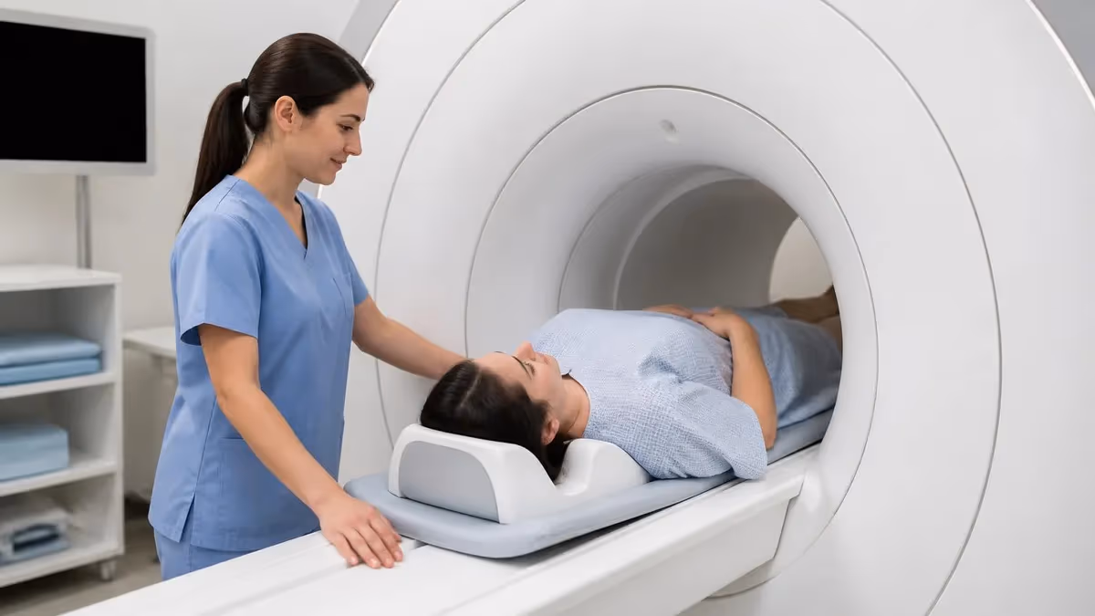
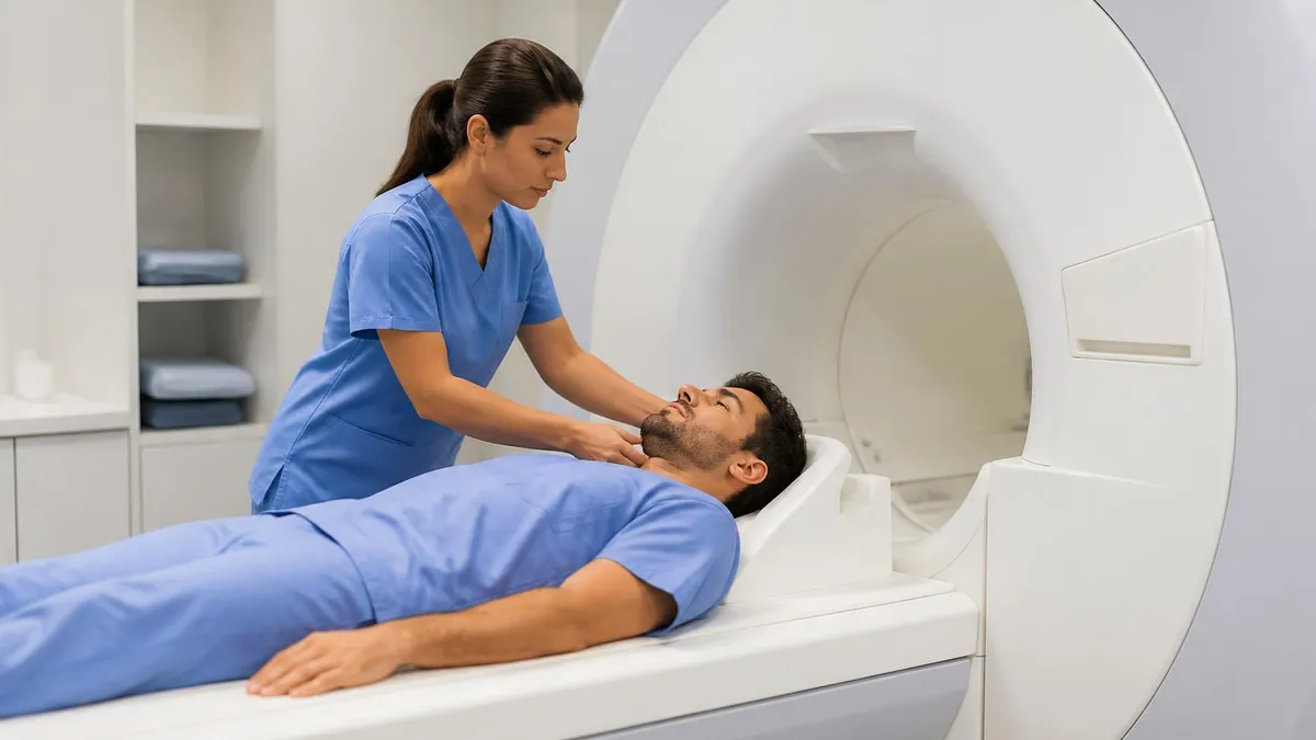
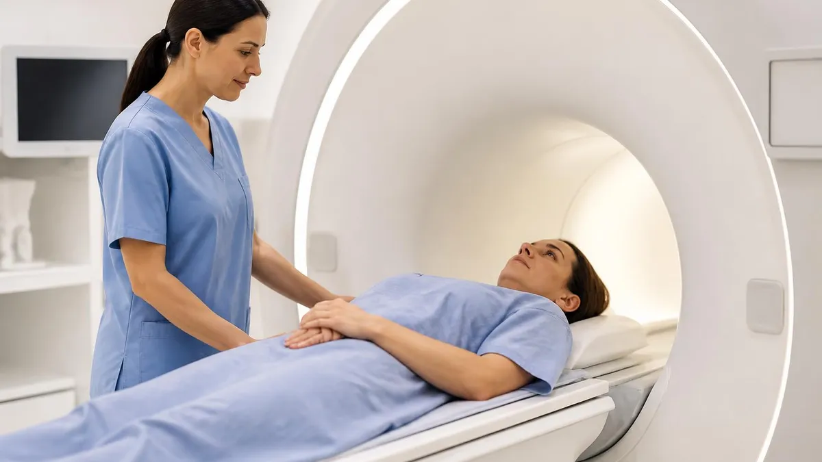
Coil selection significantly affects image quality on neck MRI. Modern phased-array neck coils wrap around the patient and provide uniform signal across the entire imaging volume. Older head coils or body coils produce inferior images because their signal-to-noise ratio drops sharply away from the coil center. Technologists must position the patient carefully, ensuring the coil is centered over the anatomy of interest and the neck is in a neutral, comfortable position to minimize motion during the 25 to 40-minute examination.
MRI Practice Test Questions
Prepare for the MRI - Magnetic Resonance Imaging exam with our free practice test modules. Each quiz covers key topics to help you pass on your first try.
MRI Knowledge
MRI Exam Questions covering Knowledge. Master MRI Test concepts for certification prep.
MRI Physics
Free MRI Practice Test featuring Physics. Improve your MRI Exam score with mock test prep.
MRI Anatomy and Pathology
MRI Test Prep for MRI Anatomy and Pathology. Practice MRI Quiz questions and boost your score.
MRI Anatomy and Positioning
MRI Questions and Answers on MRI Anatomy and Positioning. Free MRI practice for exam readiness.
MRI Contrast Agents
Free MRI Quiz on MRI Contrast Agents. MRI Exam prep questions with detailed explanations.
MRI Patient Care and Positioning
MRI Practice Questions for MRI Patient Care and Positioning. Build confidence for your MRI certification exam.
MRI Sequences by Body Part
T1-weighted images show anatomy with exceptional clarity, displaying fat as bright signal and water as dark. They are the workhorse sequence for evaluating bone marrow, subcutaneous tissues, and post-contrast enhancement. On brain MRI, T1 images delineate gray and white matter contrast beautifully, allowing assessment of cortical thickness and deep nuclei boundaries with anatomical precision that guides neurosurgical planning.
For spine imaging, T1 sagittal sequences reveal vertebral body marrow signal, helping radiologists detect metastatic disease, multiple myeloma, or marrow edema from compression fractures. Normal adult marrow contains fat and appears bright on T1; replacement by tumor or inflammation darkens the signal. T1 post-gadolinium images highlight tumor enhancement, meningeal disease, and active multiple sclerosis plaques that breach the blood-brain barrier in characteristic patterns.
MRI Imaging: Strengths and Limitations Across Body Parts
- +Superior soft tissue contrast unmatched by CT or ultrasound across all body regions
- +No ionizing radiation, safe for repeated examinations and pediatric patients
- +Multiplanar imaging in any orientation without repositioning the patient
- +Detects pathology invisible on other imaging modalities including marrow edema
- +Quantifies tissue characteristics through specialized sequences and mapping techniques
- +Visualizes vessels without iodinated contrast using time-of-flight MRA techniques
- +Provides functional information beyond anatomy through DWI, perfusion, and spectroscopy
- −Long scan times of 30–60 minutes require patient cooperation and stillness
- −Loud acoustic noise from gradient coils requires hearing protection for all patients
- −Claustrophobia affects up to 15% of patients in closed-bore systems
- −Implants and metallic foreign bodies may be contraindications or cause artifacts
- −Significantly more expensive than X-ray, CT, or ultrasound examinations
- −Motion artifact degrades image quality in restless or pediatric patients
- −Gadolinium contrast carries risks in patients with severe kidney dysfunction
Patient Preparation Checklist for MRI Neck Images and Body Scans
- ✓Complete the MRI safety screening form thoroughly, listing all surgical implants and devices
- ✓Remove all metal objects including jewelry, watches, hairpins, and dental appliances when possible
- ✓Inform the technologist about any pacemakers, cochlear implants, or aneurysm clips
- ✓Disclose pregnancy status, kidney function, and prior contrast reactions before scanning
- ✓Wear loose comfortable clothing without metal zippers, snaps, or embedded fibers
- ✓Arrive 30 minutes early to complete paperwork and change into a gown if required
- ✓Use the restroom before the scan begins, especially for pelvic or abdominal imaging
- ✓Take prescribed anti-anxiety medication if claustrophobia is a known concern
- ✓Bring prior imaging studies on CD or arrange for electronic transfer to the facility
- ✓Plan transportation home if sedation is being used during the examination
- ✓Hold breath when instructed during abdominal sequences to reduce motion blur
- ✓Communicate immediately through the squeeze ball if you feel distressed during the scan
MRI images show structure, but your symptoms tell the story
An MRI image is only as useful as the clinical context surrounding it. Up to 30% of asymptomatic adults show disc bulges or herniations on cervical MRI, yet have no pain or neurological deficit. Always interpret imaging findings alongside your physical examination and symptoms with your physician.
Reading cross-sectional anatomy on MRI requires understanding how each plane intersects the body and what structures appear in each slice. The axial plane cuts horizontally through the body like slicing a loaf of bread, displaying structures in cross section. The sagittal plane cuts vertically from front to back, separating left from right, while the coronal plane cuts vertically from side to side, separating front from back. Each plane offers unique advantages for different anatomical structures and pathology.
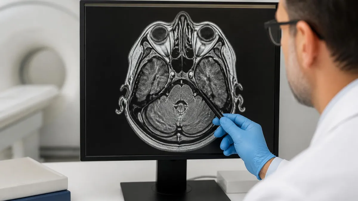
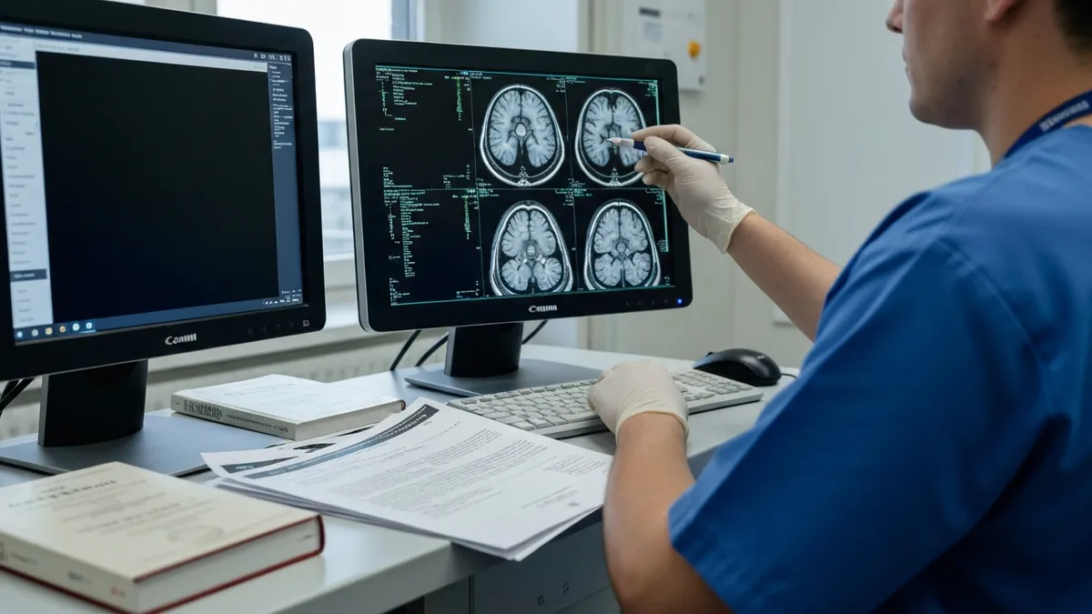
On axial brain MRI at the level of the lateral ventricles, radiologists identify the frontal horns anteriorly, the occipital horns posteriorly, and the third ventricle in the midline. Surrounding the ventricles, the caudate nucleus, putamen, globus pallidus, and thalamus form the deep gray matter structures involved in movement and sensation. Recognizing these landmarks instantly is essential for technologists planning scans and for clinicians mri brain findings during emergency neuroradiology consultations.
Sagittal cervical spine images, the foundational view for any neck MRI study, display the relationship between vertebrae, discs, and spinal cord across all seven cervical levels and the upper thoracic region. The clivus and odontoid process anchor the upper image, while the cord descends through the foramen magnum into the spinal canal. Radiologists scan systematically from top to bottom, evaluating each disc level for hydration, height, and posterior bulging, then examining the cord for signal abnormality.
Knee MRI in the sagittal plane shows the menisci as small dark triangles between the femur and tibia, the anterior and posterior cruciate ligaments traversing the joint diagonally, and the patellar tendon connecting the kneecap to the tibial tuberosity. Coronal slices reveal the medial and lateral collateral ligaments, while axial images through the patellofemoral joint assess cartilage and tracking. Each plane contributes complementary information that together produces a complete diagnostic picture.
Shoulder MRI uses oblique planes aligned with the supraspinatus tendon to optimize evaluation of the rotator cuff. Oblique coronal images parallel the supraspinatus while oblique sagittal images cut perpendicular to it. The labrum, a fibrocartilaginous ring around the glenoid, is best evaluated on axial sequences. MR arthrography injects dilute gadolinium into the joint, distending the capsule and outlining labral tears that would otherwise blend with surrounding tissues.
Abdominal MRI in the axial plane sequentially displays the liver, spleen, pancreas, kidneys, adrenal glands, and bowel loops. Coronal images provide an anatomic overview similar to a chest X-ray, useful for assessing organ size and relationships. The aorta, inferior vena cava, and portal venous system are evaluated with vascular sequences, while bile ducts and pancreatic ducts are imaged with heavily T2-weighted MRCP sequences that make fluid-filled structures appear bright against suppressed background tissue.
Pelvic MRI for prostate cancer mri staging follows a multiparametric protocol including T2-weighted high-resolution imaging, diffusion-weighted imaging, and dynamic contrast mri enhancement. Each component contributes a different aspect of tumor characterization. The PI-RADS scoring system assigns probability of clinically significant cancer based on findings across these sequences. Similar multiparametric approaches are emerging for liver, breast, and rectal cancer imaging, transforming MRI from purely anatomic into a functional diagnostic tool.
Never enter the MRI scanner room with metallic objects, electronic devices, or magnetic media. The static magnetic field is always on, even when no scan is in progress, and can launch ferromagnetic objects across the room at lethal speeds. Always complete safety screening with a qualified MRI technologist before approaching the magnet.
Physicians order MRI scans of specific body parts when they suspect conditions that other imaging modalities cannot adequately characterize. A neurologist evaluating a patient with new-onset weakness on one side of the body orders an urgent brain MRI to rule out stroke, tumor, or demyelinating disease. The diffusion-weighted sequence rapidly identifies acute infarction within minutes, while FLAIR images show chronic small vessel disease and multiple sclerosis plaques that explain longstanding subtle symptoms accumulating over years.
Orthopedic surgeons order knee MRI when a patient describes a twisting injury followed by joint locking, instability, or persistent swelling. The MRI reveals whether the meniscus has torn, whether the anterior cruciate ligament has ruptured, and whether cartilage has been damaged. Without this information, surgeons cannot plan arthroscopic repair appropriately. The same logic applies to shoulder MRI for rotator cuff tears and hip MRI for labral pathology, with details that you can explore in our shoulder mri without contrast cpt.
Primary care physicians often order cervical spine MRI for patients with persistent neck pain radiating into an arm, especially when accompanied by numbness, tingling, or weakness. These symptoms suggest nerve root compression, most commonly from disc herniation or bony stenosis. The MRI localizes the compression to a specific level, allowing targeted treatment with epidural injections, physical therapy, or surgical decompression. Without precise localization, treatment becomes guesswork that wastes time and risks unnecessary procedures.
Oncologists rely on MRI for staging, treatment planning, and surveillance across many cancer types. Breast MRI screens high-risk women and characterizes ambiguous mammographic findings. Prostate MRI identifies clinically significant tumors before biopsy. Rectal MRI determines the depth of tumor invasion and lymph node involvement, guiding decisions about neoadjuvant therapy. Liver MRI distinguishes benign hemangiomas from metastases. Each application demonstrates how anatomic and functional MRI information drives critical treatment decisions.
Emergency physicians use MRI selectively when CT scans are inconclusive or contraindicated. Pregnant patients with abdominal pain undergo MRI rather than CT to evaluate appendicitis without radiation exposure to the fetus. Patients with suspected spinal cord injury after trauma receive MRI to assess cord integrity and ligamentous stability beyond what CT can show. Stroke patients receive MRI to determine tissue viability and guide thrombolytic decisions in extended treatment windows beyond the initial four-hour window.
Rheumatologists order MRI to detect early inflammatory arthritis before joint destruction becomes visible on X-ray. Sacroiliac joint MRI identifies sacroiliitis indicating ankylosing spondylitis or other spondyloarthropathies. Hand and wrist MRI reveal early rheumatoid synovitis and bone marrow edema preceding erosion. Early diagnosis enables aggressive treatment that prevents permanent joint damage and disability across decades of patient life.
Cardiologists increasingly use cardiac MRI for tissue characterization, viability assessment, and congenital heart disease evaluation. The technique quantifies ejection fraction more accurately than echocardiography and identifies myocardial fibrosis, infiltrative diseases like amyloidosis, and inflammatory conditions like myocarditis. Cardiac MRI requires specialized expertise and ECG-gated sequences but provides information unavailable through any other noninvasive imaging modality available today.
Practical preparation for any MRI examination begins days before the appointment. Patients taking medications should clarify with their prescribing physician whether to continue or hold them on the day of the scan. Most medications including blood pressure, diabetes, and psychiatric drugs can be taken normally. However, certain procedures like prostate MRI may require antispasmodic medication to reduce bowel motion, and cardiac MRI may require beta-blockers to slow heart rate for cleaner imaging.
Dietary restrictions vary significantly by body part. Abdominal MRI typically requires four to six hours of fasting to reduce bowel gas and ensure gallbladder distension for cholangiopancreatography. Pelvic MRI for prostate or rectal evaluation may require an enema to clear the rectum. Brain, spine, and musculoskeletal MRI generally have no dietary restrictions. The scheduling staff or technologist will provide specific instructions when the appointment is confirmed in advance of the procedure.
Anxiety management deserves serious consideration for patients with claustrophobia. Modern wide-bore scanners with 70-centimeter openings reduce but do not eliminate the closed-in feeling. Patients can request prone positioning for some studies, allowing them to see out of the scanner. Eye masks paradoxically help some patients by removing visual cues of confinement. Headphones with music distract from acoustic noise and the passage of time during long examinations and improve overall patient experience.
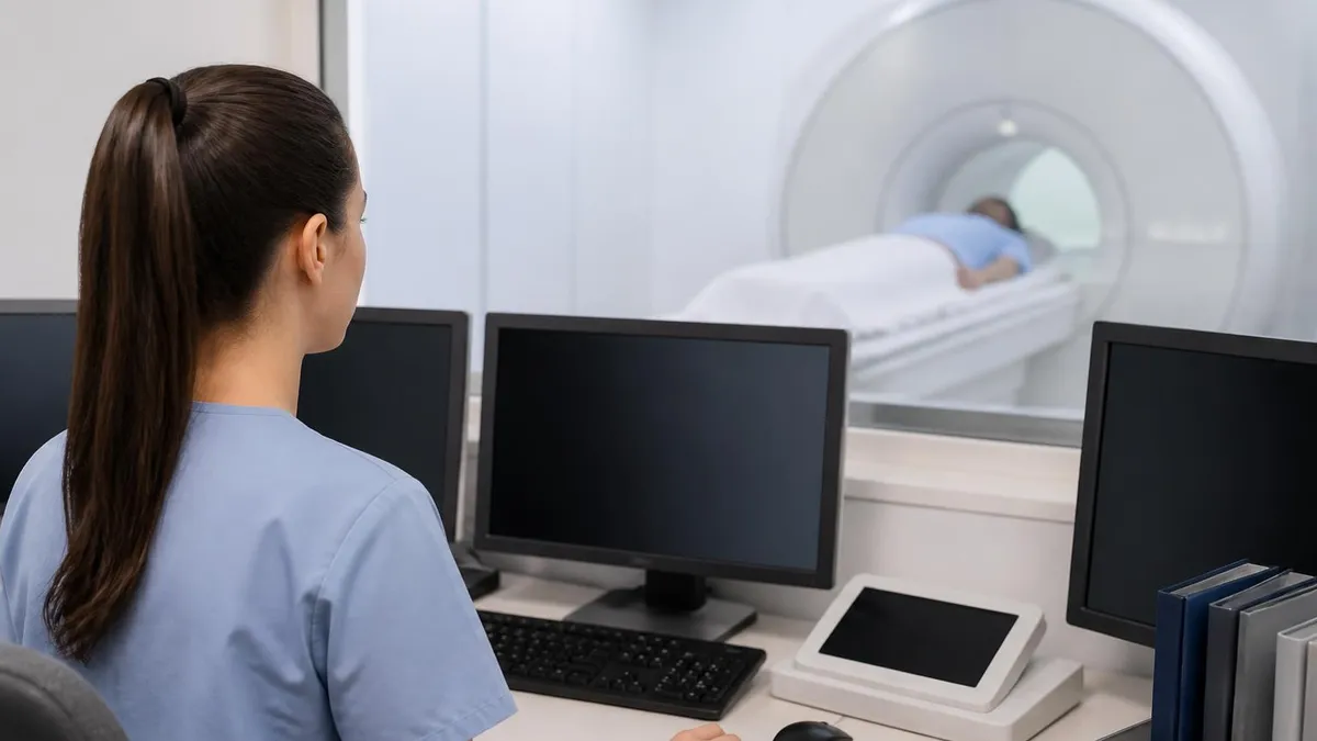
Communication during the scan is critical. Every patient holds a squeeze ball or call button connected to the technologist console. Pressing it pauses the scan and triggers immediate intervention. Patients should not hesitate to use this device for any discomfort, anxiety, or unexpected sensation. Technologists are trained to respond rapidly and can often address concerns without aborting the examination, allowing diagnostic images to be completed for the referring physician.
After the scan, patients return to normal activity immediately unless sedation was used. Gadolinium contrast may cause mild headache or nausea in some patients but rarely produces serious reactions. Patients should drink water to facilitate contrast clearance through the kidneys. Images are typically reviewed by a radiologist within 24 to 48 hours, though urgent studies are read immediately. The final report goes to the ordering physician, who communicates findings to the patient at follow-up visits.
Understanding what to expect demystifies the MRI experience and improves cooperation. Patients who know they will hear loud knocking, banging, and humming sounds tolerate the scan better than those caught by surprise. Patients who understand that the table will slide them into the magnet feet-first or head-first depending on body part imaged prepare mentally for the experience. Knowledge reduces anxiety dramatically more effectively than any medication available to outpatient imaging facilities.
For technologists and students, becoming proficient at MRI imaging across body parts takes deliberate practice with thousands of cases. Cross-sectional anatomy textbooks, online case repositories, and registry review courses build pattern recognition. Working alongside experienced radiologists during readouts accelerates learning because each case becomes a teaching moment. The combination of structured study and clinical exposure transforms novices into skilled imaging professionals capable of recognizing pathology across every region of the human body.
MRI Questions and Answers

The History of MRI: From Discovery to Modern Medicine

MRI With and Without Contrast: How It Works, What to Expect

MRI Medical Abbreviation: What MRI Stands For and Why It Matters
Shoulder MRI: What It Shows, Procedure, and Reading the Report
MRI Imaging Centers: Complete Guide to Independent Outpatient MRI Facilities
About the Author
Medical Laboratory Scientist & Clinical Certification Expert
Johns Hopkins UniversityDr. Sandra Kim holds a PhD in Clinical Laboratory Science from Johns Hopkins University and is certified as a Medical Technologist (MT) and Medical Laboratory Scientist (MLS) through ASCP. With 16 years of clinical laboratory experience spanning hematology, microbiology, and molecular diagnostics, she prepares candidates for ASCP board exams, MLT, MLS, and specialist certification tests.
Join the Discussion
Connect with other students preparing for this exam. Share tips, ask questions, and get advice from people who have been there.
View discussion (6 replies)