
Brain MRI Images: A Complete Guide to Reading, Understanding, and Interpreting Brain Scans
Learn how to read brain MRI images, understand signal intensities, 💯 identify anatomy, and recognize common pathologies. A complete guide for MRI students and...
Brain MRI images are among the most diagnostically powerful tools in modern medicine, offering unparalleled soft-tissue contrast that allows clinicians and radiologists to visualize the brain's internal structures without exposing patients to ionizing radiation. Practice with the free MRI - Magnetic Resonance Imaging practice test on PracticeTestGeeks (200+ questions with answer explanations).
Whether you are a student preparing for your ARRT registry exam, a radiologic technologist expanding your neuroimaging knowledge, or a curious patient who just received a scan, understanding how to interpret brain mri images is a skill that bridges technical expertise and clinical insight. MRI uses strong magnetic fields and radiofrequency pulses to generate images that reveal gray matter, white matter, cerebrospinal fluid, blood vessels, and lesions with exceptional clarity.
The brain is one of the most complex organs in the human body, and MRI is uniquely suited to capture that complexity. Unlike CT scans, which excel at detecting acute hemorrhage and bony pathology, brain MRI images provide superior contrast resolution for differentiating between subtle tissue types, identifying demyelinating plaques, characterizing tumors, and detecting early ischemic strokes that may be invisible on CT for the first 24 to 72 hours. The ability to acquire images in multiple planes — axial, sagittal, and coronal — without moving the patient gives MRI a distinct advantage in neurological assessment.
To truly understand brain MRI images, you must first grasp the concept of signal intensity and how it varies across different pulse sequences. On T1-weighted images, fat and subacute blood appear bright (hyperintense), while water and CSF appear dark (hypointense). On T2-weighted images, the relationship reverses: fluids and edema appear bright, while fat appears slightly less intense. FLAIR sequences suppress the CSF signal, making periventricular lesions and cortical abnormalities far easier to identify. Each sequence adds a layer of diagnostic information that, when combined, builds a comprehensive picture of brain health.
For MRI technologists and students preparing for board examinations, mastering the interpretation of brain MRI images requires not only memorizing signal characteristics but also understanding the clinical context behind each sequence. Why is diffusion-weighted imaging (DWI) ordered alongside ADC mapping for a suspected stroke? How does contrast enhancement with gadolinium help distinguish a high-grade glioma from a metastatic lesion? These questions are commonly tested on the ARRT MRI registry exam and reflect the kind of integrated thinking that separates competent technologists from exceptional ones.
Neuroanatomy is the foundation upon which brain MRI interpretation is built. You cannot identify a lesion in the corpus callosum without knowing where the corpus callosum is, what it looks like on each sequence, and what pathologies preferentially affect that structure.
Similarly, recognizing a basal ganglia infarct or a posterior fossa tumor requires confident familiarity with normal brain anatomy across all standard imaging planes. This guide will walk you through the major anatomical landmarks visible on brain MRI images, the key pulse sequences used in neuroimaging protocols, and the most common pathologies you will encounter in clinical practice and on registry examinations.
Beyond anatomy and pathology, this guide addresses the practical side of brain MRI imaging — patient preparation, positioning, common artifacts, and the technical parameters that affect image quality. Understanding why a particular sequence is prescribed, how to troubleshoot motion artifacts or susceptibility effects, and how to optimize slice thickness and field of view for pediatric versus adult brain studies are all competencies that MRI technologists are expected to demonstrate. Each of these topics is addressed in detail throughout this guide, with real-world clinical examples and examination-style insights woven into every section.
By the end of this comprehensive guide, you will have a solid, structured framework for approaching any brain MRI study with confidence. Whether your goal is to pass the ARRT MRI registry exam, improve your clinical scanning technique, or simply understand your own scan results, the knowledge presented here will serve as a durable reference. Let's begin with the numbers that define brain MRI as a discipline, then move systematically through anatomy, sequences, pathology, and practical scanning technique.
Free MRI - Magnetic Resonance Imaging Study Tools
Interactive MRI - Magnetic Resonance Imaging resources on PracticeTestGeeks, free to start:
- Free MRI Knowledge Questions and Answers — 11 questions with instant scoring and answer explanations
- MRI - Magnetic Resonance Imaging flashcards — review key terms and concepts card by card
- MRI - Magnetic Resonance Imaging study guide — topic-by-topic overview of what the exam covers
- MRI - Magnetic Resonance Imaging cheat sheet — one-page printable summary for last-minute review
Brain MRI Imaging by the Numbers
Essential MRI Pulse Sequences for Brain Imaging
The anatomical workhorse of brain MRI. Gray matter appears gray, white matter appears bright, and CSF appears dark. T1 post-contrast (with gadolinium) reveals blood-brain barrier breakdown in tumors, abscesses, and active MS lesions, making it indispensable in neuro-oncology and inflammatory disease evaluation.
Highly sensitive to pathological change. CSF and edema appear bright white, making T2 the primary sequence for detecting gliomas, demyelination, and vasogenic edema. White matter lesions from small vessel disease, trauma, or metabolic disorders light up clearly on T2, enabling early detection of subtle abnormalities.
Suppresses CSF signal so periventricular and cortical lesions are not obscured by bright fluid. FLAIR is the gold standard for detecting multiple sclerosis plaques, subarachnoid hemorrhage, leptomeningeal disease, and cortical contusions. In MS protocols, lesion burden is typically measured on FLAIR rather than standard T2.
Detects restricted water diffusion, the hallmark of acute cytotoxic edema in ischemic stroke. DWI can identify a stroke within minutes of onset, while CT remains negative for 24-72 hours. Always interpreted alongside the ADC (apparent diffusion coefficient) map to confirm true restriction versus T2 shine-through artifact.
Susceptibility-weighted imaging (SWI) and GRE sequences are exquisitely sensitive to blood products, calcium, and iron deposition. Microbleeds from cerebral amyloid angiopathy or hypertensive vasculopathy appear as dark 'blooming' foci. SWI is preferred over GRE for detecting subtle hemorrhagic transformation after ischemic stroke.
Normal brain anatomy on MRI is best understood by systematically reviewing each major structure across axial, sagittal, and coronal planes. On axial T1 images, the cerebral cortex appears as a thin gray ribbon along the outer surface of the brain, while the underlying white matter tracts — including the corona radiata, internal capsule, and corpus callosum — appear distinctly brighter due to their high myelin and lipid content.
Deep gray matter structures such as the basal ganglia (caudate nucleus, putamen, and globus pallidus), thalami, and hypothalamus appear as isointense regions relative to cortex, often separated from white matter by identifiable CSF-filled sulci and cisterns.
The corpus callosum is one of the most important landmarks in brain MRI interpretation. This large white matter commissure connects the two cerebral hemispheres and is best visualized on the midsagittal T1 image, where it appears as a curved band of bright white matter stretching from the genu anteriorly to the splenium posteriorly.
The body of the corpus callosum is the middle segment, and all three parts — genu, body, and splenium — are evaluated for thinning, signal change, or lesions. Multiple sclerosis, traumatic axonal injury, and agenesis of the corpus callosum all produce characteristic abnormalities at this landmark, and recognizing them is a core competency for registry examinations.
The ventricular system is visible on nearly every brain MRI image and provides critical anatomical orientation. The two lateral ventricles — one in each hemisphere — connect to the third ventricle via the foramina of Monro. The third ventricle sits between the thalami and communicates with the fourth ventricle through the cerebral aqueduct (aqueduct of Sylvius). On T2 and FLAIR images, CSF within the ventricles appears bright white and dark, respectively.
Hydrocephalus — enlargement of the ventricles due to impaired CSF flow or absorption — is one of the most important findings on brain MRI images, and distinguishing obstructive from communicating hydrocephalus guides treatment decisions.
The posterior fossa contains the cerebellum, pons, medulla oblongata, and the fourth ventricle. On axial images, the two cerebellar hemispheres flank the vermis centrally, and the deep cerebellar nuclei (dentate, emboliform, globose, and fastigial) are identifiable on high-resolution sequences. The brainstem — pons, midbrain, and medulla — is evaluated on sagittal and axial images for signal abnormalities, atrophy, or mass effect.
Lesions in the posterior fossa can produce dramatic symptoms including vertigo, diplopia, dysphagia, and ataxia, making accurate identification on MRI critical for patient management. Common pathologies here include posterior fossa tumors in children (medulloblastoma, ependymoma), infarcts in the PICA or AICA territories, and demyelinating plaques in the brainstem.
The cerebellopontine angle (CPA) is a cistern-filled space between the cerebellum, pons, and temporal bone that houses cranial nerves VII and VIII. Acoustic neuromas (vestibular schwannomas) are the most common masses at this location and appear as well-defined, contrast-enhancing lesions on T1 post-gadolinium images. Meningiomas and epidermoid cysts are the other two most common CPA masses, and each has characteristic signal behavior on different pulse sequences. Epidermoid cysts, for example, restrict diffusion on DWI — a key distinguishing feature from arachnoid cysts, which do not restrict.
The internal capsule is a critical white matter structure that carries motor and sensory fibers between the cortex and spinal cord. It is divided into an anterior limb, genu, and posterior limb on axial images. Infarcts involving the posterior limb of the internal capsule produce contralateral hemiparesis and are a classic finding on stroke MRI studies. The corticospinal tracts, which travel through this region, can also be delineated using diffusion tensor imaging (DTI) with fiber tractography — an advanced technique that maps white matter connectivity and is increasingly used in presurgical planning for brain tumor resections near motor pathways.
Understanding the vascular territories of the brain is essential for interpreting ischemic stroke on brain MRI images. The middle cerebral artery (MCA) supplies the lateral convexity and is the most commonly affected vessel in embolic stroke, producing infarcts in the frontal, parietal, and temporal lobes. The anterior cerebral artery (ACA) supplies the medial frontal and parietal lobes, and ACA territory infarcts cause contralateral leg weakness greater than arm weakness.
The posterior cerebral artery (PCA) supplies the occipital lobes and medial temporal structures; PCA territory infarcts characteristically cause contralateral homonymous hemianopia. Recognizing these vascular territories on DWI is a foundational skill for any MRI technologist or radiologist trainee.
MRI Practice Test Questions
Prepare for the MRI - Magnetic Resonance Imaging exam with our free practice test modules. Each quiz covers key topics to help you pass on your first try.
MRI Knowledge
MRI Exam Questions covering Knowledge. Master MRI Test concepts for certification prep.
MRI Physics
Free MRI Practice Test featuring Physics. Improve your MRI Exam score with mock test prep.
MRI Anatomy and Pathology
MRI Test Prep for MRI Anatomy and Pathology. Practice MRI Quiz questions and boost your score.
MRI Anatomy and Positioning
MRI Questions and Answers on MRI Anatomy and Positioning. Free MRI practice for exam readiness.
MRI Contrast Agents
Free MRI Quiz on MRI Contrast Agents. MRI Exam prep questions with detailed explanations.
MRI Patient Care and Positioning
MRI Practice Questions for MRI Patient Care and Positioning. Build confidence for your MRI certification exam.
Reading Brain MRI Images: T1, T2, and FLAIR Interpretation
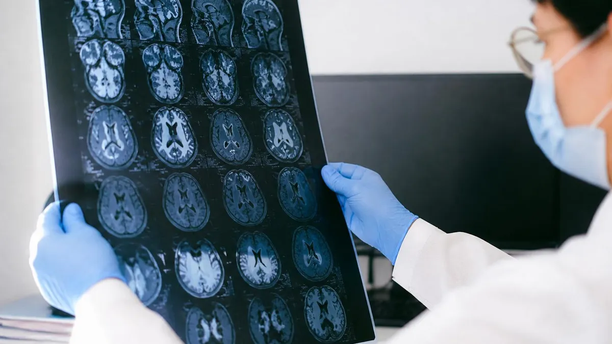
On T1-weighted brain MRI images, signal intensity correlates with tissue relaxation time. Structures with short T1 relaxation times — such as fat, proteinaceous fluid, methemoglobin in subacute blood, and gadolinium-enhanced tissue — appear hyperintense (bright white). Conversely, CSF, pure water, and most pathological tissue appear hypointense (dark). Normal white matter is brighter than gray matter on T1 because of its higher myelin and lipid content, making T1 the preferred sequence for evaluating anatomy, cortical thickness, and post-contrast enhancement patterns.
Recognizing abnormal T1 signal is crucial for identifying hemorrhagic lesions, calcifications (which can appear dark due to susceptibility), and melanin-containing metastases from melanoma, which appear characteristically T1-bright. Gadolinium-based contrast agents shorten T1 relaxation time in tissues where the blood-brain barrier is disrupted, causing those areas to enhance brightly on post-contrast T1 images. This enhancement pattern helps distinguish active MS lesions, high-grade tumors, and abscesses from stable, non-enhancing lesions like old infarcts or low-grade gliomas.
Brain MRI vs. CT Scan: Which Is Better for Brain Imaging?
- +Superior soft-tissue contrast for detecting tumors, demyelination, and early ischemia
- +No ionizing radiation — safe for repeated imaging and pediatric patients
- +Multiplanar imaging in axial, sagittal, and coronal planes without repositioning
- +Advanced sequences (DWI, MRS, fMRI) provide functional and metabolic information beyond anatomy
- +Gadolinium contrast agents have a much lower nephrotoxicity risk profile than iodinated CT contrast
- +Gold standard for evaluating posterior fossa, brainstem, and spinal cord pathology
- −Longer scan time (30–60 minutes) increases motion artifact risk, especially in uncooperative patients
- −Contraindicated in patients with certain metallic implants, pacemakers, or claustrophobia
- −Higher cost compared to CT — typically $1,000–$3,000 without insurance coverage
- −Less sensitive than CT for detecting acute hemorrhage in the hyperacute phase (first few hours)
- −Acoustic noise from gradient switching can be distressing, particularly for pediatric or anxious patients
- −Limited availability in emergency settings compared to CT scanners, which are more widely deployed
Pros and Cons at a Glance
| Pros | Cons |
|---|---|
| Superior soft-tissue contrast for detecting tumors, demyelination, and early ischemia | Contraindicated in patients with certain metallic implants, pacemakers, or claustrophobia |
| No ionizing radiation — safe for repeated imaging and pediatric patients | Less sensitive than CT for detecting acute hemorrhage in the hyperacute phase (first few hours) |
| Multiplanar imaging in axial, sagittal, and coronal planes without repositioning | Acoustic noise from gradient switching can be distressing, particularly for pediatric or anxious patients |
| Advanced sequences (DWI, MRS, fMRI) provide functional and metabolic information beyond anatomy | Limited availability in emergency settings compared to CT scanners, which are more widely deployed |
Brain MRI Interpretation Checklist: 10 Steps to a Systematic Read
- ✓Confirm patient identity, clinical indication, and the specific sequences acquired before beginning your review.
- ✓Check image quality — assess for motion artifact, field inhomogeneity, and whether all prescribed planes and sequences are present.
- ✓Review the ventricular system for size, symmetry, and CSF signal to identify hydrocephalus or ventricular effacement.
- ✓Evaluate the midline structures — corpus callosum, septum pellucidum, and falx cerebri — for displacement, lesions, or atrophy.
- ✓Systematically examine the cerebral cortex and sulci for gyral signal abnormality, cortical thickening, or leptomeningeal enhancement.
- ✓Assess the deep and periventricular white matter on FLAIR for hyperintense lesions, grading burden and distribution pattern.
- ✓Evaluate the basal ganglia, thalami, and internal capsules bilaterally for symmetry, signal change, and lacunar infarcts.
- ✓Review the posterior fossa — cerebellum, brainstem, and fourth ventricle — on axial and sagittal images for mass, signal change, or herniation.
- ✓Examine DWI and ADC maps together to identify acute ischemia, abscess, or other causes of true restricted diffusion.
- ✓Review post-contrast T1 images last to identify enhancing lesions, leptomeningeal disease, or blood-brain barrier disruption.
The ADC Map Always Confirms DWI Findings
Never diagnose acute stroke or abscess from DWI alone. True diffusion restriction shows HIGH signal on DWI and LOW signal on the ADC map. T2 shine-through appears bright on DWI but also bright on ADC — a common trap on registry examinations. Always check both maps before calling a finding positive for restricted diffusion.
Common brain pathologies visible on MRI represent the most heavily tested content area for ARRT registry candidates and the most clinically relevant knowledge for practicing MRI technologists. Ischemic stroke is one of the most frequent indications for urgent brain MRI, and its appearance changes predictably over time. In the hyperacute phase (0–6 hours), DWI shows bright restricted diffusion with a corresponding dark ADC map.
Within 24 hours, T2 signal begins to increase in the affected territory. By the subacute phase (days to weeks), the infarcted tissue develops T1 hypointensity and T2 hyperintensity with possible gyral enhancement on post-contrast images. Chronic infarcts evolve into encephalomalacic cavities that follow CSF signal on all sequences.
Multiple sclerosis (MS) is the most common inflammatory demyelinating disease of the central nervous system and produces a characteristic pattern of white matter lesions on brain MRI images. MS plaques are typically ovoid, well-defined, and oriented perpendicular to the ventricular wall — a pattern known as Dawson's fingers, best seen on sagittal FLAIR images. They preferentially involve the periventricular, juxtacortical, infratentorial, and spinal cord regions. Active lesions enhance with gadolinium, reflecting blood-brain barrier disruption. The McDonald criteria for MS diagnosis incorporate MRI findings of lesion dissemination in space and time, making accurate MRI interpretation critical for neurological diagnosis.
Brain tumors are classified by the World Health Organization (WHO) grade system, and MRI plays a central role in grading and surgical planning. Low-grade gliomas (WHO grade 1–2) typically appear as T2/FLAIR hyperintense lesions without surrounding edema or contrast enhancement. High-grade gliomas (WHO grade 3–4), including glioblastoma multiforme (GBM), show irregular ring-like contrast enhancement, surrounding vasogenic edema, mass effect, and central necrosis. The classic GBM appearance on post-contrast T1 is a thick, irregular enhancing ring surrounding a necrotic core, often crossing the corpus callosum to produce the butterfly pattern when bilateral hemispheric involvement is present.
Metastatic brain disease is the most common intracranial malignancy in adults and must be distinguished from primary brain tumors. Metastases are typically multiple, round, well-defined, cortical or corticomedullary junction lesions with surrounding vasogenic edema disproportionate to lesion size. They enhance avidly with gadolinium. Common primary sites include lung (most common in men), breast (most common in women), colon, kidney, and melanoma. Hemorrhagic metastases from melanoma, renal cell carcinoma, and choriocarcinoma show T1 hyperintensity due to blood products. Solitary metastasis can be impossible to distinguish from a primary tumor on imaging alone, and clinical history is essential.
Meningiomas are the most common benign intracranial tumors and arise from arachnoid cap cells. On MRI, they appear as extra-axial (outside the brain parenchyma), well-defined masses that are isointense to gray matter on T1 and T2 images and enhance intensely and homogeneously with gadolinium.
The dural tail sign — a linear enhancement extending from the mass along the dura — is a characteristic feature of meningioma, though it is not pathognomonic. Meningiomas can occur anywhere along the dura, with parasagittal, convexity, sphenoid wing, and posterior fossa locations being most common. They are often found incidentally and may be managed conservatively with surveillance MRI.
Traumatic brain injury (TBI) encompasses a spectrum of injuries including epidural hematoma, subdural hematoma, subarachnoid hemorrhage, contusions, and diffuse axonal injury (DAI). While CT is the first-line imaging modality for acute TBI due to its speed and sensitivity for hemorrhage, MRI — particularly SWI and FLAIR — is superior for detecting non-hemorrhagic DAI and small contusions. DAI lesions appear as punctate areas of signal abnormality at gray-white matter junctions, the corpus callosum, and the dorsolateral brainstem. SWI reveals microhemorrhages as dark blooming foci that are disproportionately larger than their true size due to susceptibility effects.
Infectious and inflammatory conditions of the brain produce a wide variety of MRI appearances. Herpes simplex encephalitis (HSE) classically involves the temporal lobes, insula, and cingulate gyri bilaterally, showing T2/FLAIR hyperintensity and restricted diffusion in the acute phase. Brain abscesses appear as ring-enhancing lesions with restricted diffusion centrally on DWI — a key distinguishing feature from necrotic tumors, which typically show peripheral (not central) restriction.
Toxoplasma encephalitis in immunocompromised patients produces multiple ring-enhancing lesions predominantly in the basal ganglia and corticomedullary junction, which can mimic CNS lymphoma. Clinical context, ancillary labs, and follow-up MRI after treatment are often required to reach a definitive diagnosis.
Gadolinium-based contrast agents (GBCAs) are generally safe but require careful patient screening. Nephrogenic systemic fibrosis (NSF) risk is associated with older linear GBCAs in patients with severe renal impairment (GFR <30 mL/min). Always check renal function, pregnancy status, and prior GBCA reactions before administering contrast. Group II macrocyclic agents (gadobutrol, gadoteridol) carry the lowest NSF risk and are preferred in renally impaired patients when contrast is clinically necessary.
Registry exam preparation for brain MRI images requires a structured, multi-pronged approach that integrates factual knowledge, visual recognition, and applied clinical reasoning. The ARRT MRI registry examination tests candidates across content areas including patient care, safety, image production, and procedures — with neuroimaging concepts appearing throughout all categories.
For the procedures section, you will be expected to identify normal anatomy on standard brain MRI images, recognize common pathological findings, understand the rationale for sequence selection, and demonstrate knowledge of gadolinium contrast indications and contraindications. Studying brain MRI systematically over several weeks, rather than cramming, produces significantly better retention of visual pattern recognition skills.
One of the most effective study strategies for brain MRI image interpretation is the use of annotated case-based learning resources. Reviewing real cases with side-by-side anatomy labels across all standard sequences helps build the mental library of normal appearances that makes pathology recognition intuitive.
Start with normal brain MRI anatomy on T1, T2, and FLAIR, ensuring you can identify every major structure — the basal ganglia, thalami, internal capsule, corpus callosum, brainstem, cerebellum, and ventricular system — before progressing to pathological cases. Using free practice resources and registry-style questions alongside image review accelerates your preparation by reinforcing conceptual knowledge with active recall.
Understanding MRI physics as it applies to brain imaging is another critical component of registry preparation. Sequence parameters — echo time (TE), repetition time (TR), inversion time (TI), flip angle, and bandwidth — directly determine image contrast and quality. A short TR and short TE produces T1 weighting; a long TR and long TE produces T2 weighting; a long TR with short TE produces a proton-density weighted image.
FLAIR uses a long inversion time (~2000 ms at 1.5T) to null CSF. SWI uses a long TE gradient echo sequence with phase post-processing. Knowing why each parameter is set the way it is — not just memorizing values — allows you to answer novel questions about image optimization on the registry exam.
Patient safety and contrast administration represent another content area where registry candidates must demonstrate mastery. For brain MRI, this includes understanding the contraindications to MRI — ferromagnetic implants, cochlear implants, certain cardiac devices, orbital foreign bodies — and the relative contraindications that require additional clinical evaluation, such as first-trimester pregnancy and some newer-generation cardiac devices.
The ACR Manual on MR Safety provides the authoritative guidance used in clinical practice and serves as the foundation for MRI safety questions on the registry. Technologists must be able to screen patients competently, recognize emergency situations, and take appropriate action when metallic objects are brought into the magnetic field environment.
Artifacts in brain MRI images are a high-yield topic for both clinical practice and registry examination. Motion artifact — blurring or ghosting in the phase-encode direction — is the most common artifact in brain MRI and is caused by patient movement or physiological pulsation from the CSF or blood. Susceptibility artifact at the skull base, dental work, and air-tissue interfaces can mimic or obscure pathology on gradient echo sequences.
Chemical shift artifact appears at fat-water interfaces and can be reduced by increasing bandwidth. Wrap-around (aliasing) artifact occurs when anatomy outside the field of view folds into the image and can be eliminated by increasing the field of view or applying saturation bands. Recognizing and troubleshooting these artifacts is a practical skill that distinguishes technically proficient technologists.
Positioning and protocol selection for brain MRI require systematic knowledge. Standard brain protocols at most institutions include axial T1, axial T2, axial FLAIR, axial DWI, axial GRE or SWI, and sagittal T1 MPRAGE. For tumor protocols, post-contrast 3D T1 sequences (MPRAGE or VIBE) in multiple planes are added. For MS protocols, sagittal FLAIR and post-contrast axial T1 are key additions.
Pediatric brain protocols often use fast spin echo T2 in multiple planes and avoid gadolinium unless there is a clear clinical indication. Advanced protocols incorporating perfusion, spectroscopy, or fMRI are added based on specific clinical questions such as tumor grading, surgical planning, or epilepsy evaluation.
The emerging role of artificial intelligence (AI) and automated post-processing tools in brain MRI interpretation is reshaping how radiologists and technologists interact with imaging data. Automated brain segmentation software can quantify cortical thickness, white matter lesion volume, and hippocampal atrophy — metrics useful for tracking neurodegeneration in Alzheimer's disease and MS.
AI-based detection algorithms can flag acute strokes on DWI with high sensitivity, enabling faster triage in stroke protocols. For MRI technologists, understanding the outputs of these tools and their limitations — including potential false positives from artifact — is increasingly important as AI becomes integrated into routine clinical workflow and, eventually, into continuing education and registry examination content.
Practical tips for optimizing brain MRI image quality begin before the patient enters the scanner room. Thorough patient preparation — including removing all metallic objects, reviewing the MRI safety screening form, explaining the procedure to reduce anxiety, and providing hearing protection — sets the stage for a cooperative scan with minimal motion artifact. For patients with claustrophobia, discussing the use of a wide-bore or open MRI system, offering a distraction technique such as music, or coordinating with the ordering physician about mild anxiolytic premedication can significantly improve scan completion rates and image quality.
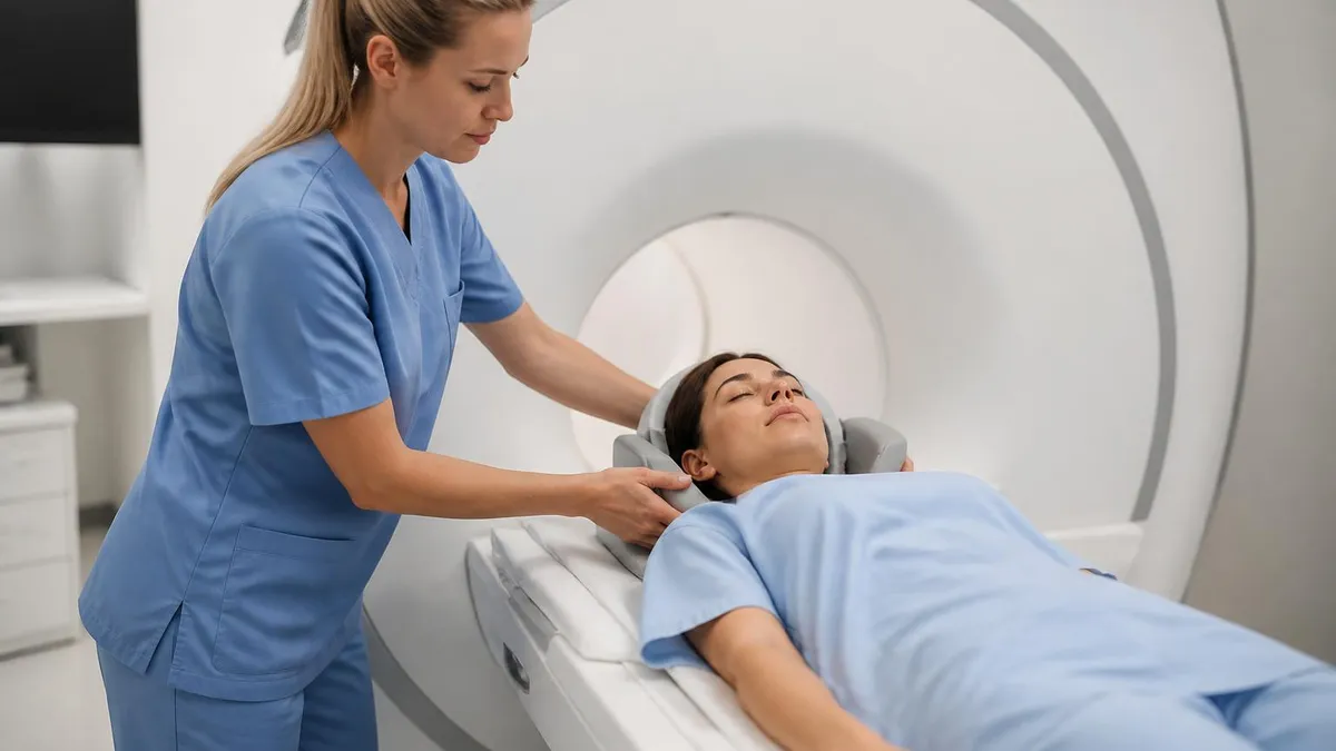
Patient positioning for brain MRI requires careful attention to alignment and coil selection. The patient lies supine with the head centered in the brain or neurovascular coil, with the head positioned so the orbitomeatal line is perpendicular to the bore axis. Padding between the head and coil minimizes movement, and foam earplugs or MRI-compatible headphones reduce acoustic discomfort from gradient noise.
For pediatric patients and sedated adults, physiological monitoring (pulse oximetry, ECG) is maintained throughout the scan using MRI-compatible equipment. Proper head immobilization is particularly critical for diffusion tensor imaging and functional MRI, where sub-millimeter motion can render advanced sequences non-diagnostic.
Field strength selection significantly affects brain MRI image quality. At 3 Tesla (T), the signal-to-noise ratio (SNR) is approximately double that of 1.5T, enabling higher spatial resolution, thinner slices, or shorter scan times for the same image quality. This advantage is most apparent in small structure visualization — hippocampal anatomy in epilepsy protocols, small MS plaques in the corpus callosum, and microbleed detection on SWI.
However, 3T also amplifies certain artifacts: susceptibility effects are more pronounced at the skull base, B1 field inhomogeneity can produce shading across large brain areas, and specific absorption rate (SAR) limitations may require longer TR values to stay within safe radiofrequency energy deposition limits.
Slice thickness and field-of-view (FOV) selection must balance spatial resolution against SNR and scan time. For standard brain protocols, axial slices of 3–5 mm with a 1 mm gap are typical, while 3D sequences like MPRAGE acquire 1 mm isotropic voxels that can be reformatted in any plane.
A FOV of 22–24 cm is standard for adult brain imaging, ensuring full coverage of the calvarium and posterior fossa without excessive wrap-around artifact. Reducing FOV to 18–20 cm for pediatric patients maintains proportional resolution while keeping scan time manageable. Phase-encode direction should be selected to direct any motion or wrap-around artifact away from the region of clinical interest.
Contrast-enhanced brain MRI with gadolinium requires precise timing and technique. For post-contrast T1 imaging, a delay of 5–10 minutes between injection and image acquisition allows adequate time for gadolinium to accumulate in enhancing lesions and for unenhanced vascular signal to diminish. The standard dose of most macrocyclic GBCAs is 0.1 mmol/kg body weight administered intravenously, typically as a slow bolus followed by a saline flush.
For brain tumor surveillance and treated MS, double-dose gadolinium (0.2 mmol/kg) was historically used to increase enhancement sensitivity, but this practice has declined significantly due to concerns about gadolinium deposition in brain tissue, particularly in the dentate nucleus and globus pallidus after repeated exposures.
Quality assurance (QA) in brain MRI programs involves regular phantom scanning to verify geometric accuracy, signal uniformity, slice thickness consistency, and contrast-to-noise ratio. ARRT-certified MRI technologists are expected to understand and participate in QA protocols as part of their professional competency.
Recognizing when scanner performance deviates from baseline — for example, when SNR drops significantly or geometric distortion increases — enables early detection of coil or gradient hardware issues before they compromise diagnostic image quality. Proactive QA prevents the need for repeat scans, reduces patient time in the scanner, and ensures that brain MRI images meet the diagnostic standard required for confident radiological interpretation.
The combination of technical proficiency, anatomical knowledge, pathology recognition, and patient safety awareness that defines expert brain MRI practice is built over time through consistent study, hands-on scanning experience, and engagement with registry-style practice questions that challenge your ability to apply knowledge under examination conditions.
Use the quiz resources linked throughout this guide to test your understanding of key concepts, review areas of weakness systematically, and build the confidence that comes from truly mastering the material. The investment you make in deeply understanding brain MRI images will serve you throughout your career as an MRI technologist and in every patient encounter that depends on your technical and clinical expertise.
Sample MRI - Magnetic Resonance Imaging Practice Questions
Try these questions from our free MRI - Magnetic Resonance Imaging practice tests. The correct answer and an explanation follow each question.
What is the effect of increasing the echo time (TE) on T2-weighted images?
- A. Increases T2 weighting by allowing more transverse relaxation
- B. Decreases T2 weighting by increasing signal
- C. Has no effect on tissue contrast
- D. Increases T1 weighting
Answer: A. Increases T2 weighting by allowing more transverse relaxation
A longer TE allows more time for T2 decay, so tissues with short T2 lose more signal, increasing contrast between tissues with different T2 values.
On MRI, cortical bone appears as which signal intensity on both T1 and T2 weighted images?
- A. High signal
- B. Intermediate signal
- C. Low signal (signal void)
- D. Variable signal
Answer: C. Low signal (signal void)
Cortical bone has very few mobile protons and produces a signal void (low signal) on both T1 and T2 weighted MRI sequences.
Which MRI technique is used to assess brain perfusion by tracking the first pass of a gadolinium bolus?
- A. Arterial Spin Labeling (ASL)
- B. Dynamic Susceptibility Contrast (DSC) perfusion
- C. Diffusion Tensor Imaging (DTI)
- D. Magnetic Resonance Spectroscopy (MRS)
Answer: B. Dynamic Susceptibility Contrast (DSC) perfusion
DSC perfusion MRI uses rapid T2*-weighted sequences to measure signal drop as gadolinium transits through the brain capillaries.
Pulsatile flow artifact from a vessel typically appears as ghost images displaced in which direction?
- A. Slice-selection direction
- B. Frequency-encoding direction
- C. Phase-encoding direction
- D. Both frequency and phase directions equally
Answer: C. Phase-encoding direction
Pulsatile flow ghosts appear in the phase-encoding direction because pulsatile variations in spin phase occur between TR intervals during phase-encode data collection.
Take the full MRI - Magnetic Resonance Imaging practice test
MRI Questions and Answers

MRI of Cervical Spine: Complete Guide to Imaging, Anatomy, Pathology, and Patient Preparation

Knee MRI Images: A Complete Guide to Reading, Understanding, and Interpreting Knee Scans
Ace Your Exam with MRI Practice Test 2026 Prep

MRI Tech School 2026 — Programs, Requirements, and What to Expect
MRI Procedure: What to Expect Before, During and After Your Scan
How this page was made: written with AI tools. Exam facts are checked against the official sources listed below. Not reviewed by a subject-matter expert. Editorial policy
Sources & references
Exam facts on this page (fees, format, dates, passing scores) are checked against these official sources; the date is our last check. Editorial policy
- arrt.org — Passing score, score release, fees, exam information · verified Oct 2026
- assets-us-01.kc-usercontent.com — Renewal, score validity, test delivery, eligibility, exam format, test dates, time limit · verified Oct 2026