Dynamic Contrast MRI: A Complete Guide to DCE-MRI Protocols, Analysis, and Clinical Applications
Learn how dynamic contrast MRI works, including DCE-MRI protocols, pharmacokinetic analysis, and clinical applications for cancer detection and staging. 🎯
Dynamic contrast MRI, also known as dynamic contrast-enhanced magnetic resonance imaging or DCE-MRI, represents one of the most powerful functional imaging techniques available to radiologists and clinicians today. This advanced imaging method goes beyond standard anatomical MRI scans by capturing detailed information about tissue perfusion and vascular characteristics in real time. By tracking the movement of a gadolinium-based contrast agent as it passes through tissues, dynamic contrast MRI provides quantitative data that helps physicians distinguish between benign and malignant lesions with remarkable accuracy across diverse clinical settings.
The fundamental principle behind dynamic contrast MRI involves rapidly acquiring multiple images of the same anatomical region before, during, and after the intravenous injection of contrast material. This time-resolved approach allows clinicians to observe how quickly tissues enhance and how rapidly the contrast agent washes out of the region of interest. These enhancement patterns are directly related to the underlying vascular architecture and permeability of the tissue, making DCE-MRI an invaluable diagnostic tool for characterizing suspicious lesions throughout the entire body.
In clinical practice, dynamic contrast MRI has become a cornerstone of breast cancer screening and diagnosis, particularly for women identified as having an elevated lifetime risk. The technique is also widely used in evaluating prostate lesions, brain tumors, liver masses, and musculoskeletal abnormalities. Its ability to provide functional information about blood flow and vascular permeability sets it apart from conventional MRI sequences that primarily depict anatomical structures without revealing the physiological behavior of tissues and ongoing disease processes at the molecular and cellular level.
Understanding the technical aspects of dynamic contrast MRI requires familiarity with several key concepts, including temporal resolution, spatial resolution, and pharmacokinetic modeling. Temporal resolution refers to how frequently images are acquired during and after the contrast injection, with faster acquisition providing more detailed time-intensity curves. Spatial resolution determines the level of anatomical detail visible in each image frame. Balancing these two competing parameters is one of the most critical decisions an MRI technologist must make when designing a DCE-MRI protocol for clinical application.
The data generated by dynamic contrast MRI examinations can be analyzed using both qualitative and quantitative methods. Qualitative analysis involves visual assessment of enhancement curves categorized into three main types that suggest benign, indeterminate, or suspicious characteristics based on their shape. Quantitative analysis applies pharmacokinetic models to extract specific parameters such as Ktrans, ve, and kep, which provide objective measurements of tissue vascularity and capillary permeability. These quantitative values are increasingly used in clinical trials and ongoing research to objectively monitor treatment response.
For MRI technologists preparing for registry examinations, a thorough understanding of dynamic contrast MRI principles is absolutely essential. Questions about DCE-MRI frequently appear on certification exams, covering topics ranging from contrast agent pharmacology and injection protocols to image acquisition timing and post-processing analysis techniques. Mastering these concepts not only prepares candidates for exam success but also ensures they can competently perform and troubleshoot dynamic contrast-enhanced examinations in their daily clinical practice across various imaging centers and hospital departments.
This comprehensive guide covers everything you need to know about dynamic contrast MRI, from the basic physics underlying contrast enhancement to advanced clinical applications and emerging research directions. Whether you are a radiology resident learning the fundamentals, an MRI technologist studying for professional certification, or a healthcare provider seeking to deepen your understanding of this powerful imaging modality, the following sections provide detailed explanations, practical tips, and evidence-based insights to support your professional development and ongoing clinical competence.
Dynamic Contrast MRI by the Numbers
Steps in Performing a Dynamic Contrast MRI Examination
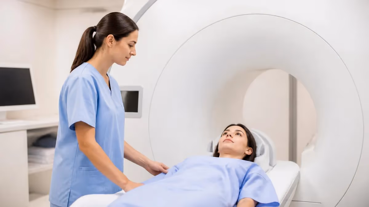
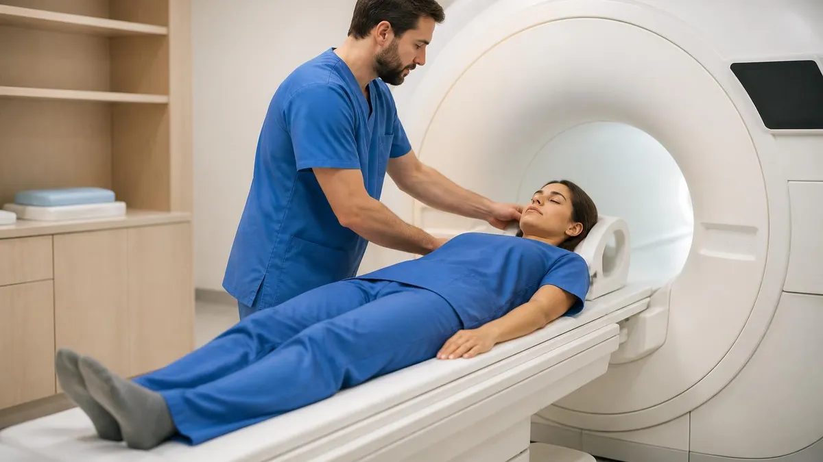
Patient Screening and Preparation
Baseline Pre-Contrast Imaging
Protocol Configuration and Contrast Loading
Dynamic Acquisition During Contrast Injection
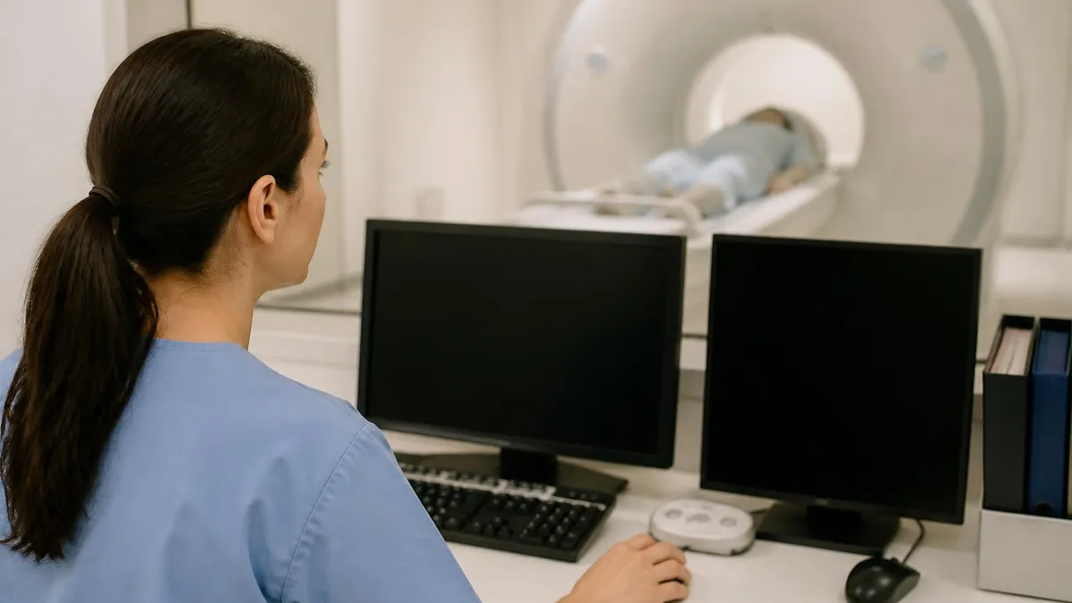
Post-Processing and Analysis
Documentation and Patient Monitoring
The acquisition protocol for dynamic contrast MRI requires careful planning and precise execution to obtain diagnostically useful data from every examination. Before the contrast injection begins, the technologist acquires baseline pre-contrast images using the same pulse sequence parameters that will be used throughout the entire dynamic series. These baseline images serve as the reference point against which all subsequent enhancement measurements are calculated and compared. Typically, a T1-weighted gradient echo sequence is selected because of its superior sensitivity to the T1-shortening effects of gadolinium-based contrast agents in biological tissues.
Timing is perhaps the most critical factor in successful dynamic contrast MRI acquisition. The contrast agent, typically administered at a standard dose of 0.1 mmol per kilogram of body weight, is injected intravenously using a power injector at a controlled rate, usually between two and three milliliters per second. The dynamic acquisition must begin simultaneously with or slightly before the contrast injection to capture the earliest phases of tissue enhancement accurately. Delays of even a few seconds can compromise the diagnostic quality of the resulting time-intensity curves significantly.
Temporal resolution requirements vary considerably depending on the clinical application and the specific anatomical region being examined. Breast DCE-MRI protocols typically employ temporal resolutions of sixty to ninety seconds per acquisition, which provides adequate sampling of the enhancement kinetics while maintaining sufficient spatial resolution for detailed lesion characterization. Prostate DCE-MRI, on the other hand, often requires much higher temporal resolution of five to fifteen seconds to accurately capture the rapid enhancement patterns that are characteristic of prostate carcinoma compared to normal tissue.
The total duration of a dynamic contrast MRI acquisition typically ranges from five to ten minutes, during which the scanner continuously acquires image volumes at regular intervals throughout the enhancement and washout phases. This results in a four-dimensional dataset consisting of three spatial dimensions plus the time dimension. The number of temporal phases acquired depends on both the temporal resolution setting and the total scan duration chosen. Most clinical protocols capture between ten and forty temporal phases, providing enough data points to construct diagnostically meaningful time-intensity curves.
Post-processing of dynamic contrast MRI data begins with motion correction, which is particularly important for body applications where respiratory motion can cause significant misregistration between temporal phases. Subtraction images, created by subtracting the pre-contrast baseline from each post-contrast phase, help isolate areas of true enhancement from background signal and inherently bright structures. Color-coded overlay maps can then be generated to display the enhancement kinetics at each pixel location, providing an efficient visual summary of the entire dynamic dataset that radiologists can rapidly interpret during reading sessions.
Pharmacokinetic analysis represents the most sophisticated level of dynamic contrast MRI post-processing available in clinical practice. Models such as the Tofts model and the extended Tofts model fit the observed time-intensity data to mathematical equations that describe the exchange of contrast agent between the blood plasma and the extravascular extracellular space. The resulting parameters, including the volume transfer constant Ktrans and the extravascular extracellular volume fraction ve, provide quantitative biomarkers that correlate with tissue angiogenesis, vascular permeability, and overall tumor biology characteristics.
Quality assurance for dynamic contrast MRI protocols should include regular phantom testing to verify temporal resolution accuracy, spatial resolution performance, and signal-to-noise ratios across all phases of the dynamic acquisition. Technologists should also verify that the power injector is properly calibrated and that the injection rate and contrast volume are accurately programmed for each individual patient examination. Documentation of protocol parameters and any deviations from standard acquisition settings is important for ensuring reproducibility and maintaining compliance with institutional and national accreditation requirements.
MRI Practice Test Questions
Prepare for the MRI - Magnetic Resonance Imaging exam with our free practice test modules. Each quiz covers key topics to help you pass on your first try.
MRI Knowledge
MRI Exam Questions covering Knowledge. Master MRI Test concepts for certification prep.
MRI Physics
Free MRI Practice Test featuring Physics. Improve your MRI Exam score with mock test prep.
MRI Anatomy and Pathology
MRI Test Prep for MRI Anatomy and Pathology. Practice MRI Quiz questions and boost your score.
MRI Anatomy and Positioning
MRI Questions and Answers on MRI Anatomy and Positioning. Free MRI practice for exam readiness.
MRI Contrast Agents
Free MRI Quiz on MRI Contrast Agents. MRI Exam prep questions with detailed explanations.
MRI Patient Care and Positioning
MRI Practice Questions for MRI Patient Care and Positioning. Build confidence for your MRI certification exam.
Analysis Methods for Dynamic Contrast MRI Data
Qualitative analysis of dynamic contrast MRI involves visual assessment of time-intensity curves generated from regions of interest placed over suspicious lesions. These curves are classified into three standard types based on their morphology. Type I curves show persistent enhancement that continues to increase throughout the acquisition, a pattern most commonly associated with benign processes such as fibroadenomas in breast imaging or benign prostatic hyperplasia in prostate evaluation settings.
Type II curves demonstrate a plateau pattern where the signal intensity rises rapidly during the initial enhancement phase and then levels off, neither increasing nor decreasing significantly during the delayed phases. This pattern is considered indeterminate and may represent either benign or malignant pathology depending on associated morphologic features. Type III curves exhibit washout kinetics where the signal intensity increases rapidly then decreases below the plateau level, and this pattern carries the highest suspicion for malignancy across most organ systems evaluated with DCE-MRI.
Advantages and Limitations of Dynamic Contrast MRI
- +Provides functional information about tissue vascularity beyond anatomical imaging
- +Achieves sensitivity exceeding ninety percent for invasive breast cancer detection
- +Enables quantitative biomarkers for objective treatment response monitoring
- +No ionizing radiation exposure compared to CT perfusion studies
- +Superior soft tissue contrast resolution for lesion characterization
- +Supports pharmacokinetic modeling for research and clinical trial endpoints
- −Requires intravenous gadolinium contrast agent with associated safety risks
- −Lower temporal resolution compared to CT perfusion imaging techniques
- −Susceptible to motion artifacts especially in abdominal applications
- −Pharmacokinetic analysis requires specialized software and expertise
- −Longer examination times compared to standard non-dynamic MRI protocols
- −Contraindicated in patients with severe renal insufficiency due to NSF risk
Dynamic Contrast MRI Quality Assurance Checklist
- ✓Verify power injector calibration and contrast agent expiration date before each examination.
- ✓Confirm patient renal function with eGFR results obtained within the required timeframe.
- ✓Establish reliable intravenous access and test line patency with a saline flush.
- ✓Program correct contrast dose based on patient body weight at 0.1 mmol per kilogram.
- ✓Set temporal resolution appropriate for the specific clinical application and anatomical region.
- ✓Acquire pre-contrast baseline images using identical sequence parameters as the dynamic series.
- ✓Synchronize contrast injection start with the beginning of the dynamic acquisition sequence.
- ✓Monitor real-time image reconstruction to confirm proper enhancement timing during acquisition.
- ✓Apply motion correction algorithms before generating subtraction and parametric maps.
- ✓Document contrast agent type, volume, injection rate, and any patient adverse reactions.
Enhancement Curve Types Are the Foundation of DCE-MRI Interpretation
The three standard enhancement curve types — persistent (Type I), plateau (Type II), and washout (Type III) — form the basis for clinical interpretation of dynamic contrast MRI across virtually all organ systems. A Type III washout curve carries the highest suspicion for malignancy, with positive predictive values exceeding 70% in breast imaging. Understanding these patterns is essential for both clinical practice and MRI registry examination preparation.
Clinical applications of dynamic contrast MRI span virtually every organ system and continue to expand as new research validates its diagnostic utility in previously unexplored areas. In breast imaging, DCE-MRI has achieved sensitivity rates exceeding ninety percent for the detection of invasive breast cancer, making it the most sensitive imaging modality currently available for breast cancer screening. The American Cancer Society and other major organizations recommend annual breast MRI screening for women with a lifetime breast cancer risk of twenty percent or greater, underscoring the established clinical importance of this technique.
In mri of prostate dynamic contrast MRI has become an integral component of the multiparametric MRI protocol defined by the Prostate Imaging Reporting and Data System, commonly known as PI-RADS. While diffusion-weighted imaging and T2-weighted sequences serve as the primary diagnostic sequences, DCE-MRI provides supplementary information that can upgrade the assessment of suspicious lesions located in the peripheral zone. Rapid and focal enhancement in the peripheral zone is considered a positive DCE finding under current guidelines and increases the likelihood that a detected lesion represents clinically significant prostate cancer warranting biopsy.
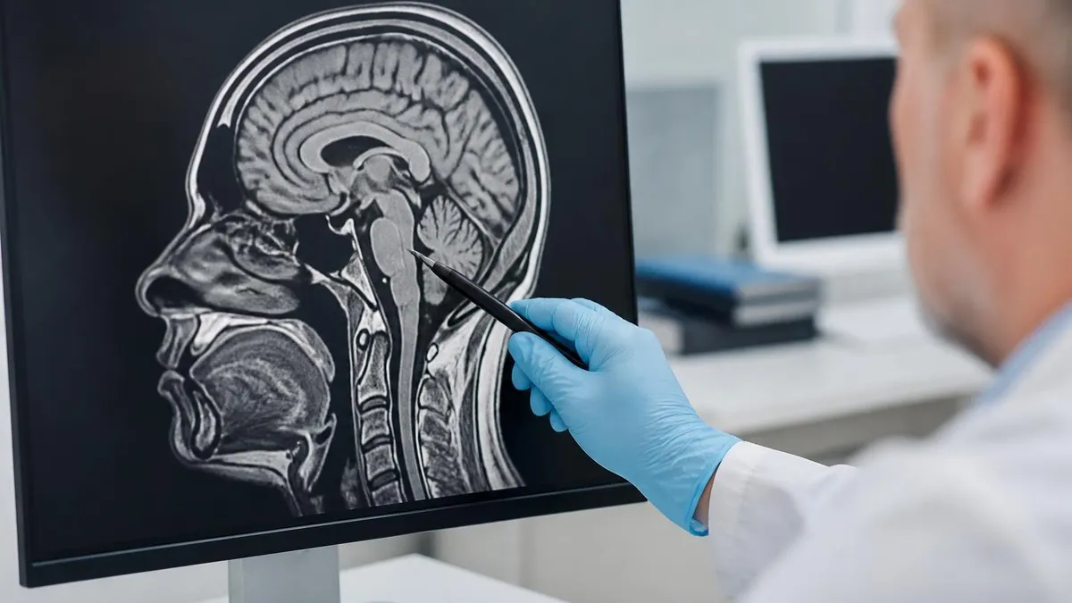
Neuro-oncology represents another major application area for dynamic contrast MRI in contemporary clinical practice. In brain tumor evaluation, DCE-MRI helps differentiate between true tumor recurrence and treatment-related changes such as pseudoprogression or radiation necrosis, which can appear remarkably similar on conventional contrast-enhanced sequences. The ability to quantify blood-brain barrier permeability through pharmacokinetic analysis provides objective data that guides critical treatment decisions and helps neuro-oncologists determine whether to continue, modify, or fundamentally change therapeutic strategies for patients with primary brain tumors or cerebral metastases.
Liver imaging with dynamic contrast MRI follows a unique protocol that takes full advantage of the dual blood supply of the liver from both the hepatic artery and the portal vein. The dynamic acquisition captures arterial phase, portal venous phase, and delayed phase images that reveal characteristic enhancement patterns for different categories of liver lesions. Hepatocellular carcinoma, for example, typically shows intense arterial phase enhancement followed by washout in the portal venous and delayed phases, a pattern formally recognized by the Liver Imaging Reporting and Data System as a major diagnostic criterion.
Musculoskeletal applications of dynamic contrast MRI include the evaluation of soft tissue masses, assessment of synovial inflammation in rheumatoid arthritis, and monitoring of treatment response in bone and soft tissue tumors undergoing therapy. The enhancement characteristics of musculoskeletal lesions on DCE-MRI can help differentiate between aggressive malignant tumors that demonstrate rapid early enhancement and more indolent benign lesions that show slower and more gradual enhancement patterns over time. This information is particularly valuable when biopsy planning is needed or when ongoing treatment response assessment is clinically required.
Cardiac dynamic contrast MRI, often referred to as myocardial perfusion imaging, evaluates blood flow to the heart muscle during both pharmacologic stress and rest conditions. First-pass perfusion imaging captures the initial transit of gadolinium through the myocardium, revealing perfusion defects that indicate significant coronary artery disease. Late gadolinium enhancement imaging, acquired ten to fifteen minutes after contrast administration, demonstrates areas of myocardial scar or fibrosis. Together these complementary techniques provide comprehensive assessment of both ischemia and myocardial viability, guiding important clinical decisions about revascularization procedures.
Emerging applications of dynamic contrast MRI include the evaluation of treatment response in patients undergoing antiangiogenic therapy, where early changes in Ktrans values can predict therapeutic efficacy before changes in tumor size become apparent on conventional imaging. Active research is also exploring the use of DCE-MRI in evaluating inflammatory bowel disease activity, assessing placental function during pregnancy, and characterizing complex renal lesions. As machine learning algorithms are increasingly applied to DCE-MRI data analysis, automated interpretation tools promise to significantly improve both the efficiency and diagnostic accuracy of clinical interpretation workflows.
All patients must be screened for renal insufficiency before receiving gadolinium-based contrast agents for dynamic contrast MRI. Patients with an estimated glomerular filtration rate below 30 mL/min are at increased risk for nephrogenic systemic fibrosis. Use Group II macrocyclic agents when contrast administration is essential in at-risk patients, and document the risk-benefit discussion in the medical record.
Contrast agents used in dynamic contrast MRI are predominantly gadolinium-based compounds that shorten the T1 relaxation time of nearby water protons, producing characteristically bright signal on T1-weighted images. Several different gadolinium chelates are approved for clinical use in the United States, including gadopentetate dimeglumine, gadobutrol, gadoterate meglumine, and gadobenate dimeglumine. Each agent has slightly different relaxivity characteristics, molecular weight, and biodistribution properties that can influence the enhancement patterns observed on dynamic imaging sequences and must be carefully considered when selecting an appropriate agent for each examination.
Safety considerations for gadolinium-based contrast agents have evolved significantly over the past two decades of clinical experience and research. The recognition of nephrogenic systemic fibrosis as a serious complication in patients with severe renal insufficiency led to major changes in clinical screening protocols and contrast agent selection practices across all imaging centers. Current guidelines require assessment of renal function using estimated glomerular filtration rate before administering any gadolinium-based agent, with specific restrictions on certain agents in patients whose eGFR falls below thirty milliliters per minute per body surface area.
Gadolinium deposition in brain tissue has emerged as another important safety concern that continues to influence clinical practice and drive ongoing research. Published studies have demonstrated that repeated administration of linear gadolinium chelates can lead to measurable deposits of gadolinium in certain brain structures, particularly the dentate nucleus and globus pallidus, visible as increased signal intensity on unenhanced T1-weighted images. While no clinical symptoms have been definitively attributed to these deposits in humans, regulatory agencies have responded by restricting the use of certain linear gadolinium agents in favor of more stable macrocyclic formulations.
Allergic-like reactions to gadolinium-based contrast agents occur less frequently than reactions to iodinated contrast agents used in computed tomography examinations, but they remain an important safety consideration for all MRI departments. Mild reactions including nausea, urticaria, and headache occur in approximately one to two percent of patients receiving gadolinium contrast material. Severe anaphylactoid reactions are rare but can be life-threatening, occurring in approximately one to ten per one hundred thousand administrations. MRI departments must maintain appropriate emergency medications and equipment and ensure all personnel are properly trained in managing reactions.
Power injector technology plays a crucial role in dynamic contrast MRI by ensuring consistent and reproducible contrast delivery across examinations performed on different patients and different days. Modern MRI-compatible power injectors allow precise control of injection rate, volume, and timing parameters, with typical flow rates ranging from one to five milliliters per second depending on the specific clinical application and patient factors. A saline flush of twenty to thirty milliliters is typically administered immediately after the contrast bolus to ensure complete delivery of the agent into the central venous circulation effectively.
Patient preparation for dynamic contrast MRI examinations includes standard MRI screening for absolute and relative contraindications such as ferromagnetic implants, cardiac pacemakers, and cochlear implants. Additionally, patients must be assessed for current renal function status and any history of prior gadolinium contrast reactions. Intravenous access must be established before the patient enters the scanner room, and the patency of the IV line should be verified with a test injection of normal saline. Clear and empathetic communication with patients about what to expect during the contrast injection helps reduce anxiety and minimize motion-related artifacts.
Documentation requirements for dynamic contrast MRI examinations include accurately recording the specific contrast agent used, the total volume administered, the injection rate, and any adverse reactions observed during or after the examination. The American College of Radiology recommends monitoring patients for at least thirty minutes after gadolinium administration to observe for any delayed adverse reactions. Technologists should be thoroughly familiar with their department-specific protocols for contrast reaction management and know the exact location of emergency equipment including supplemental oxygen, epinephrine auto-injectors, and intravenous diphenhydramine supplies.
Optimizing dynamic contrast MRI examinations in daily clinical practice requires careful attention to several practical details that can significantly impact diagnostic quality and patient outcomes. First and foremost, ensure that the patient is positioned comfortably and that adequate immobilization devices are used to minimize voluntary and involuntary motion during the entire acquisition period. For breast DCE-MRI specifically, proper coil placement and breast positioning are essential for obtaining uniform fat suppression and consistent image quality across all temporal phases of the dynamic series throughout the complete examination.
Selecting the appropriate pulse sequence parameters for dynamic contrast MRI involves balancing competing demands for temporal resolution, spatial resolution, and anatomic coverage volume. Three-dimensional spoiled gradient echo sequences with short repetition times and short echo times are the standard choice for most clinical DCE-MRI applications across body regions. Parallel imaging techniques such as GRAPPA or SENSE can be employed to accelerate acquisition speed and improve temporal resolution without unacceptably sacrificing spatial coverage. Newer acceleration techniques including compressed sensing and simultaneous multi-slice acquisition offer additional options that are becoming increasingly available on modern scanners.
Fat suppression techniques are particularly important in dynamic contrast MRI because enhancing tissue must be clearly distinguishable from inherently high-signal fat on T1-weighted imaging sequences. Chemical shift-based fat suppression methods, including Dixon techniques and spectral presaturation with inversion recovery, are commonly employed in clinical practice. Dixon-based methods offer the distinct advantage of providing both water-only and fat-only images from a single acquisition, which can be especially useful for identifying fat-containing lesions and improving the conspicuity of small enhancing lesions against a uniformly suppressed fat background.
Troubleshooting common problems in dynamic contrast MRI examinations is an essential practical skill for MRI technologists working in busy clinical environments. Contrast timing failures, where the dynamic acquisition begins too late or too early relative to the actual contrast arrival time, can significantly compromise diagnostic quality. Monitoring the real-time image display during the early phases of the acquisition helps identify timing issues that may require immediate adjustment or protocol modification. If the first post-contrast phase shows no enhancement in expected vascular structures, the technologist should immediately consider whether a contrast delivery failure has occurred.
Motion artifacts represent one of the most significant technical challenges in dynamic contrast MRI, particularly for abdominal and thoracic applications where respiratory motion is completely unavoidable during the extended acquisition period. Breath-hold techniques can effectively reduce respiratory artifacts but limit the available scan time per temporal phase acquisition. Navigator-gated and free-breathing approaches with retrospective motion correction algorithms provide alternative strategies that allow longer acquisition windows at the expense of more complex image reconstruction processes. Communicating clearly with patients about specific breathing instructions before beginning the dynamic acquisition significantly helps ensure cooperation and compliance.
Continuing education in dynamic contrast MRI is essential for maintaining clinical competence as the field continues to evolve rapidly with new technical developments and clinical applications. Professional organizations including the International Society for Magnetic Resonance in Medicine and the Section for Magnetic Resonance Technologists offer comprehensive educational resources, hands-on workshops, and annual scientific meetings that cover the latest advances in DCE-MRI techniques and their clinical applications. Online learning platforms and peer-reviewed journals publish regular updates on emerging research findings and evolving clinical best practices in this field.
Preparing for MRI registry examination questions about dynamic contrast MRI requires focused and systematic study of several key topic areas. Candidates should thoroughly understand the basic pharmacology of gadolinium-based contrast agents, the principles of T1 relaxation and contrast enhancement mechanisms, the technical parameters that affect temporal and spatial resolution, and the clinical significance of different enhancement curve types. Practicing with sample examination questions and reviewing detailed case studies that illustrate typical and atypical enhancement patterns will reinforce understanding and build examination-day confidence for achieving certification success and long-term clinical excellence.
MRI Questions and Answers

MRI Medical Abbreviation: What MRI Stands For and Why It Matters

Knee MRI Images: A Complete Guide to Reading, Understanding, and Interpreting Knee Scans

Noise of MRI Machine: Why MRI Scanners Are So Loud and What to Expect
Is Nickel Titanium MRI Compatible? A Complete Guide to MRI Safety Materials
What to Expect During MRI: Your Complete Patient Guide to the Scanning Process
About the Author
Medical Laboratory Scientist & Clinical Certification Expert
Johns Hopkins UniversityDr. Sandra Kim holds a PhD in Clinical Laboratory Science from Johns Hopkins University and is certified as a Medical Technologist (MT) and Medical Laboratory Scientist (MLS) through ASCP. With 16 years of clinical laboratory experience spanning hematology, microbiology, and molecular diagnostics, she prepares candidates for ASCP board exams, MLT, MLS, and specialist certification tests.
Join the Discussion
Connect with other students preparing for this exam. Share tips, ask questions, and get advice from people who have been there.
View discussion (6 replies)