Multiparametric MRI for Prostate Cancer: What Every Patient and Clinician Should Know
Learn how multiparametric MRI detects prostate cancer with high accuracy. Understand mpMRI sequences, PI-RADS scoring, and what to expect during the scan. 📝
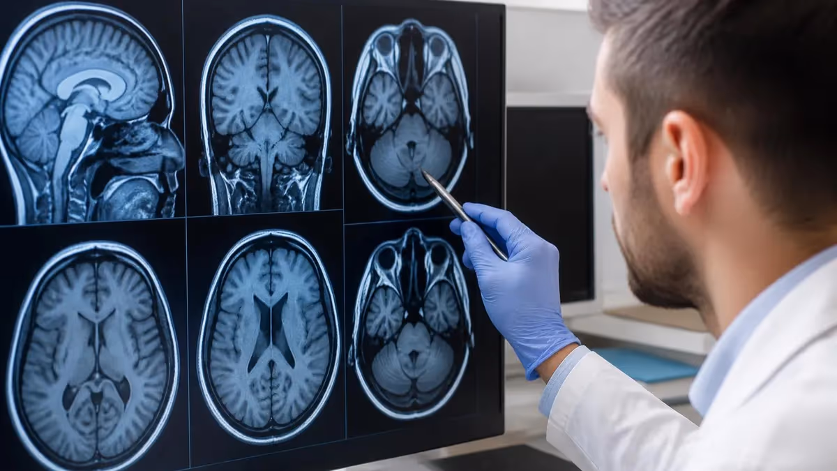
Multiparametric MRI prostate cancer detection has transformed the way urologists and radiologists identify, characterize, and manage suspicious lesions within the prostate gland. Unlike conventional imaging approaches that rely on a single contrast mechanism, multiparametric MRI combines several complementary pulse sequences into one comprehensive examination, delivering far greater diagnostic accuracy than any individual technique could achieve alone. For men facing elevated PSA levels or abnormal digital rectal exam findings, mpMRI has become the recommended first-line imaging study before biopsy.
The American Urological Association and European Association of Urology now endorse multiparametric MRI before initial biopsy in most clinical scenarios. This paradigm shift reflects over a decade of high-quality evidence demonstrating that mpMRI reduces unnecessary biopsies by approximately twenty-eight percent while simultaneously improving detection of clinically significant cancers. Patients benefit from fewer invasive procedures, lower complication rates, and more precisely targeted tissue sampling when biopsy ultimately remains indicated after imaging review.
At its core, a multiparametric MRI examination acquires at least two distinct pulse sequences that probe fundamentally different tissue properties. T2-weighted imaging reveals anatomical detail and the characteristic zonal architecture of the prostate, while diffusion-weighted imaging measures the random motion of water molecules to identify areas of abnormally high cellular density that often correspond to malignant tissue. Dynamic contrast-enhanced imaging tracks gadolinium uptake patterns over time, adding another functional layer to the overall diagnostic picture.
The standardized reporting framework known as PI-RADS, or Prostate Imaging Reporting and Data System, assigns each suspicious lesion a score from one through five based on the estimated probability of clinically significant cancer. PI-RADS version 2.1, published in 2019, refined assessment criteria for both the transition zone and peripheral zone, improving inter-reader agreement and overall diagnostic consistency across institutions of varying experience levels. Radiologists worldwide now use this shared language to communicate findings clearly to referring urologists.
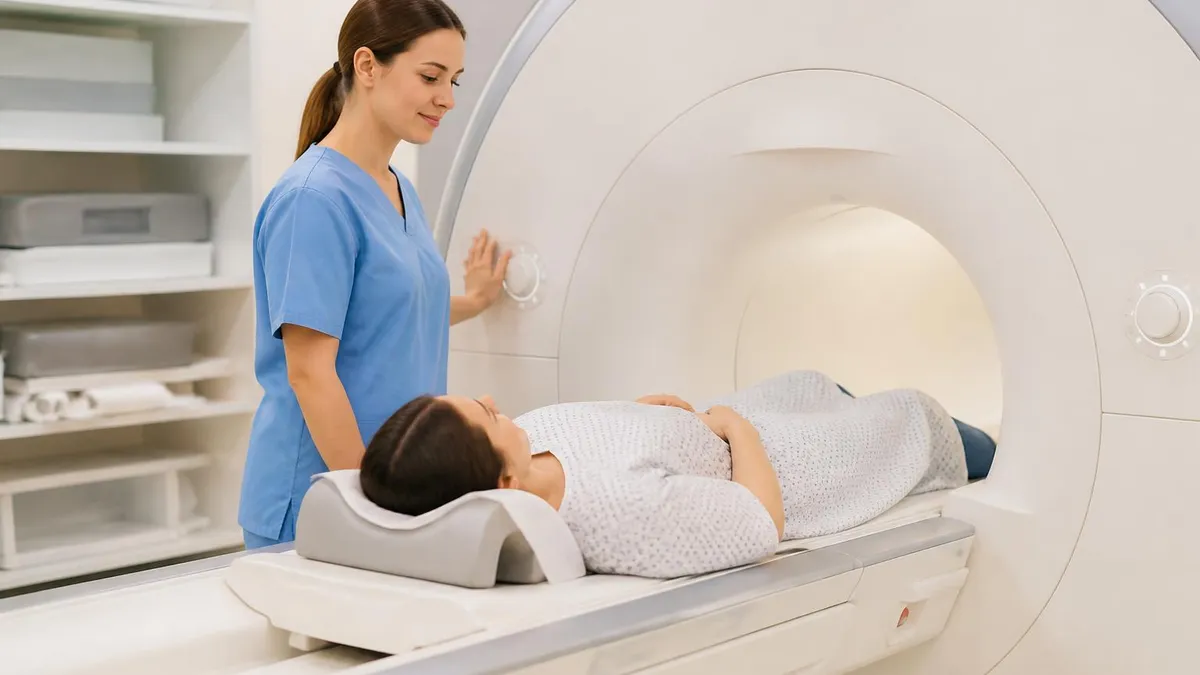
Many patients understandably feel anxious about undergoing an MRI examination, but the multiparametric protocol typically adds only ten to fifteen additional minutes beyond a standard pelvic MRI scan duration. Modern three-Tesla scanners offer higher signal-to-noise ratios and substantially improved spatial resolution, which translates directly to sharper images and more confident lesion characterization. Some facilities also offer an endorectal coil for enhanced image quality, although advances in phased-array surface coil technology have made this option increasingly unnecessary.
Beyond initial diagnosis, multiparametric MRI plays a vital role in active surveillance programs for men with low-risk prostate cancer who choose careful monitoring over immediate treatment. Serial mpMRI examinations can detect progression in lesion size or imaging characteristics, prompting timely clinical intervention before the cancer advances to a potentially incurable stage. This monitoring approach spares thousands of men each year from surgery or radiation side effects that may never have been clinically necessary.
Understanding the fundamentals of multiparametric MRI empowers both clinicians and patients to make better-informed decisions at every stage of the prostate cancer journey. From initial screening and diagnosis through treatment planning and post-therapy surveillance, mpMRI has established itself as an indispensable tool in modern urologic oncology that continues to evolve rapidly with advances in artificial intelligence, quantitative imaging biomarkers, and radiomics-driven precision medicine approaches.
Multiparametric MRI Prostate Cancer by the Numbers
How Multiparametric MRI Works: Core Sequences and Techniques
Provides high-resolution anatomical maps of prostate zonal architecture, depicting the peripheral zone, transition zone, and central gland with exceptional soft-tissue contrast to reveal structural abnormalities and capsular integrity.
Measures random Brownian motion of water molecules within tissue. Cancer restricts diffusion due to high cellularity, appearing bright on high b-value images and dark on apparent diffusion coefficient maps for quantitative assessment.
Tracks gadolinium contrast agent uptake and washout patterns over time. Malignant lesions demonstrate early arterial enhancement and rapid washout reflecting tumor angiogenesis, serving as a tiebreaker for equivocal peripheral zone findings.
All sequences feed into the Prostate Imaging Reporting and Data System framework, producing a composite score from one through five that communicates cancer probability to the urologist in a standardized, reproducible format across institutions.
The PI-RADS scoring system provides a standardized framework for interpreting multiparametric MRI findings and communicating the likelihood of clinically significant prostate cancer to the referring physician. Each identified lesion receives a score between one and five, where PI-RADS 1 indicates very low probability and PI-RADS 5 indicates very high probability of harboring aggressive disease. This structured approach replaced the subjective narrative descriptions that previously dominated radiology reports and frequently left urologists uncertain about the most appropriate next clinical step.
In the peripheral zone, which accounts for approximately seventy percent of all prostate cancers diagnosed, diffusion-weighted imaging serves as the dominant sequence for determining the PI-RADS assessment category. Radiologists evaluate the apparent diffusion coefficient map for focal areas of markedly restricted diffusion that appear conspicuously hypointense against the normal glandular background. Lesions measuring fifteen millimeters or greater with severely restricted diffusion typically receive a PI-RADS 5 designation, while smaller or less conspicuous findings receive correspondingly lower scores based on size and signal intensity criteria.
Transition zone assessment follows a fundamentally different paradigm, with T2-weighted imaging serving as the dominant sequence rather than diffusion-weighted imaging for this anatomical region. Because the transition zone naturally contains benign prostatic hyperplasia nodules that can closely mimic cancer on diffusion sequences, the morphological characteristics visible on T2-weighted images become paramount for accurate assessment. Lenticular or irregularly shaped homogeneous lesions that obscure normal zonal boundaries raise clinical concern, particularly when they measure larger than fifteen millimeters in greatest dimension.
Dynamic contrast-enhanced imaging plays a supporting role within the PI-RADS framework, primarily serving to upgrade equivocal PI-RADS 3 lesions identified in the peripheral zone to a higher suspicion category. When a lesion demonstrates focal early enhancement that corresponds spatially to a diffusion abnormality, the radiologist may upgrade the assessment from PI-RADS 3 to PI-RADS 4. This upgrading mechanism captures important cases where diffusion findings alone are borderline but the additional vascular signature strongly supports a higher clinical suspicion for underlying malignancy.
Research published over the past several years has validated the predictive accuracy of PI-RADS scoring across diverse patient populations and institutional settings worldwide. Studies consistently demonstrate that PI-RADS 4 and 5 lesions harbor clinically significant cancer in approximately fifty-five to eighty percent of cases, depending on the study population and the definition of clinical significance applied. Conversely, PI-RADS 1 and 2 lesions carry a negative predictive value exceeding ninety percent, providing substantial reassurance for patients who receive favorable imaging results.
Inter-reader variability remains an acknowledged challenge with the PI-RADS system, particularly for PI-RADS 3 assessments that fall squarely in the diagnostic gray zone between clearly benign and clearly suspicious findings. Subspecialty-trained radiologists with dedicated prostate MRI experience generally achieve higher concordance rates and superior diagnostic performance compared to general body imaging radiologists without focused training. This expertise gradient underscores the importance of having multiparametric MRI examinations interpreted at centers with sufficient annual case volume and fellowship-trained subspecialty readers.
Ongoing efforts to refine PI-RADS continue with active discussions around version 2.2 and potential integration of artificial intelligence-assisted scoring into clinical workflows. Machine learning algorithms trained on thousands of annotated prostate MRI cases have shown considerable promise in matching or even exceeding the diagnostic performance of expert human readers for lesion detection and characterization. These computational tools may ultimately help standardize interpretation quality across institutions and meaningfully reduce the inter-reader variability that currently limits the system's reliability.
MRI Practice Test Questions
Prepare for the MRI - Magnetic Resonance Imaging exam with our free practice test modules. Each quiz covers key topics to help you pass on your first try.
MRI Knowledge
MRI Exam Questions covering Knowledge. Master MRI Test concepts for certification prep.
MRI Physics
Free MRI Practice Test featuring Physics. Improve your MRI Exam score with mock test prep.
MRI Anatomy and Pathology
MRI Test Prep for MRI Anatomy and Pathology. Practice MRI Quiz questions and boost your score.
MRI Anatomy and Positioning
MRI Questions and Answers on MRI Anatomy and Positioning. Free MRI practice for exam readiness.
MRI Contrast Agents
Free MRI Quiz on MRI Contrast Agents. MRI Exam prep questions with detailed explanations.
MRI Patient Care and Positioning
MRI Practice Questions for MRI Patient Care and Positioning. Build confidence for your MRI certification exam.
mpMRI Sequences Explained: T2, DWI, and DCE in Detail
T2-weighted imaging provides the anatomical foundation of every multiparametric MRI examination by depicting the zonal anatomy of the prostate gland with exceptional soft-tissue contrast and spatial resolution. The peripheral zone appears hyperintense on T2-weighted images due to its glandular fluid content, while prostate cancer typically manifests as a focal hypointense area disrupting the normal bright signal pattern. This sequence is acquired in multiple orthogonal planes to provide comprehensive three-dimensional assessment of each lesion's location, size, and spatial relationship to the prostatic capsule and adjacent structures.
Transition zone evaluation relies heavily on T2-weighted morphological assessment because diffusion-weighted imaging frequently produces false-positive findings in this anatomically complex region. Radiologists systematically search for lenticular-shaped homogeneous hypointense lesions with indistinct margins that erase the normal heterogeneous architectural features characteristic of benign prostatic hyperplasia nodules. Noncircumscribed lesions measuring greater than fifteen millimeters demonstrating homogeneous moderate hypointensity warrant elevated concern for clinically significant cancer and typically receive a PI-RADS 4 or higher assessment category designation.
Advantages and Limitations of Multiparametric MRI for Prostate Cancer
- +Detects clinically significant prostate cancer with over 90% sensitivity, far exceeding systematic biopsy alone
- +Reduces unnecessary biopsies by approximately 28%, sparing patients from invasive procedures and complications
- +Provides precise lesion localization for MRI-targeted biopsy, improving tissue sampling accuracy and diagnostic yield
- +Supports active surveillance monitoring without repeated biopsies through serial noninvasive imaging assessments
- +Enables treatment planning by mapping tumor extent, capsular involvement, and neurovascular bundle relationship
- +High negative predictive value of 90%+ for PI-RADS 1-2 gives patients reliable reassurance when scans are negative
- −Higher upfront cost than ultrasound-guided biopsy, typically ranging from $1,000 to $3,000 depending on facility
- −Reader expertise significantly affects diagnostic accuracy, with less experienced radiologists producing variable results
- −PI-RADS 3 equivocal scores create clinical uncertainty and may still require biopsy for definitive tissue diagnosis
- −Claustrophobia affects 5-10% of patients and may prevent completion of the full examination without sedation
- −Gadolinium contrast agents carry rare risks including allergic reactions and nephrogenic systemic fibrosis in renal impairment
- −Cannot reliably detect very small cancers below 5mm or low-grade tumors that produce minimal diffusion restriction
Multiparametric MRI Patient Preparation Checklist
- ✓Fast for four hours before your scheduled scan to reduce bowel motion artifacts on imaging.
- ✓Drink moderate fluids to maintain comfortable bladder distention without overdistension during the exam.
- ✓Complete the MRI safety screening questionnaire disclosing all implants, devices, and surgical hardware.
- ✓Inform staff of any kidney disease or renal impairment before gadolinium contrast administration.
- ✓Remove all metallic jewelry, watches, hearing aids, and removable dental work before entering the scanner.
- ✓Bring prior PSA results and any previous prostate biopsy pathology reports to your appointment.
- ✓Request anxiolytic medication from your physician at least one week before if you experience claustrophobia.
- ✓Wait at least six weeks after any prior prostate biopsy before scheduling your multiparametric MRI.
- ✓Arrive fifteen minutes early to complete registration paperwork and change into a hospital gown.
- ✓Ask your urologist whether an endorectal coil or antispasmodic injection will be used during your examination.
mpMRI Before Biopsy Saves Patients from Unnecessary Procedures
The PROMIS and PRECISION trials demonstrated that performing multiparametric MRI before prostate biopsy detects 20% more clinically significant cancers while reducing detection of clinically insignificant tumors by nearly half. For every 100 men undergoing the MRI-first pathway, approximately 28 avoid unnecessary biopsy entirely, and those who do proceed receive more accurate, targeted sampling that improves diagnostic confidence and treatment planning.
Clinical studies evaluating multiparametric MRI for prostate cancer detection have consistently demonstrated superior performance compared to systematic biopsy alone, particularly for identifying clinically significant tumors classified as Gleason grade group two or higher. The landmark PROMIS trial published in 2017 established that mpMRI achieved a sensitivity of ninety-three percent for clinically significant cancer, far exceeding the forty-eight percent sensitivity of standard transrectal ultrasound-guided systematic biopsy performed without any prior imaging guidance to direct needle placement toward suspicious areas.
The subsequent PRECISION trial provided even more compelling clinical evidence by demonstrating that MRI-targeted biopsy detected twenty percent more clinically significant cancers than systematic biopsy while simultaneously reducing the detection of clinically insignificant cancers by nearly half. This remarkable dual benefit directly addressed a longstanding criticism of prostate cancer screening, specifically the pervasive concern that widespread PSA testing leads to substantial overdiagnosis and overtreatment of indolent tumors that would never cause meaningful clinical harm during the patient's natural lifetime.
Negative predictive value represents one of the most clinically useful performance metrics for multiparametric MRI, because a negative or low-suspicion scan can safely defer biopsy for many patients with borderline clinical findings. When mpMRI yields a PI-RADS 1 or 2 result, fewer than ten percent of patients harbor clinically significant cancer at subsequent confirmatory biopsy, and even fewer develop aggressive disease during extended clinical follow-up observation periods. This exceptionally high negative predictive value allows urologists to confidently recommend watchful monitoring rather than immediate invasive tissue sampling.
MRI-ultrasound fusion biopsy has emerged as the overwhelmingly preferred technique for sampling lesions identified on multiparametric MRI, combining the superior soft-tissue contrast of MRI with the real-time procedural guidance capability of transrectal or transperineal ultrasound. Fusion technology platforms overlay preprocedural MRI images onto live ultrasound images during the biopsy procedure, enabling the operator to precisely target each suspicious lesion while also performing systematic sampling of gland regions not specifically highlighted by the prior MRI examination. This combined approach maximizes overall diagnostic yield.
The diagnostic accuracy of multiparametric MRI depends significantly on several interrelated technical and clinical factors including magnetic field strength, acquisition protocol adherence, and individual reader expertise and training background. Three-Tesla scanners consistently outperform 1.5-Tesla systems for most prostate imaging applications, offering meaningfully improved spatial resolution and superior diffusion-weighted image quality that enhances lesion conspicuity against surrounding normal tissue. Institutions that rigorously follow standardized acquisition protocols aligned with published PI-RADS technical recommendations achieve substantially more reproducible results.
False positive findings on multiparametric MRI do occur and most commonly result from acute or chronic prostatitis, post-biopsy hemorrhage artifacts, or benign prostatic hyperplasia nodules that closely mimic cancer characteristics on one or more imaging sequences. Clinicians should carefully consider the patient's complete clinical history, including any recent prostate procedures, urinary tract infections, or antibiotic treatments, when interpreting ambiguous or equivocal MRI findings. Scheduling mpMRI at least six weeks after any prostate biopsy substantially reduces hemorrhage-related artifacts that can degrade image quality and obscure genuine lesions.
The cost-effectiveness of incorporating multiparametric MRI into the standard diagnostic pathway has been rigorously evaluated in multiple health economic analyses conducted across different healthcare systems worldwide. These analyses generally conclude that the upfront cost of adding MRI to the workup is offset by substantial reductions in unnecessary biopsies, fewer biopsy-related complications such as urosepsis and significant hemorrhage, and decreased downstream costs associated with overdiagnosis and overtreatment. For the average patient presenting with elevated PSA, mpMRI adds meaningful clinical value while maintaining or reducing overall expenditure.
If you have undergone a recent prostate biopsy, wait a minimum of six weeks before scheduling your multiparametric MRI examination. Post-biopsy hemorrhage creates T1-bright signal artifacts that can obscure true lesions and produce false-positive findings on diffusion-weighted imaging, potentially leading to inaccurate PI-RADS scoring and inappropriate clinical decisions. Discuss optimal timing with your urologist to ensure maximum diagnostic accuracy.
After completing your multiparametric MRI examination, results are typically available within two to five business days depending on the institution and whether the interpreting radiologist subspecializes specifically in prostate imaging. Your urologist will carefully review the complete report alongside your clinical history, PSA trends, and physical examination findings to determine the most appropriate next clinical step, which may include targeted biopsy, enrollment in an active surveillance program, or continued observation with repeat PSA testing performed at carefully defined intervals.
Patients who receive a PI-RADS 4 or PI-RADS 5 result will most likely proceed to MRI-targeted biopsy, frequently performed using an MRI-ultrasound fusion platform that enables precise sampling of each identified lesion along with systematic cores obtained from the remainder of the gland. Transperineal biopsy approaches have gained considerable clinical traction in recent years due to significantly lower infection rates compared to the traditional transrectal route, with serious sepsis rates dropping from approximately two to four percent down to well below one percent with the transperineal technique.
For patients who receive a PI-RADS 3 or otherwise equivocal imaging result, the clinical decision-making process becomes considerably more nuanced and depends heavily on individual patient risk factors and clinical context. Men with elevated PSA density, a strong family history of prostate cancer, or African American heritage may benefit from proceeding directly to biopsy even with an indeterminate MRI finding. Conversely, men with reassuringly low PSA density and no additional established risk factors can reasonably consider short-interval clinical follow-up with repeat PSA and possible repeat imaging within twelve to eighteen months.
Active surveillance candidates represent a growing patient population for whom serial multiparametric MRI provides exceptional clinical value, enabling truly noninvasive monitoring of known low-grade prostate cancer without requiring repeated invasive biopsies over time. Current evidence-based active surveillance protocols typically recommend confirmatory biopsy within twelve months of initial diagnosis, followed by periodic mpMRI examinations performed every one to two years to systematically monitor for radiographic progression. Any changes in lesion size, PI-RADS score, or the appearance of new lesions may trigger repeat biopsy to evaluate upgrading.
Treatment planning also benefits substantially from the detailed anatomical and functional information provided by multiparametric MRI before definitive therapy is undertaken. Surgeons performing robotic-assisted radical prostatectomy routinely use mpMRI findings to assess the presence of extracapsular extension, seminal vesicle invasion, and neurovascular bundle involvement, all of which critically influence the surgical approach and nerve-sparing decisions that directly affect postoperative functional outcomes including urinary continence and sexual potency preservation.
Post-treatment surveillance with multiparametric MRI allows clinicians to detect local recurrence after radical prostatectomy or radiation therapy, often months or years before PSA levels rise sufficiently to trigger clinical concern through standard biochemical monitoring alone. Recurrent tumor in the prostatectomy bed or within the previously irradiated gland characteristically shows diffusion restriction and early contrast enhancement patterns that experienced subspecialty radiologists can identify with high diagnostic confidence, enabling timely salvage interventions when clinically appropriate.
Emerging applications of multiparametric MRI extend into the promising field of focal therapy guidance, where ablative treatments such as high-intensity focused ultrasound or cryotherapy are directed precisely at individual cancer lesions rather than the entire prostate gland. This targeted focal approach preserves urinary continence and sexual function while achieving cancer control rates that rival whole-gland therapy for carefully selected patients with unifocal disease. MRI-guided focal therapy represents a genuine paradigm shift toward precision oncology in prostate cancer management.
Preparing for your multiparametric MRI examination requires careful attention to several practical details that can meaningfully influence image quality and overall diagnostic accuracy of the completed study. Most imaging facilities recommend a minimum four-hour fast before the scheduled scan to reduce bowel peristalsis artifacts that degrade image quality, particularly affecting the diffusion-weighted sequences that are most susceptible to motion-related signal distortion. Patients should also void their bladder to a comfortable level approximately one hour before the examination to achieve moderate bladder distention without uncomfortable overdistension.
Inform your imaging team about any metallic implants, cardiac devices, or prior surgical hardware well before entering the MRI suite, as certain devices remain absolutely contraindicated in the powerful magnetic field environment of three-Tesla scanners. Modern MRI-conditional pacemakers and orthopedic joint replacements are generally considered safe for scanning at clinical field strengths, but verification against the specific manufacturer's compatibility documentation is mandatory before proceeding. Additionally, alert the technologist to any history of renal impairment because gadolinium-based contrast agents require adequate kidney function for safe administration and physiological clearance.
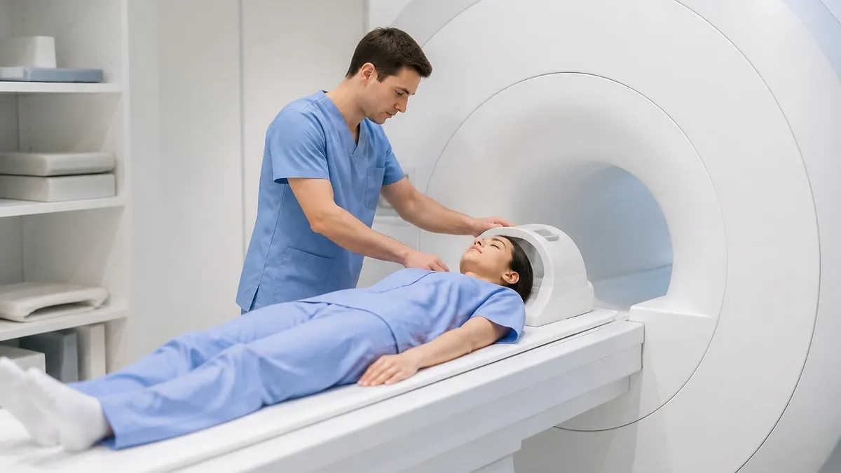
During the examination itself, the patient lies supine on the scanner table with a phased-array surface coil carefully positioned over the pelvic region for optimal signal reception from the prostate gland. The entire multiparametric acquisition protocol typically requires thirty to forty-five minutes of total scan time, during which the patient must remain as motionless as possible to minimize motion-related artifacts that degrade diagnostic quality. Some facilities routinely administer an antispasmodic agent such as glucagon or hyoscine butylbromide to reduce bowel peristalsis motion, though this practice varies by institution.
Claustrophobia affects approximately five to ten percent of patients undergoing MRI examinations and can significantly interfere with completing the full study if not addressed proactively before the scheduled appointment. Wide-bore scanner designs measuring seventy centimeters in diameter, noise-canceling headphones with patient-selected music, and mild oral anxiolytic medications prescribed in advance can collectively help anxious patients tolerate the examination comfortably throughout its duration. Continuous communication with the technologist through the built-in intercom system provides additional reassurance during scanning.
Reviewing your mpMRI results with a urologist who thoroughly understands the nuances of PI-RADS scoring and MRI-targeted biopsy techniques is essential for translating imaging findings into clinically appropriate action plans. Consider seeking care at a center that performs a high annual volume of prostate MRI examinations, as institutional experience correlates strongly with both diagnostic accuracy and patient clinical outcomes over time. Academic medical centers and specialized prostate cancer programs performing more than one hundred prostate mpMRI examinations annually consistently demonstrate superior reader performance and lower missed cancer rates.
Insurance coverage for multiparametric MRI of the prostate has expanded significantly in recent years as major clinical practice guidelines increasingly endorse its routine use before initial prostate biopsy for appropriate candidates. Most Medicare and commercial insurance plans now provide coverage for prebiopsy mpMRI when appropriately ordered by a urologist for clinically indicated reasons, though specific prior authorization requirements and patient cost-sharing obligations vary substantially by payer and plan type. Patients should verify individual coverage details with their insurance provider before scheduling.
Looking ahead, the integration of artificial intelligence into multiparametric MRI interpretation promises to further improve diagnostic consistency and substantially reduce the inter-reader variability that currently limits standardization across institutions of different expertise levels. AI-powered algorithms can automatically highlight suspicious regions on images, generate quantitative biomarker maps, and provide preliminary PI-RADS assessment scores that serve as an independent computational second opinion for the interpreting radiologist. As these sophisticated tools receive regulatory clearance and accumulate clinical validation data, they will likely become standard components of the multiparametric MRI workflow.
MRI Questions and Answers

MRI Medical Abbreviation: What MRI Stands For and Why It Matters

Knee MRI Images: A Complete Guide to Reading, Understanding, and Interpreting Knee Scans

Noise of MRI Machine: Why MRI Scanners Are So Loud and What to Expect
Is Nickel Titanium MRI Compatible? A Complete Guide to MRI Safety Materials
What to Expect During MRI: Your Complete Patient Guide to the Scanning Process
About the Author
Medical Laboratory Scientist & Clinical Certification Expert
Johns Hopkins UniversityDr. Sandra Kim holds a PhD in Clinical Laboratory Science from Johns Hopkins University and is certified as a Medical Technologist (MT) and Medical Laboratory Scientist (MLS) through ASCP. With 16 years of clinical laboratory experience spanning hematology, microbiology, and molecular diagnostics, she prepares candidates for ASCP board exams, MLT, MLS, and specialist certification tests.
Join the Discussion
Connect with other students preparing for this exam. Share tips, ask questions, and get advice from people who have been there.
View discussion (6 replies)