
Water T2 MRI and MRI Imaging Sequences: The Complete Guide 2026 October
Master water T2 MRI and all major imaging sequences. Learn how T1, T2, FLAIR, and more work — with real clinical examples. ✅ Study guide inside.
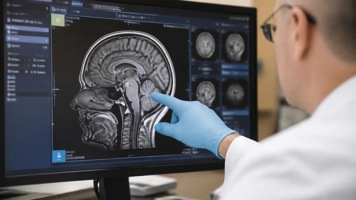
Understanding water T2 MRI is one of the most fundamental skills any MRI technologist, radiologist, or student must develop. T2-weighted imaging exploits the fact that free water molecules — found in cerebrospinal fluid, edema, and most pathological tissues — have extremely long T2 relaxation times, causing them to appear brilliantly bright on T2-weighted images. This property makes T2 one of the most clinically sensitive sequences in the entire MRI toolkit, capable of detecting lesions that would be invisible on other sequences.
MRI imaging sequences are far more than simple on/off switches. Each sequence is a precisely engineered radiofrequency pulse program that manipulates how hydrogen protons relax after being excited by the magnetic field. The radiographer selects sequences based on what tissue contrast is needed: T1 excels at anatomy and post-contrast enhancement, T2 reveals fluid and pathology, FLAIR suppresses CSF to unmask periventricular lesions, and diffusion-weighted imaging captures acute ischemia within minutes of onset. Mastering these sequences is the core competency of MRI practice.
For students preparing for the ARRT MRI board exam or the ARMRIT certification, imaging sequences represent one of the highest-yield topic areas. Questions regularly test your ability to predict signal intensity based on tissue characteristics, choose the correct sequence for a clinical scenario, and explain the physics behind relaxation times. A thorough understanding of mri imaging sequences — from spin echo to gradient echo to inversion recovery — will give you a decisive advantage on exam day.
This guide walks through every major MRI sequence category used in clinical practice. We cover the underlying physics of T1 and T2 relaxation, explain how pulse sequence parameters like TR, TE, and TI are manipulated to produce different tissue contrasts, and discuss the clinical indications for each sequence type. Whether you are scanning a brain for multiple sclerosis plaques or evaluating a knee meniscus for tears, the right sequence selection makes all the difference between a diagnostic and a non-diagnostic exam.
Beyond the physics, we also address practical considerations: how to recognize artifacts specific to each sequence type, how field strength affects signal and contrast, and how modern sequences like PROPELLER, SPACE, and Dixon fat suppression have expanded the diagnostic range of MRI. Clinical examples, signal intensity rules, and memory aids are woven throughout so that abstract concepts translate into real scanner decisions.
The article is structured to mirror how a study guide should build knowledge — starting with relaxation physics, then moving through spin echo, gradient echo, inversion recovery, and advanced sequences, before tackling artifacts, optimization strategies, and board-exam tips. Each section ends with a practical takeaway you can apply immediately, whether at the console or in an exam question. By the end, you will be able to look at any MRI image and confidently identify the sequence type from tissue signal characteristics alone.
Whether you are a first-year MRI student, a veteran technologist cross-training to a new modality, or a radiologist resident reviewing fundamentals, this comprehensive guide provides the depth and clarity needed to master MRI imaging sequences in 2026 and beyond. Read on to build the kind of sequence knowledge that separates good technologists from great ones.
MRI Imaging Sequences by the Numbers
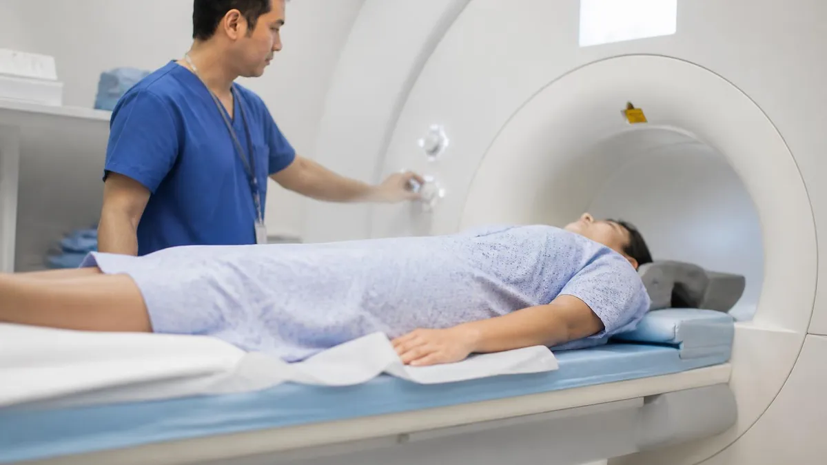
The Four Core MRI Sequence Families
The gold standard for T1 and T2 contrast. SE uses a 90° excitation pulse followed by a 180° refocusing pulse. FSE acquires multiple echoes per TR using an echo train, dramatically reducing scan time while preserving T2 contrast for clinical imaging.
Uses a flip angle less than 90° and a gradient reversal instead of a 180° RF pulse. Faster than spin echo and sensitive to magnetic susceptibility, making it ideal for detecting hemorrhage, calcification, and cartilage. T2* weighting is unique to GRE.
Adds a 180° inverting pulse before the standard spin echo to null signal from specific tissues. STIR nulls fat signal; FLAIR nulls CSF signal. The inversion time (TI) parameter determines which tissue is suppressed based on its T1 relaxation time.
Acquires an entire image from a single RF excitation using rapid gradient switching. Enables diffusion-weighted imaging (DWI) and fMRI with sub-second scan times. Highly susceptible to geometric distortion and N/2 ghosting artifacts at air-tissue interfaces.
The spin echo sequence remains the backbone of clinical MRI, and understanding it thoroughly is non-negotiable for anyone working with magnetic resonance imaging. In a standard spin echo, the scanner first applies a 90-degree radiofrequency pulse that tips the net magnetization vector into the transverse plane. The protons then begin to dephase due to both true T2 relaxation and local magnetic field inhomogeneities. A 180-degree refocusing pulse applied at time TE/2 reverses this dephasing and produces an echo at time TE, effectively canceling the effect of static field inhomogeneities but not true T2 decay.
The power of spin echo lies in how TR and TE control tissue contrast. A short TR (typically 300–600 ms) and short TE (10–20 ms) produces T1-weighted images because tissues with short T1 relaxation times — such as fat and white matter — recover their longitudinal magnetization quickly and appear bright.
A long TR (2,000 ms or more) combined with a long TE (80–120 ms) produces T2-weighted images. Here, tissues with long T2 relaxation times — most notably water T2 MRI contrast in CSF and edematous tissue — appear brightest. Proton density weighting uses a long TR with a short TE, minimizing both T1 and T2 effects to reflect actual hydrogen proton concentration.
Fast Spin Echo (FSE), also called Turbo Spin Echo (TSE) by Siemens, revolutionized clinical MRI by dramatically cutting scan times. Instead of collecting a single echo per TR, FSE collects multiple echoes — the echo train length (ETL) — by applying a series of 180-degree refocusing pulses. Each echo is phase-encoded differently and fills a different k-space line. An ETL of 8 means eight times fewer TR periods are needed, reducing a 10-minute SE T2 brain to under two minutes. The tradeoff is blurring on high-ETL acquisitions and altered contrast due to T2 decay across the echo train.
One subtle but critical difference between standard SE and FSE involves fat signal. In FSE, fat appears brighter on T2-weighted images than it does in conventional SE. This happens because the multiple 180-degree pulses in FSE reduce the J-coupling effect between coupled methylene protons in lipid molecules, artificially prolonging their effective T2 and causing fat to appear brighter. This has important clinical consequences: a FSE T2 of a spine may show epidural fat as very bright, potentially mimicking a fluid collection. Radiologists and technologists must recognize this artifact to avoid misinterpretation.
Half-Fourier Acquisition Single-shot Turbo Spin Echo (HASTE), known as SSFSE on GE platforms, takes FSE to the extreme by collecting the entire image in a single shot. By acquiring only slightly more than half of k-space and using mathematical reconstruction to fill the rest, HASTE produces images in under a second. This makes it invaluable for uncooperative patients, pediatric imaging without sedation, and fetal MRI. Signal-to-noise ratio and resolution suffer compared to multi-shot FSE, but the ability to freeze motion is clinically irreplaceable for abdominal and fetal applications.
3D FSE sequences such as SPACE (Siemens), CUBE (GE), or VISTA (Philips) represent the modern evolution of spin echo imaging. These sequences acquire a volumetric slab using variable flip angle refocusing pulses, which reduces specific absorption rate (SAR) and enables very high echo train lengths. The resulting isotropic voxels — often 0.8 to 1 mm on a side — allow reformatting in any plane without loss of resolution. 3D FSE is now the preferred technique for inner ear imaging, brachial plexus MRI, knee cartilage evaluation, and neurovascular imaging, offering diagnostic detail that 2D acquisitions simply cannot match.
Signal-to-noise ratio (SNR) in spin echo sequences scales with voxel volume, the square root of the number of signal averages, and field strength. At 3T compared to 1.5T, spin echo sequences theoretically double in SNR, allowing faster scan times, thinner slices, or higher resolution for the same scan time.
In practice, 3T spin echo imaging requires careful management of SAR — the energy deposited in tissue — because the 180-degree refocusing pulses are energy-intensive. Parallel imaging techniques like GRAPPA and SENSE are routinely used to reduce SAR, cut scan time, and maintain the SNR advantages of higher field strength in clinical FSE protocols.
Gradient Echo, Inversion Recovery & EPI Sequences Explained
Gradient recalled echo sequences replace the 180-degree refocusing pulse with a gradient reversal, making them significantly faster than spin echo. By using flip angles less than 90 degrees — often 10–40 degrees — GRE sequences allow much shorter TR values without saturating the signal, enabling rapid 2D and 3D acquisitions. T1-weighted GRE (such as FLASH, SPGR, or T1-FFE) is the standard for dynamic contrast-enhanced liver imaging and post-gadolinium brain scans. The absence of the refocusing pulse means GRE images are sensitive to T2* effects rather than true T2, making them exquisitely sensitive to blood products, calcification, and air-tissue interfaces.
Susceptibility-weighted imaging (SWI) is an advanced GRE-based sequence that combines magnitude and phase information to maximize sensitivity to paramagnetic substances. SWI can detect microhemorrhages, venous blood, iron deposition, and calcification with far greater sensitivity than conventional GRE or CT. Balanced SSFP sequences (TrueFISP, FIESTA, bFFE) are a special GRE variant where the gradient moments are fully balanced, producing extremely high SNR with mixed T1/T2 contrast. These sequences are the preferred choice for cardiac cine imaging, inner ear anatomy, and fetal brain surveys where high contrast between fluid and soft tissue is needed in very short scan times.
T2-Weighted vs T1-Weighted Sequences: Clinical Trade-offs
- +T2 sequences detect edema, inflammation, and most pathological tissue with very high sensitivity
- +Water T2 MRI contrast highlights CSF, making it ideal for myelography and cisternography
- +T2 FLAIR suppresses CSF to unmask lesions at brain-fluid interfaces that standard T2 misses
- +T2 requires no gadolinium contrast agent, reducing cost and eliminating nephrogenic risks
- +FSE T2 sequences provide excellent SNR with clinically acceptable scan times of 2–4 minutes
- +T2 contrast is strong to slight patient motion in many body regions due to inherent tissue contrast
- −T2 sequences have poor anatomical landmark definition compared to T1 in many regions
- −Fat appears bright on FSE T2 and can obscure adjacent pathology without fat suppression
- −Long TR required for T2 weighting increases overall scan time per sequence
- −T2 cannot reliably differentiate between tumor, edema, and gliosis — all appear bright
- −EPI-based T2* sequences suffer from distortion at air-tissue and bone-tissue interfaces
- −FLAIR T2 signal can be artifactually improved near CSF pulsation and oxygen administration
MRI Sequence Selection Checklist for Clinical Practice
- ✓Confirm the clinical question before choosing sequences — anatomy vs. pathology detection vs. characterization.
- ✓Select T1-weighted SE or GRE for post-gadolinium imaging and anatomical landmark definition.
- ✓Use water T2 MRI (FSE T2) as the primary pathology-sensitive sequence in brain, spine, and MSK exams.
- ✓Add FLAIR to every brain protocol to detect periventricular and cortical lesions near CSF.
- ✓Include DWI in all brain exams and any exam where abscess, stroke, or high-grade tumor is suspected.
- ✓Apply STIR instead of chemical-shift fat saturation when imaging near air-tissue interfaces (chest wall, neck).
- ✓Choose GRE or SWI sequences when hemorrhage, calcification, or iron deposition is on the differential.
- ✓Use 3D isotropic acquisitions (SPACE, CUBE, MP-RAGE) when multiplanar reformats are clinically needed.
- ✓Verify TR and TE settings match the intended contrast weighting before beginning any scan.
- ✓Document sequence parameters, field strength, and coil selection in every scan protocol sheet.
The Water T2 Rule: Bright Means Pathology Until Proven Otherwise
On any T2-weighted MRI image, free water appears bright because of its extremely long T2 relaxation time. This single principle — that water is bright on T2 — is the foundation of MRI pathology detection. Virtually every clinically significant process (tumor, edema, infarction, demyelination, infection) involves increased tissue water content, making T2 the most sensitive single sequence for detecting disease. When a lesion is bright on T2, your next step is to characterize it with T1, FLAIR, DWI, and contrast enhancement patterns.
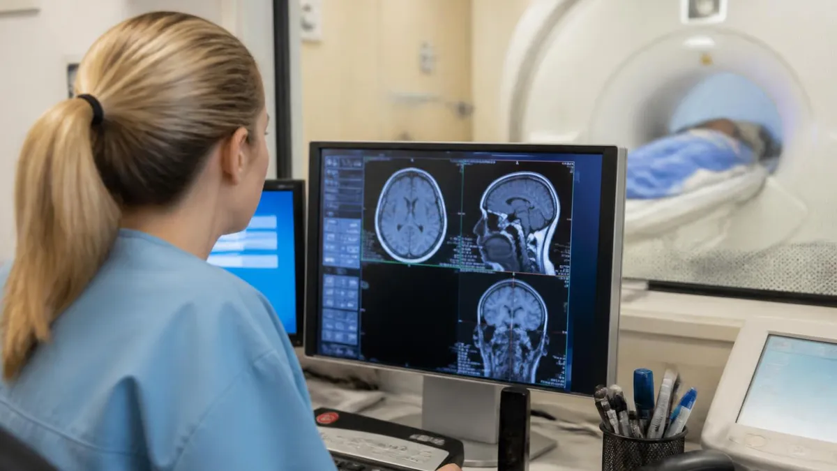
Clinical MRI protocol design is a specialty unto itself, and understanding how imaging sequences are applied across different body regions is what separates a technically competent technologist from a clinically valuable one. In neuroimaging, the standard brain protocol at most institutions includes a sagittal T1 (often MP-RAGE or FLAIR T1), axial FLAIR, axial T2, axial DWI with ADC mapping, and axial T2* or SWI for hemorrhage detection. Post-gadolinium axial and sagittal T1 sequences are added for tumor, infection, or inflammatory protocols. Each sequence answers a different diagnostic question, and omitting any one of them can result in a missed diagnosis.
Spine MRI protocols are built around two fundamental views: sagittal and axial planes, each with T1 and T2 weighting. The sagittal T2 is arguably the most important single sequence in spine MRI — it surveys the entire length of the cord and thecal sac, reveals disc herniations as dark intrusions into bright CSF, identifies cord signal abnormalities as bright intramedullary lesions, and shows vertebral body marrow edema as bright T2 signal replacing the normally dark fatty marrow on T1.
The axial T2 then provides cross-sectional detail of foraminal narrowing, facet joint disease, and lateral disc pathology at individual levels identified on the sagittal survey.
Musculoskeletal MRI relies heavily on fat suppression in combination with T2 weighting to maximize the conspicuity of bone marrow edema, joint effusions, tendon tears, and ligament injuries. STIR sequences provide uniform fat suppression across large anatomical areas like the pelvis or entire femur and are preferred when evaluating for occult fractures or marrow infiltrative disease. Proton density fat-suppressed sequences with intermediate TE values (30–50 ms) are the workhorses for knee meniscus and cartilage evaluation, providing the signal-to-noise ratio and contrast resolution needed to characterize internal derangement of the joint with the precision required for surgical planning.
Abdominal and pelvic MRI introduces the challenges of respiratory motion, peristalsis, and vascular pulsation, each of which demands specific sequence strategies. Breath-hold T1 GRE sequences (VIBE, LAVA, THRIVE) with and without fat suppression are the standard for liver lesion characterization during dynamic contrast phases.
In-phase and opposed-phase T1 GRE sequences exploit the chemical shift between water and fat protons to detect intracellular lipid in hepatocellular adenoma and adrenal adenoma — one of the few truly pathognomonic MRI findings. T2 sequences with respiratory triggering or navigator echo correction provide the motion-free images needed for biliary and pancreatic duct evaluation in MRCP (magnetic resonance cholangiopancreatography).
Cardiac MRI is perhaps the most sequence-demanding subspecialty in all of radiology. Cine balanced SSFP sequences provide high-contrast, high-temporal-resolution movies of cardiac function, enabling precise measurement of ejection fraction, wall motion abnormalities, and valve morphology. T1 mapping with modified Look-Locker inversion recovery (MOLLI) quantifies myocardial T1 values to detect diffuse fibrosis, edema, and amyloid infiltration. Late gadolinium enhancement (LGE) using phase-sensitive inversion recovery (PSIR) sequences images the distribution of gadolinium in infarcted or fibrotic myocardium, where delayed washout makes scar tissue bright against nulled normal myocardium — the gold standard for myocardial viability assessment.
Breast MRI protocols combine high spatial resolution 3D T1 GRE with fat suppression for dynamic contrast-enhanced (DCE) imaging with T2 sequences for morphological characterization of background parenchymal enhancement and cyst assessment. The kinetic curve analysis — initial rapid enhancement followed by washout in malignant lesions versus persistent enhancement in benign lesions — depends on precise timing of the contrast injection relative to the acquisition of the contrast phase.
DWI is increasingly added to breast protocols as a non-contrast biomarker of tumor cellularity, with malignant lesions showing restricted diffusion and low ADC values that complement the morphological and kinetic information from DCE imaging.
Prostate MRI has undergone standardization through the PI-RADS (Prostate Imaging Reporting and Data System) framework, which assigns scoring criteria to T2-weighted, DWI, and DCE sequences. T2 is the dominant sequence for the peripheral zone and transition zone, depicting the normal zonal anatomy and identifying suspicious low-T2 signal lesions.
DWI with high b-values (1,400–2,000 s/mm²) provides the most specific non-invasive indicator of clinically significant prostate cancer, with high-grade tumors showing markedly restricted diffusion. Multiparametric prostate MRI (mpMRI) combining T2, DWI, and DCE has transformed prostate cancer diagnosis, enabling targeted fusion biopsy of PI-RADS 4 and 5 lesions with far higher detection rates than systematic random biopsy.
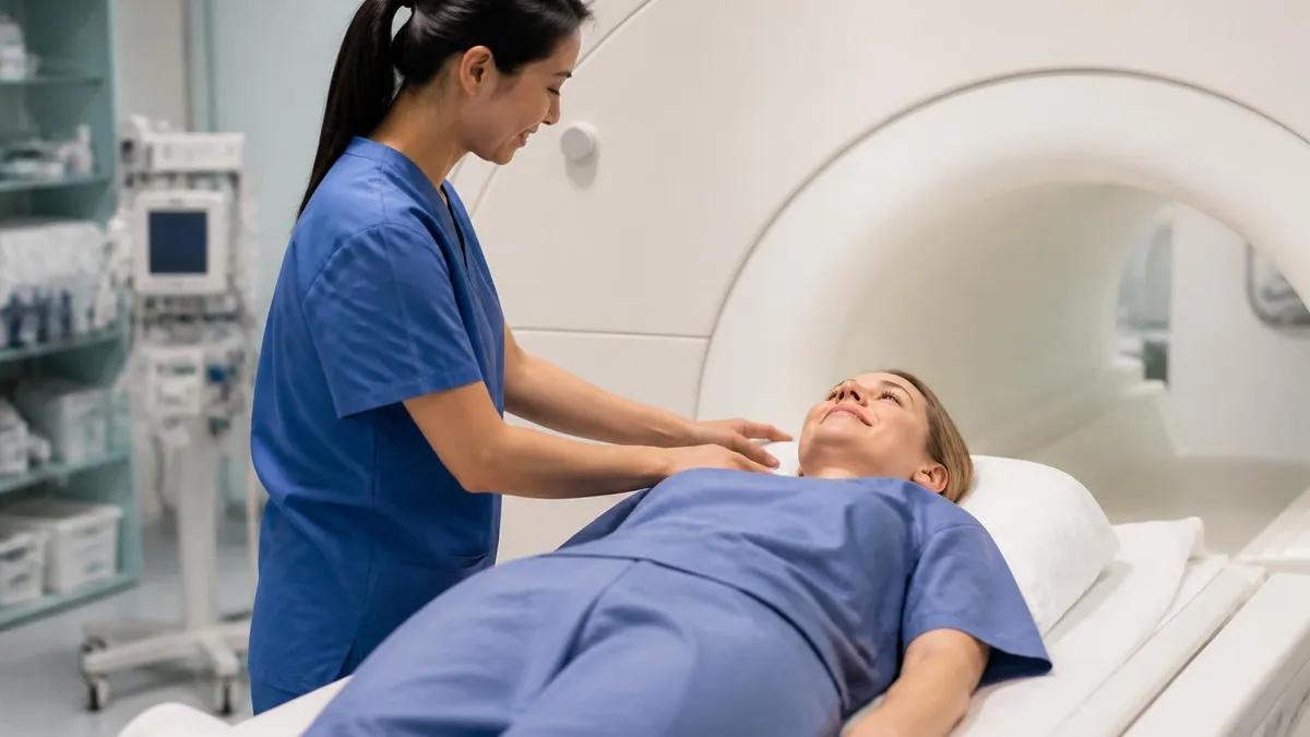
Patients receiving supplemental oxygen via nasal cannula or mask during an MRI exam may show artifactual T2/FLAIR signal hyperintensity in the CSF and subarachnoid spaces, mimicking leptomeningeal disease or subarachnoid hemorrhage. This occurs because dissolved oxygen shortens T2 relaxation time of CSF, reducing CSF nulling by FLAIR. Always document oxygen delivery in the scan report and consider scanning room air when leptomeningeal pathology is a diagnostic concern. This artifact is frequently tested on board examinations.
MRI artifacts are unavoidable in clinical practice, and the ability to recognize, explain, and correct them is a core competency tested on every MRI board examination. Motion artifact is the most common quality issue encountered at the scanner, manifesting as ghosting in the phase-encoding direction on spin echo sequences and blurring on gradient echo sequences.
The ghosts arise because periodic motion — breathing, cardiac pulsation, CSF flow — creates consistent phase errors during k-space filling. Phase oversampling, saturation bands placed over the chest or abdomen, respiratory triggering, and cardiac gating are the primary tools for motion artifact reduction, each suited to different clinical scenarios and anatomical regions.
Chemical shift artifact occurs at fat-water interfaces because fat and water protons precess at slightly different frequencies — approximately 220 Hz apart at 1.5T and 440 Hz at 3T. This frequency difference causes fat to be misregistered in the frequency-encoding direction, producing a bright or dark band at fat-water interfaces such as the renal cortex-perirenal fat boundary, the optic nerve sheath, and intervertebral disc margins.
Increasing bandwidth reduces chemical shift artifact at the cost of SNR, while fat suppression sequences eliminate it entirely. The chemical shift misregistration direction and magnitude are essential to know for any image quality question on the ARRT MRI board exam.
Magnetic susceptibility artifacts arise from local distortions of the magnetic field near materials with different magnetic susceptibility than tissue — including metallic implants, surgical clips, dental amalgam, gas-filled bowel, and even deoxygenated blood. In spin echo sequences, the 180-degree refocusing pulse partially compensates for susceptibility-induced dephasing, making SE sequences more strong near metal than GRE. EPI sequences are most severely affected, because the long echo train allows susceptibility-induced phase errors to accumulate, producing geometric distortion and signal pile-up or drop-out. Increasing bandwidth, using shorter echo spacings, and applying distortion correction algorithms are standard mitigation strategies in clinical EPI protocols.
Truncation artifact (Gibbs ringing) appears as alternating bright and dark bands parallel to high-contrast interfaces — most notably at the cord-CSF boundary in cervical spine MRI — and is caused by representing a sharp signal discontinuity with a limited number of Fourier terms. The cure is to increase the matrix size in the affected direction, which acquires more k-space data and reduces the Gibbs ringing bandwidth.
Alternatively, spatial filtering at the reconstruction stage smooths the artifact at the cost of slight image blurring. Understanding that Gibbs ringing is a k-space truncation phenomenon, not a pathological cord signal change, prevents false positive diagnosis of syringomyelia or myelopathy on cervical spine exams.
Parallel imaging techniques — GRAPPA, SENSE, and their vendor-specific implementations — accelerate MRI acquisitions by undersampling k-space and using the spatially distinct sensitivity profiles of phased-array coil elements to reconstruct missing data. The acceleration factor R determines how much k-space is skipped: R=2 halves scan time but introduces a √R noise penalty and characteristic central artifact.
When parallel imaging is pushed beyond the coil's geometry factor (g-factor) limit, the reconstruction fails and a distinctive noise amplification artifact appears in predictable image regions. For the board exam, understand that GRAPPA reconstruction artifacts appear as structured noise in the image center, while SENSE artifacts appear as fold-over from the edges of the reduced field of view.
Fat suppression failures are a practical daily challenge at the scanner. Spectral fat saturation (ChemSat, FATSAT) applies a frequency-selective RF pulse to saturate fat proton signal before the imaging sequence. This technique fails when field homogeneity is poor — at air-tissue interfaces, in obese patients, and at field strengths below 1T — because the fat and water spectral peaks overlap or the saturation pulse is off-resonance.
STIR is the preferred fat suppression alternative in these cases because it relies on T1-based nulling rather than frequency selectivity, making it field homogeneity independent. Dixon water-fat separation methods, which acquire in-phase and opposed-phase images and mathematically separate the contributions, provide the most strong fat suppression but require careful sequence timing to acquire echoes at the correct phase angles.
Board examinees should also be familiar with zipper artifacts, which appear as discrete lines across the image in the frequency-encoding direction and are caused by radiofrequency interference from outside sources penetrating the Faraday cage. Checking that all penetration panel filters are intact, doors are fully closed, and no contraband electronic devices are present in the scan room resolves most zipper artifacts.
Cross-excitation artifact occurs when closely spaced slices are excited simultaneously due to imperfect RF slice profiles, causing signal loss and image degradation; it is corrected by increasing the slice gap or using interleaved slice acquisition schemes that prevent adjacent slices from being excited in sequence.
Preparing for the ARRT MRI board examination requires a systematic approach to imaging sequences that goes beyond memorizing signal intensities. The exam tests your ability to apply physics principles to clinical scenarios, interpret image findings, select appropriate protocols, and troubleshoot artifacts. High-yield topics consistently appearing on MRI board exams include the relationship between TR, TE, and tissue contrast; inversion recovery pulse sequence design; the physics of k-space filling and spatial frequency; parallel imaging fundamentals; and the signal behavior of gadolinium-based contrast agents at different vascular phases.
A reliable study strategy is to master signal intensity tables first. Create a grid with tissue types — fat, water, cortical bone, acute blood, subacute blood, gadolinium-enhanced tissue, flowing blood — across the columns, and sequence types — T1 SE, T2 FSE, FLAIR, STIR, GRE in-phase, GRE out-of-phase, SWI, DWI high b-value, ADC map — across the rows. Filling in each cell with bright, dark, or intermediate forces you to reason from first principles rather than passively reading. This grid becomes your most powerful board exam reference document, especially for the blood product evolution questions that appear repeatedly.
Time management on the ARRT MRI exam is critical. The examination contains 200 questions to be completed in three hours, giving you approximately 54 seconds per question. Questions involving sequence physics or artifact identification tend to take longer than factual recall questions, so pace yourself by spending no more than 90 seconds on any single question before making your best choice and marking it for review. Skipping genuinely uncertain questions and returning at the end is a proven strategy for maximizing your score, as the exam does not penalize wrong answers.
Practice questions should be integrated from the first week of study, not reserved for the final weeks. Working through test questions immediately after studying each sequence type cements the material through active retrieval, the most powerful encoding mechanism known to cognitive science.
Focus on understanding why wrong answers are wrong, not just why correct answers are correct — this dual analysis dramatically improves your ability to handle novel question phrasings on the actual exam. Multiple free and paid practice question banks exist for MRI registry preparation, and using at least two different sources exposes you to a broader range of question styles and topic emphases.
Understanding MRI safety is interwoven with sequence knowledge on the board exam. Specific absorption rate (SAR) is directly relevant to pulse sequence selection: spin echo sequences with multiple 180-degree pulses at high field strength generate substantially more SAR than GRE sequences with low flip angles.
The FDA limits SAR to 4 W/kg averaged over the whole body and 8 W/kg per gram of tissue for the head. When scanning patients with implanted devices, selecting sequences with lower SAR — longer TR, lower flip angle, reduced echo train length — may be required to keep RF energy deposition within safe limits while still obtaining diagnostic images.
Contrast agent physics is another high-yield topic. Gadolinium-based contrast agents (GBCAs) shorten T1 relaxation time of adjacent water protons through an inner-sphere relaxation mechanism, causing T1 shortening and signal enhancement on T1-weighted sequences. The degree of T1 shortening depends on the local gadolinium concentration, the relaxivity of the specific agent, and the field strength.
At very high concentrations — such as in the urinary collecting system or a renal cyst — gadolinium can paradoxically decrease signal on T1 by also shortening T2 and T2*, a phenomenon called T2 shortening or T2 blooming that can be misinterpreted as a non-enhancing lesion. Recognizing this dose-dependent behavior is a classic board exam trap question.
Finally, the most effective long-term preparation strategy is hands-on time at the MRI scanner. Reading about k-space is valuable; watching k-space fill in real time on a scanner console is significant. If you are a practicing technologist, deliberately change one parameter per scan (TR, TE, flip angle, ETL) and observe the resulting contrast change before restoring the clinical protocol.
If you are a student, ask your clinical site supervisors to walk you through protocol modifications on a phantom. This experiential learning creates durable conceptual understanding that no amount of passive reading can fully replicate, and it is precisely the depth of understanding that separates candidates who score in the top percentiles from those who barely pass.
Sample MRI - Magnetic Resonance Imaging Practice Questions
Try these questions from our free MRI - Magnetic Resonance Imaging practice tests. The correct answer and an explanation follow each question.
The difference between the hydrogen's periodic frequency in fat and water is known as
- A. SPIN DENSITY
- B. CHEMICAL SHIFT
- C. FOURIER TRANSFORM
- D. FID
Answer: B. CHEMICAL SHIFT
Chemical shift refers to the slight difference in the resonant frequency of protons when they are in different chemical environments, such as those found in fat versus water molecules. This frequency difference arises because electron shielding around protons varies with their molecular bonds, causing them to experience slightly different effective magnetic fields. This phenomenon is exploited in MRI to differentiate tissues but can also lead to artifacts if not properly accounted for.
Inversion recovery sequences null a specific tissue when the readout pulse is applied at time TI equal to:
- A. 0.693 × T1 of the tissue (T1 × ln2)
- B. 2 × T1 of the tissue
- C. T1/2 of the tissue
- D. 0.5 × TE
Answer: A. 0.693 × T1 of the tissue (T1 × ln2)
The longitudinal magnetization of a tissue crosses zero at TI = T1 × ln(2) ≈ 0.693 × T1; applying the readout at this moment nulls that tissue's signal.
SSFP (steady-state free precession) sequences such as TrueFISP/FIESTA produce contrast that is proportional to which tissue property ratio?
- A. T1/T2
- B. T2/T1
- C. TE/T2*
- D. TR/T1
Answer: B. T2/T1
SSFP signal is proportional to T2/T1 ratio; tissues with high T2/T1 (like blood and fluids) appear very bright, making SSFP ideal for cardiac cine and vascular imaging.
Fast spin-echo (FSE/TSE) sequences acquire multiple lines of k-space per TR using which approach?
- A. A single 90° pulse followed by multiple 180° refocusing pulses (echo train)
- B. Multiple 90° pulses with a single 180° refocusing pulse
- C. Rapidly toggling the frequency-encoding gradient
- D. Using a series of partial flip-angle pulses
Answer: A. A single 90° pulse followed by multiple 180° refocusing pulses (echo train)
FSE uses an echo train (RARE principle): after a 90° excitation, a series of 180° pulses generates multiple spin echoes, each phase-encoded differently, filling multiple k-space lines per TR.
Take the full MRI - Magnetic Resonance Imaging practice test
MRI Questions and Answers
DWI MRI: Diffusion-Weighted Imaging Explained (2026 Guide)
Side Effects of Brain MRI: Understanding Risks, Safety, and What Every Patient Should Know
MRI Lumbar Spine Without Contrast: Complete Guide to the Scan, What It Shows, and How to Prepare
MRI Technologist Jobs: Salary, Demand, and Career Path Guide
MRI History: From Nuclear Resonance to Modern Scanners
About the Author

Medical Laboratory Scientist & Clinical Certification Expert
Johns Hopkins UniversityDr. Sandra Kim holds a PhD in Clinical Laboratory Science from Johns Hopkins University and is certified as a Medical Technologist (MT) and Medical Laboratory Scientist (MLS) through ASCP. With 16 years of clinical laboratory experience spanning hematology, microbiology, and molecular diagnostics, she prepares candidates for ASCP board exams, MLT, MLS, and specialist certification tests.
Join the Discussion
Connect with other students preparing for this exam. Share tips, ask questions, and get advice from people who have been there.
View discussion (8 replies)