Prostate MRI Procedure: What to Expect Before, During, and After 2026 June
Complete guide to the prostate MRI procedure — prep, what happens in the scanner, how to read results, and what radiologists look for.
The prostate mri procedure has become one of the most important diagnostic tools in urology, allowing radiologists to visualize prostate tissue in extraordinary detail without radiation. Unlike a traditional biopsy taken blindly, a prostate MRI creates high-resolution, multi-parametric images that reveal tumor location, size, and aggressiveness — giving physicians a roadmap before any tissue is sampled. Understanding exactly what happens during this exam reduces anxiety and helps patients prepare correctly, which directly affects image quality and diagnostic accuracy.
Prostate MRI is performed using a technique called multiparametric MRI, or mpMRI, which combines at least three distinct imaging sequences: T2-weighted imaging for anatomical detail, diffusion-weighted imaging (DWI) to assess how freely water molecules move through tissue, and dynamic contrast-enhanced (DCE) imaging to track blood flow patterns. Cancerous tissue tends to restrict water diffusion and show abnormal enhancement — two characteristics the scanner can detect long before a patient feels any symptoms. The combination of these sequences gives clinicians a composite picture far more informative than any single scan.
Prostate cancer is the second most common cancer diagnosed in American men, with roughly 300,000 new cases expected in the United States in 2026. The shift toward MRI-guided or MRI-fusion biopsy has dramatically improved detection rates for clinically significant cancers while reducing the number of unnecessary biopsies for low-risk disease. Major urology guidelines from the American Urological Association now recommend mpMRI before repeat biopsy in men with a prior negative result and a persistently elevated PSA, recognizing MRI's ability to identify anterior and apical lesions that transrectal ultrasound often misses.
Despite its growing role, many patients arrive at their scan appointment with little information about what the procedure involves. Some are surprised by the length — a full multiparametric study typically runs 45 to 75 minutes. Others are unaware that certain prostate MRI protocols require an endorectal coil, a small device inserted into the rectum to improve signal reception near the gland. Knowing whether your facility uses an endorectal coil versus a surface phased-array coil exclusively can help you prepare mentally and physically for the experience.
Preparation instructions vary by institution but generally include a bowel preparation the night before, a period of abstinence from ejaculation to reduce intraluminal fluid in the seminal vesicles, and in many centers a two-day high-fiber diet to reduce gas artifacts. Following these instructions precisely is not optional — motion artifacts from bowel peristalsis or gas distension can obscure critical structures and force a repeat study. Your radiologist or ordering urologist should provide a written preparation sheet, and if one is not offered, you should ask for it explicitly.
Radiologists report prostate MRI findings using a standardized scoring system called PI-RADS, which stands for Prostate Imaging Reporting and Data System. Scores range from 1 (very low probability of clinically significant cancer) to 5 (very high probability). A PI-RADS score of 4 or 5 typically triggers a biopsy recommendation, often targeted using MRI-ultrasound fusion technology to guide the needle precisely into the suspicious zone. Understanding this scoring system before your appointment allows you to engage more productively with your urologist when discussing next steps.
This guide walks through every stage of the prostate MRI procedure — from the first phone call to schedule the appointment through receiving your final radiology report. Whether you are facing this scan for the first time or preparing to repeat one after active surveillance, each section below provides the practical, evidence-based information you need to navigate the process confidently and make the most of what modern MRI technology offers.
Prostate MRI by the Numbers
Preparing for Your Prostate MRI: Step-by-Step
5–7 Days Before: Review Medications
2 Days Before: Dietary Adjustment
24–48 Hours Before: Ejaculatory Abstinence
Evening Before: Bowel Preparation
Morning of Scan: Hydration and Arrival
At the Scanner: Safety Screening
Understanding the imaging sequences used in a prostate MRI helps patients interpret their reports and have more meaningful conversations with their care team. The multiparametric protocol is the gold standard recommended by the American College of Radiology and the European Society of Urogenital Radiology, and it is composed of three functional layers that each interrogate tissue biology from a different angle. Together they create a composite picture that far exceeds the information available from any single sequence or from transrectal ultrasound alone.
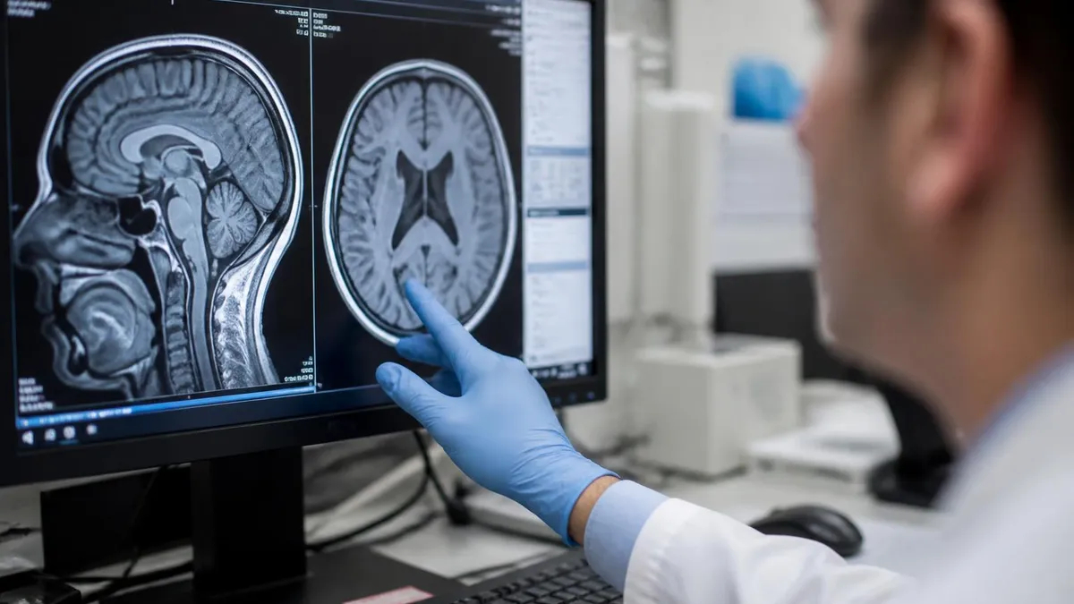
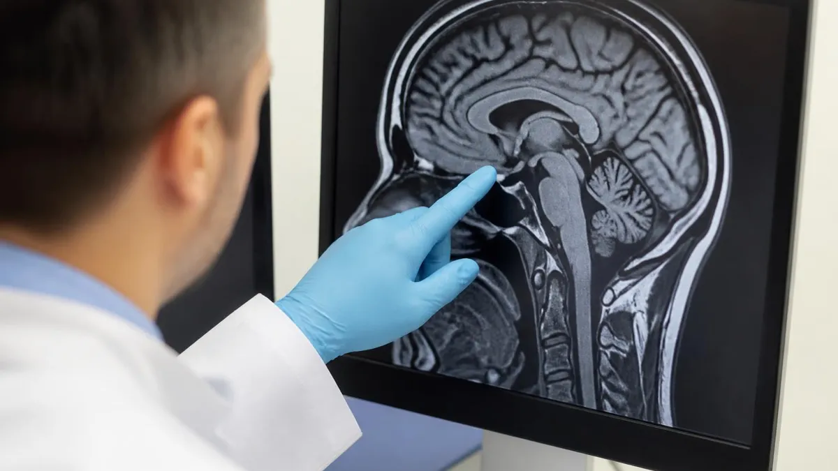
T2-weighted imaging forms the anatomical foundation of every prostate MRI. In healthy peripheral zone tissue, T2 signal is characteristically bright due to high water content within the glandular architecture. Prostate cancer disrupts this architecture, replacing normal glands with dense stromal cells that produce a dark, low-signal region on T2. Radiologists use T2 images to map the prostate's zonal anatomy — distinguishing the peripheral zone from the transition zone and central gland — and to assess whether any suspicious lesion extends beyond the prostatic capsule into periprostatic fat or the seminal vesicles.
Diffusion-weighted imaging measures how rapidly water molecules move through tissue at the cellular level. Cancer cells pack closely together, reducing extracellular space and restricting water diffusion. This restriction appears as high signal intensity on DWI images and as a low apparent diffusion coefficient, or ADC, value on quantitative maps. ADC values below approximately 900 µm²/s in the peripheral zone are strongly associated with Gleason grade 3+4 or higher disease. DWI is particularly powerful because it requires no contrast agent and provides objective, quantifiable data rather than purely visual assessment.
Dynamic contrast-enhanced imaging captures the rapid influx of gadolinium-based contrast agent into prostate tissue after intravenous injection. Cancerous tissue recruits abnormal blood vessels through a process called angiogenesis, and these vessels tend to show early, rapid enhancement followed by early washout — a pattern distinct from benign tissue. DCE adds incremental value primarily in equivocal cases where T2 and DWI findings are borderline (PI-RADS 3). Many radiologists treat DCE as a tiebreaker rather than a primary diagnostic sequence, reflecting its role in the PI-RADS v2.1 scoring algorithm.
Some advanced centers add a fourth sequence called magnetic resonance spectroscopic imaging, or MRSI, which measures metabolite ratios within the prostate. Elevated choline-to-citrate ratios indicate increased cellular membrane turnover, a hallmark of malignancy. While MRSI adds specificity in research settings, it is technically demanding and not yet part of routine clinical protocols at most U.S. imaging centers. Patients should not expect MRSI unless their facility has specifically mentioned it as part of the protocol.
Field strength matters significantly in prostate MRI. A 3 Tesla scanner provides approximately twice the signal-to-noise ratio of a 1.5 Tesla unit for the same acquisition time, which translates to sharper images and better lesion conspicuity. The American Urological Association recommends 3T when available, though high-quality 1.5T exams using an endorectal coil can be diagnostically equivalent in experienced hands. When scheduling your exam, it is worth asking the scheduling coordinator which field strength the facility uses and whether the interpreting radiologist has subspecialty training in genitourinary MRI.
The radiologist's experience interpreting prostate MRI is arguably as important as the scanner itself. Studies consistently show that PI-RADS scoring is subject to significant inter-reader variability among general radiologists but highly consistent among fellowship-trained genitourinary radiologists. If your facility does not have a dedicated GU MRI radiologist on staff, ask your urologist whether a second read from an academic medical center or teleradiology service specializing in prostate MRI would be appropriate, particularly before committing to a targeted biopsy or treatment decision.
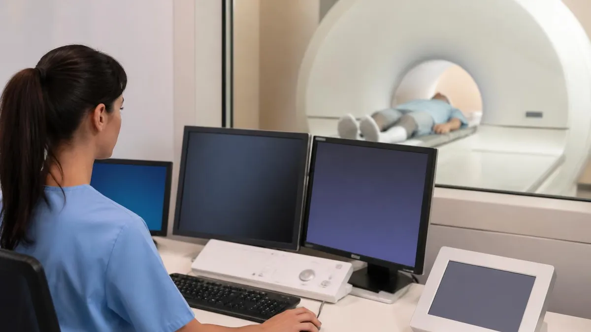
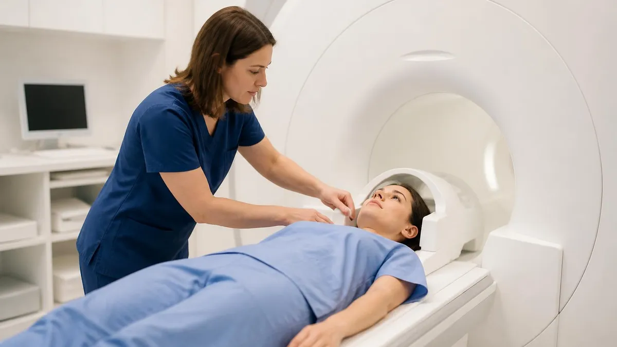
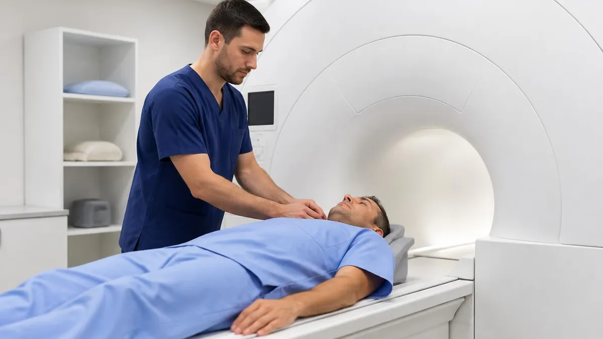
During the Prostate MRI Scan: What You Will Experience
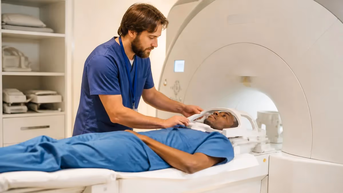
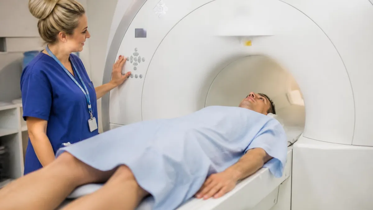
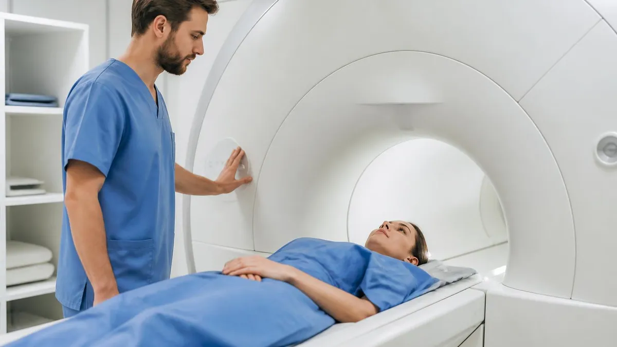
After completing safety screening, you will change into a hospital gown and lie on the MRI table in a supine position with your feet entering the bore first. The technologist will position a surface phased-array coil over your pelvis to maximize signal reception from the prostate region. If your protocol includes an endorectal coil, a lubricated balloon-tipped device approximately the diameter of a finger will be gently inserted into the rectum and inflated with a small amount of air to maintain contact with the prostate wall — most patients describe mild pressure rather than pain.
Once positioned, the table slides into the bore of the magnet, typically placing your pelvis and abdomen within the imaging volume. Foam earplugs or MRI-compatible headphones are provided because the gradient coils produce loud banging and clicking sounds during pulse sequences — noise levels can reach 100 to 110 decibels. Many centers offer music through the headphones to make the 45 to 75 minutes more comfortable. A squeeze bulb emergency call device is always placed in your hand so you can alert the technologist if you feel unwell at any moment.
Prostate MRI: Benefits and Limitations
- +No ionizing radiation — safe for repeat examinations during active surveillance
- +Detects clinically significant cancers missed by transrectal ultrasound-guided biopsy
- +Enables MRI-fusion targeted biopsy with greater diagnostic yield per core
- +Accurately maps tumor location, size, and extracapsular extension for surgical planning
- +Reduces unnecessary biopsies in men with PI-RADS 1–2 findings
- +Standardized PI-RADS v2.1 reporting enables consistent communication across institutions
- −Higher cost than ultrasound — can range from $1,000 to $3,500 without insurance
- −Not widely available in rural or community settings with GU-specialized radiologists
- −Endorectal coil insertion is uncomfortable and not accepted by all patients
- −Motion artifacts from bowel gas can degrade image quality and necessitate repeat scanning
- −PI-RADS scoring has moderate inter-reader variability among non-subspecialized radiologists
- −Gadolinium contrast carries a small but real risk for patients with impaired renal function
Prostate MRI Procedure Day-of Checklist
- ✓Complete bowel preparation (enema or suppository) as directed by your facility the morning of the scan.
- ✓Avoid eating a large meal within 2 hours of your appointment to minimize bowel peristalsis.
- ✓Wear comfortable, metal-free clothing — avoid underwire bras, belts with metal buckles, and jeans with rivets.
- ✓Bring a complete list of all implants, including pacemakers, joint replacements, surgical clips, or dental work.
- ✓Arrive at least 20 minutes early to complete MRI safety screening paperwork before your scheduled slot.
- ✓Inform the technologist of any prior allergic reaction to gadolinium contrast agents before the IV is placed.
- ✓If you take metformin, confirm with your physician whether it should be held after contrast administration.
- ✓Bring your insurance card, photo ID, and any prior prostate MRI images or reports for comparison.
- ✓Request that the technologist explain the squeeze-bulb emergency call device before entering the bore.
- ✓Arrange transportation home if you have been prescribed oral anxiolytic medication for claustrophobia.
PI-RADS 3 Is Not a Green Light — It Is a Gray Zone
A PI-RADS score of 3 indicates equivocal findings where the probability of clinically significant cancer is neither low nor high. Many patients misinterpret this as reassuring, but approximately 20–30% of PI-RADS 3 lesions harbor Gleason grade group 2 or higher disease on targeted biopsy. Your urologist should factor PSA density, biopsy history, and family risk into the decision — never rely on a single PI-RADS number without clinical context.
Receiving a prostate MRI report for the first time can be bewildering. The document is written in technical radiological language and may reference anatomy you have never heard of, such as the neurovascular bundles, the anterior fibromuscular stroma, or the ejaculatory duct complex. Learning to read the key sections of the report empowers you to ask better questions and participate more meaningfully in decisions about whether to proceed to biopsy, active surveillance, or further imaging.
Every structured prostate MRI report should include several core elements: a technical section describing the field strength, sequences performed, and whether an endorectal coil was used; a findings section describing each identified lesion with its location, size, and signal characteristics on each sequence; a PI-RADS score for each lesion; and an impression section that synthesizes the findings into a clinical recommendation. Some reports also include a prostate volume measurement and a calculated PSA density (PSA divided by prostate volume in mL), which helps stratify the significance of borderline findings.
Lesion location is described using a standardized anatomical sector map developed by the PI-RADS steering committee. The prostate is divided into 36 sectors organized by zone (peripheral, transition, central, anterior fibromuscular stroma), gland level (base, mid, apex), and laterality (left, right, midline). Your report will use terms like "left mid peripheral zone" or "right base transition zone" to precisely localize a finding. Understanding these terms helps you correlate the report with the images if you review them with your urologist on a workstation.
Extraprostatic extension, or EPE, is one of the most clinically significant findings a prostate MRI can identify. When tumor cells breach the prostatic capsule, they enter the periprostatic fat where surgical margins become harder to achieve. Radiologists assess EPE by looking for capsular bulge, irregular capsular margin, obliteration of the rectoprostatic angle, and direct visualization of tumor signal outside the gland. Seminal vesicle invasion, a related finding, is classified as T3b disease and is associated with higher rates of biochemical recurrence after treatment.
The report may also comment on pelvic lymph nodes, though conventional MRI has relatively low sensitivity for detecting microscopic nodal metastases. Nodes larger than 8 to 10 mm in short-axis diameter, or those with irregular borders and heterogeneous signal, are flagged as suspicious. For patients with high-risk disease, PSMA PET-CT has largely supplanted conventional cross-sectional imaging for nodal and distant staging, and your urologist may recommend this examination in addition to or instead of a standard prostate MRI in certain clinical scenarios.
When reviewing your report with your urologist, the critical question is not just "what was found" but "what is the clinical significance given my PSA, PSA density, prior biopsy history, and family risk profile?" A PI-RADS 4 lesion measuring 8 mm in a patient with a PSA density of 0.20 ng/mL/mL and a prior negative biopsy is a very different clinical scenario from the same score in a patient with PSA density of 0.06 ng/mL/mL and a prior positive biopsy in a distant location.
Your physician integrates all of these variables — the MRI report alone does not make treatment decisions.
If your MRI report identifies suspicious findings, the next step is usually an MRI-ultrasound fusion targeted biopsy. In this procedure, the radiologist's annotated MRI images are co-registered with real-time transrectal ultrasound during the biopsy procedure, allowing the urologist to navigate a needle directly to the PI-RADS 4 or 5 lesion identified on MRI. Studies consistently show that targeted fusion biopsy detects approximately 30% more clinically significant cancers and 30% fewer insignificant cancers compared to systematic 12-core biopsy alone, making the pre-biopsy MRI a transformative addition to the prostate cancer diagnostic pathway.

Inadequate bowel preparation is the single most common cause of non-diagnostic prostate MRI. Gas and stool in the rectum create susceptibility artifacts that obscure the peripheral zone — exactly where the majority of clinically significant prostate cancers arise. If you are uncertain about any preparation instruction, call the MRI facility the day before your appointment rather than guessing. A repeat scan not only costs time and money but may delay a diagnosis.
After the scan is complete, the technologist will remove the endorectal coil if one was used, and you will be free to dress and return to normal activities almost immediately. Unlike procedures involving sedation or biopsy, there is no recovery period required after an uncomplicated prostate MRI. If intravenous gadolinium contrast was administered, drinking extra fluids over the following several hours helps the kidneys clear the agent efficiently, though this is a general wellness recommendation rather than a clinical requirement for patients with normal renal function.
Your images will be sent to the interpreting radiologist, who typically issues a report within 24 to 72 hours depending on the urgency classification and facility workload. If your MRI was ordered as urgent due to a rapidly rising PSA or a clinical suspicion of high-grade disease, most facilities can prioritize the read. Ask your ordering urologist to specify the clinical urgency on the requisition form rather than assuming a standard turnaround applies to your case.
Once the report is ready, your urologist will contact you to discuss findings and recommend next steps. In many practices this is done via a secure patient portal message followed by a telephone or in-person consultation. Do not attempt to interpret the report in isolation before speaking with your physician — the combination of imaging findings and your broader clinical picture is what drives decisions, and a radiologist's report is a consultation to your ordering physician, not a direct patient communication designed to stand alone.
If a targeted biopsy is recommended based on your PI-RADS findings, the procedure itself is usually scheduled within two to four weeks at most academic centers. The wait can feel agonizing, but this timeline reflects appropriate scheduling of equipment, pathology staffing, and physician availability rather than clinical indifference. During this interval, continuing your normal activities, including moderate exercise and a healthy diet, is entirely appropriate and will not influence biopsy results.
For men already on active surveillance for low-risk prostate cancer, the prostate MRI procedure plays a recurring role in monitoring disease stability. Most active surveillance protocols include mpMRI at enrollment and then every one to three years depending on institutional protocol and any changes in PSA kinetics. A new or enlarging lesion on follow-up MRI, or a PI-RADS upgrade from 2 to 3 or higher, typically triggers a repeat targeted biopsy to reassess Gleason grade and guide continued management decisions.
It is also worth asking about MRI-guided biopsy as an alternative to MRI-ultrasound fusion in some centers. In-bore MRI-guided biopsy is performed entirely within the MRI scanner using MRI-compatible needles, offering the highest possible targeting accuracy since there is no registration error from ultrasound co-registration. This technique is more time-consuming and expensive, and requires a facility with an interventional MRI suite, but may be appropriate for very small lesions in technically challenging locations such as the anterior apex where ultrasound visualization is poor.
Following the full diagnostic pathway — from elevated PSA through prostate MRI to targeted biopsy — represents the current best practice for prostate cancer detection in the United States. This approach, endorsed by the National Comprehensive Cancer Network, the American Urological Association, and the American Cancer Society, has shifted the field away from systematic blind biopsies toward a precision diagnosis model in which treatment decisions are grounded in comprehensive biological and anatomical data rather than statistical probability alone.
Making the most of your prostate MRI procedure requires active preparation, informed participation, and thoughtful follow-through after results arrive. The patients who get the highest diagnostic value from their scans are not passive recipients — they arrive prepared, communicate clearly with the technologist, and engage their urologist with specific, targeted questions about what the report means for their individual risk profile and treatment options. The following practical guidance consolidates the most important actions across each phase of the process.
Before your appointment, verify three things: the field strength of the scanner (3T is preferred), whether a subspecialized genitourinary radiologist will interpret the study, and whether the facility's radiologists use structured PI-RADS v2.1 reporting. These factors correlate strongly with diagnostic accuracy and directly affect the clinical usefulness of the report your urologist receives. If the answer to any of these questions is unsatisfactory, asking for a referral to a center with greater MRI prostate volume is entirely reasonable and routinely done at academic medical centers.
On the day of the scan, your primary job is to follow all preparation instructions precisely and to communicate openly with the technologist about any concerns. If you feel claustrophobic as the table slides into the bore, tell the technologist immediately — most facilities can pause and reposition you, adjust room lighting, or offer a cool cloth for the forehead. Attempting to endure severe anxiety in silence often leads to motion artifacts that compromise the entire study, whereas a brief pause almost never prolongs the total examination by more than a few minutes.
After the scan, track when your report is expected and follow up with your urologist's office if you have not heard within the promised timeframe. Radiology reports occasionally sit in an ordering physician's inbox for days without triggering outreach, particularly in busy practices. Proactive follow-up is not impatient — it is appropriate self-advocacy in a healthcare system where communication gaps happen regularly. Keep a copy of your report and your prior MRI images on a personal flash drive or patient portal download for future reference and second opinions.
If your results are equivocal or you receive a PI-RADS 3 score, consider requesting a multidisciplinary tumor board review before committing to a biopsy or watchful waiting. At major cancer centers, prostate cancer cases are routinely reviewed by urologists, radiation oncologists, medical oncologists, and genitourinary radiologists together. This collective expertise often yields a more nuanced recommendation than a single urologist reviewing the report alone. Tumor board access is not reserved for established cancer patients — a suspicious MRI finding is an appropriate trigger for multidisciplinary consultation.
For technologists, radiology residents, and MRI students studying for certification exams, the prostate MRI protocol represents an ideal case study for applying multiparametric imaging principles. Understanding why each sequence contributes unique information — T2 for anatomy, DWI for cellularity, DCE for vascularity — reinforces the broader principle that multiparametric approaches always outperform single-sequence acquisitions when the goal is tissue characterization rather than simple anatomical mapping. This principle extends to breast, liver, cardiac, and musculoskeletal MRI applications as well.
Regular practice with clinical MRI scenarios, including prostate imaging questions, is the most effective way to consolidate this knowledge for professional examinations. Working through registry-style questions that test both the physical principles behind each sequence and the clinical significance of findings builds the dual competency that board exams and clinical practice both demand. The quiz resources linked throughout this article are designed to help you bridge theoretical knowledge and applied clinical reasoning, preparing you to interpret and discuss prostate MRI findings with confidence in any professional setting.
MRI Questions and Answers
About the Author
Medical Laboratory Scientist & Clinical Certification Expert
Johns Hopkins UniversityDr. Sandra Kim holds a PhD in Clinical Laboratory Science from Johns Hopkins University and is certified as a Medical Technologist (MT) and Medical Laboratory Scientist (MLS) through ASCP. With 16 years of clinical laboratory experience spanning hematology, microbiology, and molecular diagnostics, she prepares candidates for ASCP board exams, MLT, MLS, and specialist certification tests.
Join the Discussion
Connect with other students preparing for this exam. Share tips, ask questions, and get advice from people who have been there.
View discussion (4 replies)