MRI of Prostate Cancer: Understanding Multiparametric Imaging for Detection, Staging, and Treatment Planning
📗 Learn how MRI of prostate cancer aids detection, PI-RADS scoring, biopsy guidance, and staging. Essential guide for MRI technologists and patients.
MRI of prostate cancer has become the gold standard imaging modality for detecting, characterizing, and staging malignancies within the prostate gland. Unlike conventional ultrasound-guided approaches, multiparametric MRI combines multiple contrast mechanisms to visualize suspicious lesions with remarkable clarity and specificity. Radiologists and urologists rely on prostate MRI to guide clinical decisions ranging from initial biopsy targeting to active surveillance monitoring over many years. Understanding how this technology works empowers both imaging professionals and patients to appreciate its critical role in modern oncological care and treatment planning.
Prostate cancer affects approximately one in eight men during their lifetime, making it the most commonly diagnosed non-skin malignancy among American males. The American Cancer Society estimates roughly 299,000 new cases annually in the United States, with early detection significantly improving long-term survival outcomes and quality of life. MRI has fundamentally transformed the diagnostic pathway by enabling clinicians to identify clinically significant cancers that warrant immediate treatment while reducing the overdetection of indolent, low-risk tumors that may never cause symptoms or harm during a patient's remaining lifetime.
The evolution from standard transrectal ultrasound-guided systematic biopsy to MRI-targeted approaches represents one of the most important advances in urological imaging over the past two decades. Multiparametric MRI, commonly abbreviated as mpMRI, combines T2-weighted imaging, diffusion-weighted imaging, and dynamic contrast-enhanced sequences to create a comprehensive picture of prostate tissue architecture and cellular behavior. Each sequence contributes unique information about cellular density, water molecule movement, and vascular perfusion patterns within suspicious regions of the gland.
The Prostate Imaging Reporting and Data System, known as PI-RADS, provides a standardized framework for interpreting prostate MRI examinations consistently across institutions worldwide. Currently in its second major revision as PI-RADS version 2.1, this scoring system assigns each identified lesion a score from one to five based on the likelihood of clinically significant cancer being present. Scores of four and five indicate high probability and typically prompt targeted biopsy, while lower scores may warrant continued monitoring or alternative clinical management strategies.
For MRI technologists, understanding the specific protocols and imaging sequences used in prostate cancer evaluation is essential for producing diagnostic-quality examinations that radiologists can interpret with confidence. Proper patient preparation, coil positioning, and sequence optimization directly impact image quality and diagnostic accuracy in meaningful ways. Field strength selection between 1.5 Tesla and 3 Tesla systems also influences spatial resolution and signal-to-noise characteristics. Technologists who master these technical parameters contribute significantly to the overall success of the prostate cancer detection pathway.
Patients preparing for a prostate MRI examination often have questions about what to expect during the scan, how long the entire procedure takes, and whether the results will definitively determine the presence or absence of cancer in their gland. While prostate MRI provides highly valuable diagnostic information, it functions as one component within a broader clinical evaluation that includes PSA blood testing, digital rectal examination, and potentially tissue biopsy for pathological confirmation. The integration of these multiple data points guides urologists toward optimal treatment recommendations.
This comprehensive guide explores every aspect of MRI in prostate cancer evaluation, from the underlying physics of multiparametric imaging sequences to the clinical interpretation of PI-RADS scores and their direct impact on patient management decisions. Whether you are an MRI technologist seeking to deepen your technical knowledge, a radiology student preparing for certification examinations, or a healthcare professional wanting to understand current best practices, this resource covers the essential concepts you need to master regarding prostate cancer imaging with magnetic resonance technology.
Prostate Cancer MRI by the Numbers
The Prostate MRI Examination Process
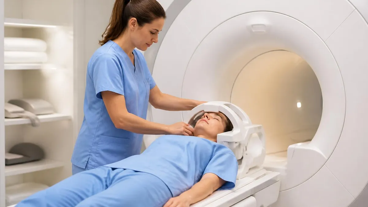
Patient Preparation and Screening
Positioning and Coil Placement
Scout Imaging and Shimming
T2-Weighted Acquisition
Diffusion-Weighted and DCE Imaging
Post-Processing and Interpretation
Multiparametric MRI of the prostate relies on the combined interpretation of several distinct imaging sequences, each designed to highlight different tissue characteristics that help distinguish malignant from benign pathology within the gland. T2-weighted imaging forms the anatomical backbone of the examination, providing excellent soft tissue contrast that allows radiologists to evaluate the zonal anatomy of the prostate gland in fine detail. The peripheral zone, where approximately seventy percent of prostate cancers originate, normally appears as high signal intensity on T2-weighted images, making low-signal suspicious lesions readily identifiable.
Diffusion-weighted imaging has emerged as arguably the most important sequence in the multiparametric protocol for detecting clinically significant prostate cancer with high sensitivity. This technique measures the random Brownian motion of water molecules within tissue, with areas of restricted diffusion appearing as bright signal on high b-value images and corresponding dark signal on apparent diffusion coefficient maps. Malignant prostate tissue exhibits significantly restricted diffusion compared to normal glandular tissue because densely packed cancer cells physically impede water molecule movement within the extracellular space.
Dynamic contrast-enhanced imaging involves the intravenous administration of gadolinium-based contrast agent followed by rapid serial image acquisition to evaluate tissue vascularity and perfusion patterns over time. Prostate cancers typically demonstrate early arterial enhancement with rapid washout, reflecting the abnormal neovascularity that develops as tumors recruit new blood vessel growth through the process of angiogenesis. While DCE imaging plays a supporting rather than dominant role in the PI-RADS scoring system, it provides valuable confirmatory information when T2-weighted and diffusion-weighted findings are equivocal.
Field strength selection significantly impacts the quality of prostate MRI examinations, with both 1.5 Tesla and 3 Tesla systems capable of producing diagnostic images when properly optimized for this specific application. Three Tesla magnets offer inherently higher signal-to-noise ratios, which translate into improved spatial resolution and better lesion conspicuity, particularly for small tumors measuring less than one centimeter in maximum diameter. However, 3T systems also amplify susceptibility artifacts and may produce greater geometric distortion on diffusion-weighted sequences, requiring careful shimming and parameter adjustment by experienced technologists.
The role of endorectal coils in prostate MRI has evolved considerably as external phased-array coil technology has improved in recent years. At 3 Tesla field strength, most centers now achieve diagnostic image quality using surface coils alone, eliminating the patient discomfort associated with endorectal coil insertion and simplifying the overall workflow. However, some institutions still employ endorectal coils at 1.5 Tesla to compensate for the lower intrinsic signal-to-noise ratio, and certain research protocols may utilize them at higher field strengths for maximum spatial resolution.
Patient preparation for prostate MRI typically includes a period of fasting for approximately four hours before the examination and may involve administration of an antispasmodic agent such as glucagon to reduce bowel peristalsis that can degrade image quality through motion artifacts. Patients are also generally advised to avoid ejaculation for several days before the scan, as seminal vesicle distension improves the assessment of potential tumor invasion into these important structures. A small-volume fleet enema may be recommended to reduce rectal gas and stool that create susceptibility artifacts.
Scan duration for a complete multiparametric prostate MRI examination typically ranges from thirty to forty-five minutes, depending on the specific protocol employed and whether additional sequences are acquired for research or specialized clinical investigation. Technologists must ensure consistent patient positioning throughout the examination and actively monitor for motion artifacts that can compromise diagnostic quality. Proper instruction and communication with the patient before and during the scan helps minimize anxiety-related movement and ensures the highest possible image quality for accurate radiological interpretation.
MRI Practice Test Questions
Prepare for the MRI - Magnetic Resonance Imaging exam with our free practice test modules. Each quiz covers key topics to help you pass on your first try.
MRI Knowledge
MRI Exam Questions covering Knowledge. Master MRI Test concepts for certification prep.
MRI Physics
Free MRI Practice Test featuring Physics. Improve your MRI Exam score with mock test prep.
MRI Anatomy and Pathology
MRI Test Prep for MRI Anatomy and Pathology. Practice MRI Quiz questions and boost your score.
MRI Anatomy and Positioning
MRI Questions and Answers on MRI Anatomy and Positioning. Free MRI practice for exam readiness.
MRI Contrast Agents
Free MRI Quiz on MRI Contrast Agents. MRI Exam prep questions with detailed explanations.
MRI Patient Care and Positioning
MRI Practice Questions for MRI Patient Care and Positioning. Build confidence for your MRI certification exam.
PI-RADS Scoring System for Prostate MRI
PI-RADS scores of one and two indicate very low and low likelihood of clinically significant prostate cancer, respectively. Lesions receiving these scores demonstrate imaging characteristics that are overwhelmingly consistent with normal prostate tissue or common benign conditions such as benign prostatic hyperplasia nodules. For peripheral zone lesions, this means no restricted diffusion is observed on DWI sequences, while transition zone lesions show typical benign hyperplastic nodular features on T2-weighted imaging without suspicious diffusion abnormality.
Clinical management for PI-RADS 1 and 2 lesions generally does not require targeted biopsy, and patients may return to routine PSA screening at intervals determined by their overall risk profile and clinical history. However, clinicians must weigh MRI findings alongside other clinical parameters including PSA density, patient age, family history, and prior biopsy results when making management decisions. Some patients with persistently elevated PSA levels despite low PI-RADS scores may still undergo systematic biopsy to rule out sampling error or MRI-occult disease.

Advantages and Limitations of Prostate MRI
- +Superior soft tissue contrast enables detailed visualization of prostate zonal anatomy and lesion characteristics
- +No ionizing radiation exposure, making it safe for repeated surveillance imaging over extended periods
- +Multiparametric assessment combines anatomical, functional, and vascular information in a single examination
- +Guides targeted biopsy to specific lesions, improving cancer detection while reducing unnecessary sampling
- +Supports active surveillance monitoring by tracking lesion stability or progression over time
- +Reduces overdetection and overtreatment of clinically insignificant low-grade prostate cancers
- −Higher cost compared to transrectal ultrasound, with examinations typically ranging from $1,000 to $3,000
- −Limited availability of subspecialty-trained genitourinary radiologists experienced in PI-RADS interpretation
- −Claustrophobia can prevent some patients from completing the full multiparametric examination protocol
- −Susceptibility to motion artifacts from bowel peristalsis and patient movement during lengthy scan times
- −Gadolinium contrast administration carries rare risks including nephrogenic systemic fibrosis in renal impairment
- −Interpretation variability between radiologists can result in inconsistent PI-RADS scoring across institutions
Prostate MRI Quality Assurance Checklist
- ✓Verify patient has completed MRI safety screening and has no contraindicated implants or devices.
- ✓Confirm patient followed preparation instructions including fasting and bowel preparation protocols.
- ✓Center the pelvic phased-array coil precisely over the pubic symphysis before starting the examination.
- ✓Perform manual shimming centered on the prostate to minimize susceptibility artifacts on diffusion-weighted images.
- ✓Acquire T2-weighted images in all three orthogonal planes with adequate spatial resolution.
- ✓Include at least two low b-values and one high b-value of 1400 or greater for diffusion imaging.
- ✓Generate apparent diffusion coefficient maps and verify they correspond accurately to diffusion-weighted acquisitions.
- ✓Administer gadolinium contrast at the correct dose and timing for dynamic contrast-enhanced sequences.
- ✓Review all images for motion artifacts before releasing the patient from the scanner.
- ✓Document prostate gland volume, PI-RADS scores, and lesion locations in a structured radiology report.
MRI-Targeted Biopsy Detects More Significant Cancers
Research consistently demonstrates that MRI-targeted biopsy detects approximately 30% more clinically significant prostate cancers compared to standard systematic biopsy alone, while simultaneously reducing the detection of clinically insignificant cancers by up to 50%. This means fewer men undergo unnecessary treatment for indolent tumors while more men with dangerous cancers receive timely diagnosis and appropriate care.
MRI-guided prostate biopsy represents a significant advancement over traditional systematic biopsy approaches, offering superior detection rates for clinically significant cancers while reducing the unnecessary sampling of benign tissue throughout the gland. Three distinct targeting methods have been developed, each with specific advantages depending on institutional resources, operator expertise, and individual clinical requirements. Cognitive fusion relies on the operator's mental registration of MRI findings to guide ultrasound-directed sampling, while software-based fusion platforms provide real-time image overlay for more precise targeting of identified suspicious lesions.
Software-based MRI-ultrasound fusion biopsy has become the predominant approach at many academic medical centers and specialized urology practices across the United States in recent years. Platforms such as UroNav, Artemis, and BioJet import the diagnostic MRI dataset and use electromagnetic or mechanical tracking systems to register the stored MRI images with live transrectal ultrasound during the biopsy procedure. This technology allows urologists to visualize the exact location of PI-RADS lesions superimposed on the real-time ultrasound image and direct biopsy needles precisely into specific targeted areas.
Direct in-bore MRI-guided biopsy is performed with the patient positioned inside the MRI scanner, using MRI-compatible needle guides and intermittent imaging to confirm needle placement within the target lesion in real time. This approach offers the highest spatial accuracy for targeting small or difficult-to-reach lesions but requires specialized equipment, increased procedure time, and dedicated MRI scanner availability that limits throughput. Most institutions reserve in-bore biopsy for patients with previously negative systematic biopsy results who have persistent clinical suspicion based on elevated PSA levels or high PI-RADS scores.
The transperineal approach to prostate biopsy has gained substantial momentum in recent years, driven by growing concerns about infection risks associated with transrectal needle passage through the rectal wall into the prostate gland. Transperineal biopsy routes the needle through the perineal skin between the scrotum and anus, virtually eliminating the risk of serious post-procedure sepsis that affects approximately two to four percent of transrectal biopsy patients annually. Many major urological centers have now transitioned entirely to transperineal techniques, combining this safer access route with MRI-fusion targeting technology.
Systematic biopsy, which samples twelve or more predetermined locations throughout the prostate gland regardless of MRI findings, continues to play a complementary role alongside targeted sampling in current clinical practice. Current guidelines from the National Comprehensive Cancer Network recommend performing both targeted cores from MRI-identified lesions and systematic cores from standard template locations to maximize cancer detection rates. Studies show that combining targeted and systematic approaches identifies approximately ten to fifteen percent more clinically significant cancers compared to either technique performed independently.
The accuracy of MRI-targeted biopsy depends critically on the quality of the preceding diagnostic MRI examination and the experience of the radiologist interpreting the images and assigning PI-RADS scores to identified lesions. Institutions that perform high volumes of prostate MRI examinations and maintain robust quality assurance programs consistently achieve better cancer detection rates than lower-volume centers with less specialized expertise. Radiologist subspecialization in genitourinary imaging, participation in multidisciplinary tumor boards, and correlation of imaging findings with surgical pathology specimens all contribute to improved diagnostic performance.
Following biopsy, pathological analysis of tissue cores provides the definitive Gleason score or International Society of Urological Pathology grade group that determines cancer aggressiveness and guides subsequent treatment planning decisions. MRI findings and biopsy pathology results are integrated during multidisciplinary case conferences where urologists, radiologists, radiation oncologists, and medical oncologists collaborate to develop individualized treatment recommendations for each patient. This team-based approach ensures that the wealth of information provided by multiparametric MRI is fully leveraged in every aspect of patient management.
Patients with severe renal impairment (eGFR below 30 mL/min) face increased risk of nephrogenic systemic fibrosis from gadolinium-based contrast agents. Always verify renal function before administering contrast for dynamic contrast-enhanced prostate MRI sequences. Some biparametric protocols omit contrast entirely while maintaining adequate diagnostic accuracy for PI-RADS assessment.
Prostate MRI plays an essential role in local staging of confirmed prostate cancer, providing critical information about extraprostatic extension, seminal vesicle invasion, and potential involvement of adjacent anatomical structures including the neurovascular bundles, bladder neck, and external urethral sphincter. Accurate local staging directly influences treatment selection because patients with organ-confined disease may be candidates for nerve-sparing radical prostatectomy, while those with extracapsular extension may require wider surgical margins or consideration of radiation-based treatment alternatives to achieve optimal long-term oncological outcomes.
T2-weighted imaging serves as the primary sequence for evaluating extraprostatic extension, with radiologists looking for disruption of the normally smooth, low-signal prostatic capsule and extension of tumor signal intensity into the periprostatic fat. Signs of extracapsular extension include irregular capsular bulging, obliteration of the rectoprostatic angle, and direct visualization of tumor tissue beyond the confines of the prostate gland itself. Detecting extraprostatic extension preoperatively allows surgeons to modify their surgical approach, potentially sacrificing the ipsilateral neurovascular bundle to achieve negative surgical margins at the transgression site.
Seminal vesicle invasion represents an advanced stage of local disease that significantly impacts prognosis and treatment planning for affected patients. On MRI, normal seminal vesicles demonstrate high T2 signal due to their fluid content, while tumor invasion manifests as focal low T2 signal within the seminal vesicle lumen accompanied by restricted diffusion and enhancement on contrast-enhanced sequences. The presence of seminal vesicle invasion upstages the cancer to at least stage T3b in the TNM classification system and typically indicates a higher risk of lymph node metastasis and biochemical recurrence following treatment.
Active surveillance has become an increasingly accepted management strategy for men diagnosed with low-risk prostate cancer, and MRI plays a central monitoring role in these patients over extended follow-up periods. Serial MRI examinations performed at regular intervals, typically annually or biannually, allow clinicians to monitor for radiographic progression that might indicate upgrading from low-risk to intermediate-risk disease warranting definitive treatment intervention. MRI stability over multiple years provides reassurance that the cancer remains indolent, while new or enlarging PI-RADS lesions prompt confirmatory targeted biopsy to assess for grade progression.
Radiation therapy planning increasingly incorporates MRI data to improve target delineation and treatment precision for prostate cancer patients undergoing external beam radiotherapy or brachytherapy seed implantation. MRI-based contouring of the prostate gland and any identifiable dominant intraprostatic lesions allows radiation oncologists to deliver focal dose escalation to areas of known cancer while maintaining standard doses to the remainder of the gland. This approach, known as focal boost radiotherapy, has shown promising results in clinical trials by improving local tumor control without significantly increasing radiation toxicity.
Whole-body MRI and prostate-specific membrane antigen PET-CT represent complementary imaging modalities used for detecting distant metastatic disease in men with high-risk prostate cancer presentations. While standard multiparametric prostate MRI focuses on local staging within the pelvis, whole-body diffusion-weighted MRI can screen the entire skeleton and lymph node chains for metastatic deposits throughout the body. PSMA PET-CT has demonstrated superior sensitivity for detecting small-volume metastatic disease, particularly in regional pelvic lymph nodes, and is increasingly used alongside prostate MRI to provide comprehensive staging information.
The integration of artificial intelligence and machine learning into prostate MRI interpretation represents an exciting frontier that promises to enhance diagnostic accuracy and reduce variability between radiologists at different institutions. Computer-aided detection algorithms trained on thousands of annotated prostate MRI examinations can identify suspicious regions and generate preliminary PI-RADS assessments that radiologists then review and refine based on their clinical expertise. While these tools currently serve as decision support rather than autonomous diagnostic systems, ongoing research suggests they may eventually help standardize interpretation quality across all institutions.
Preparing patients effectively for prostate MRI examinations requires clear and thorough communication about what the procedure involves and how they can contribute to obtaining optimal image quality throughout the scan. Technologists should explain the importance of remaining perfectly still throughout the entire examination, describe the expected duration of approximately thirty-five to forty-five minutes, and proactively address common concerns about claustrophobia and scanner noise levels. Providing patients with earplugs or noise-canceling headphones, a squeeze ball for emergency communication, and blankets for comfort helps reduce anxiety and minimize motion artifacts.
Positioning the patient correctly within the MRI scanner is fundamental to achieving high-quality prostate images that radiologists can interpret with confidence and accuracy. The patient should be placed supine with arms positioned at their sides or crossed comfortably over their chest, and the pelvic phased-array coil must be centered precisely over the pubic symphysis to ensure optimal signal coverage of the entire prostate gland. Verification scout images should be reviewed carefully to confirm that the prostate is centered within the imaging field of view before proceeding with the full multiparametric acquisition.
Shimming optimization deserves particular attention when performing prostate MRI because the air-filled rectum adjacent to the posterior prostate surface creates significant magnetic susceptibility gradients that can severely distort diffusion-weighted images. Manual shimming adjustments centered on the prostate gland may be necessary to minimize these effects, especially at 3 Tesla field strength where susceptibility artifacts are inherently amplified compared to lower field systems. Some institutions use small-volume rectal gel to displace air and improve field homogeneity, though this practice adds preparation time and may cause minor patient discomfort during the examination.
Selecting appropriate diffusion-weighted imaging parameters is crucial for detecting clinically significant prostate cancer, as the choice of b-values directly affects lesion conspicuity and apparent diffusion coefficient calculation accuracy. Current PI-RADS version 2.1 guidelines recommend acquiring at least two b-values below eight hundred seconds per square millimeter plus a high b-value of at least fourteen hundred seconds per square millimeter. Many institutions acquire a calculated high b-value image using exponential extrapolation from the acquired lower b-value data to maintain adequate signal-to-noise ratio while achieving the desired diffusion contrast for lesion detection.
Quality assurance programs for prostate MRI should include regular phantom testing to verify system performance metrics, ongoing correlation of imaging findings with surgical pathology results, and periodic review of PI-RADS scoring consistency among all interpreting radiologists. Institutions performing prostate MRI should systematically track their positive predictive value for PI-RADS 4 and 5 lesions, cancer detection rates stratified by PI-RADS category, and inter-reader agreement metrics to identify specific opportunities for improvement. These quality metrics help maintain high diagnostic standards and provide objective evidence of institutional imaging performance for accreditation purposes.
Technologists preparing for MRI certification examinations should focus on understanding the physics underlying each sequence in the multiparametric protocol and how specific technical parameters affect diagnostic image quality in clinical practice. Knowledge of T2 contrast mechanisms, diffusion-weighted imaging principles including b-value selection and ADC map generation, and dynamic contrast-enhanced acquisition timing are all testable topics that relate directly to clinical prostate MRI practice. Reviewing published PI-RADS technical standards provides an excellent framework for understanding the minimum acceptable imaging requirements for diagnostic prostate examinations.
Continuing education in prostate MRI remains essential as protocols, technology, and clinical guidelines continue to evolve rapidly in this dynamic subspecialty field. The transition from PI-RADS version 2.0 to version 2.1 introduced important refinements in transition zone lesion assessment criteria and clarified the specific role of dynamic contrast-enhanced imaging in the overall scoring algorithm. Technologists and radiologists who stay current with published literature, attend relevant professional conferences, and participate in case-based learning activities maintain the expertise necessary to deliver state-of-the-art prostate cancer imaging services to patients and referring clinicians.
MRI Questions and Answers

MRI Medical Abbreviation: What MRI Stands For and Why It Matters

Knee MRI Images: A Complete Guide to Reading, Understanding, and Interpreting Knee Scans

Noise of MRI Machine: Why MRI Scanners Are So Loud and What to Expect
Is Nickel Titanium MRI Compatible? A Complete Guide to MRI Safety Materials
What to Expect During MRI: Your Complete Patient Guide to the Scanning Process
About the Author
Medical Laboratory Scientist & Clinical Certification Expert
Johns Hopkins UniversityDr. Sandra Kim holds a PhD in Clinical Laboratory Science from Johns Hopkins University and is certified as a Medical Technologist (MT) and Medical Laboratory Scientist (MLS) through ASCP. With 16 years of clinical laboratory experience spanning hematology, microbiology, and molecular diagnostics, she prepares candidates for ASCP board exams, MLT, MLS, and specialist certification tests.
Join the Discussion
Connect with other students preparing for this exam. Share tips, ask questions, and get advice from people who have been there.
View discussion (6 replies)