
MRI Interpretation 2026 October
MRI interpretation guide: 🏆 how radiologists read scans, sequences (T1, T2, FLAIR, DWI), blood aging, nerve root compression, neurosyphilis. 2026 October.
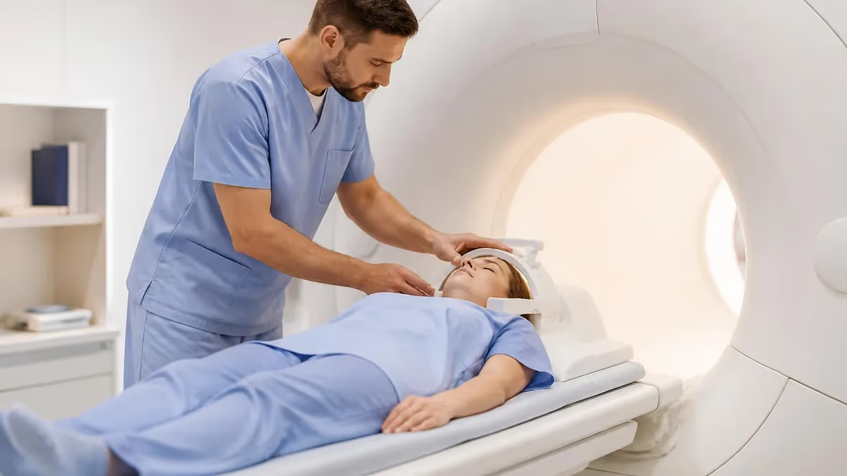
What does MRI interpretation involve?
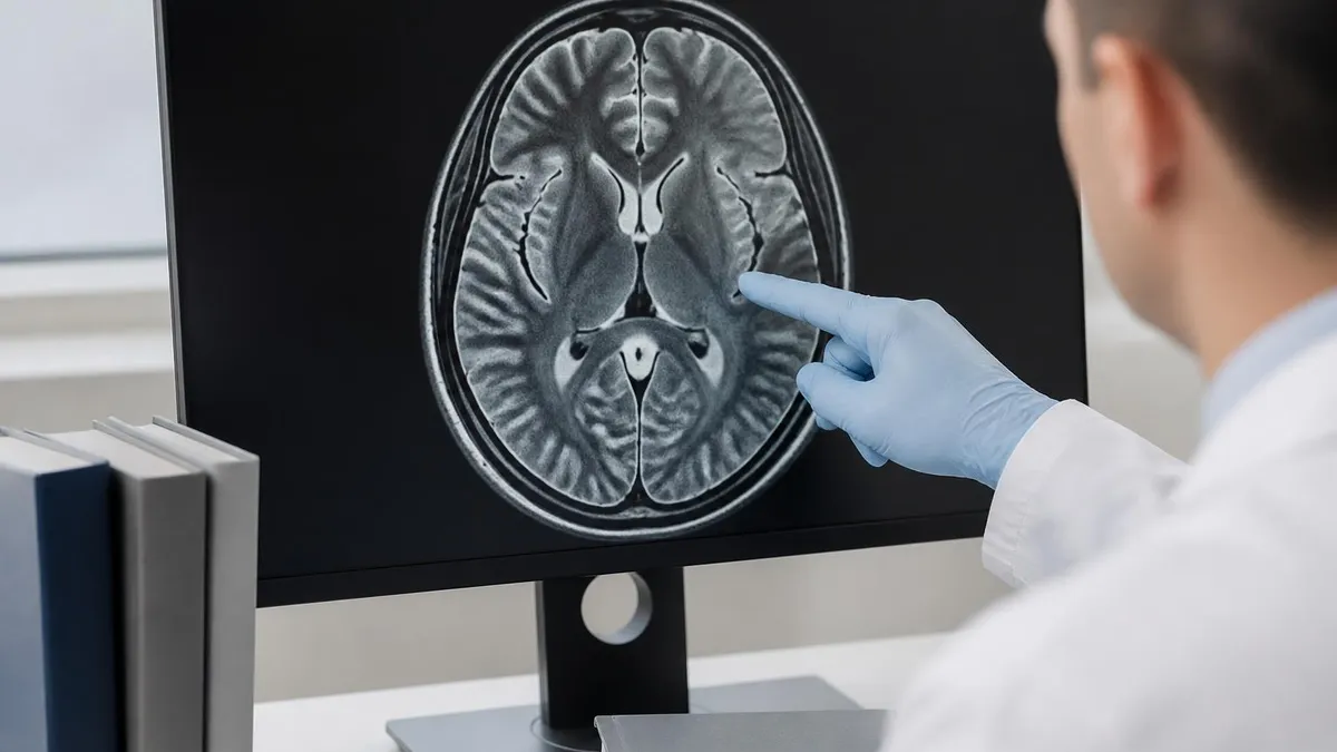
A board-certified radiologist reviews each sequence (T1, T2, FLAIR, DWI, STIR), maps anatomy, identifies pathology, compares with prior studies, and writes an impression. Reports follow ACR templates so referring clinicians see a predictable structure every time.
Who Actually Reads Your MRI
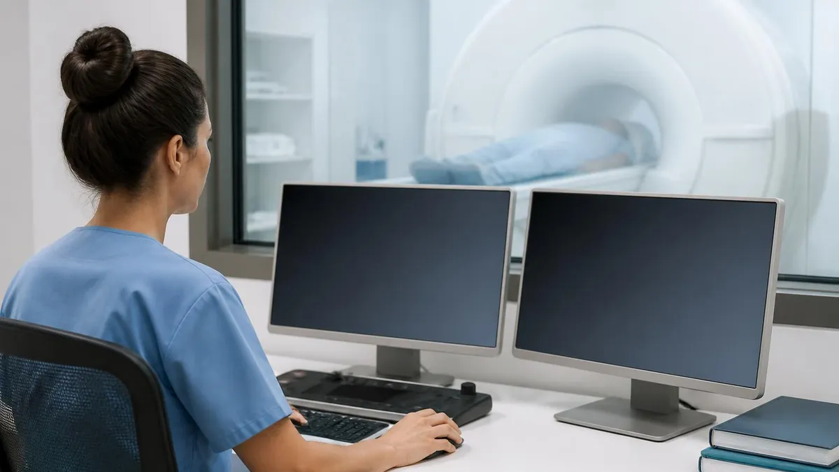
Short answer: a radiologist. Not the tech who scanned you. Not the front-desk staff. A physician who finished medical school, four years of radiology residency, and usually a one-year fellowship in a sub-specialty like neuroradiology, musculoskeletal, or body imaging.
Board certification matters. In the United States that means passing the American Board of Radiology Core Exam and a Certifying Exam after residency. Sub-specialty fellowships add another layer — most academic centers route brain scans to neuroradiologists and joints to MSK readers. That's why your knee MRI report often comes from a different person than your brain MRI report, even at the same hospital.
You'll sometimes see two names on the report. The first is the attending who finalized it. The second might be a resident or fellow who drafted the preliminary read overnight. The attending always signs off — that's the legal author. Worth knowing if you're tracking who to call back with questions.
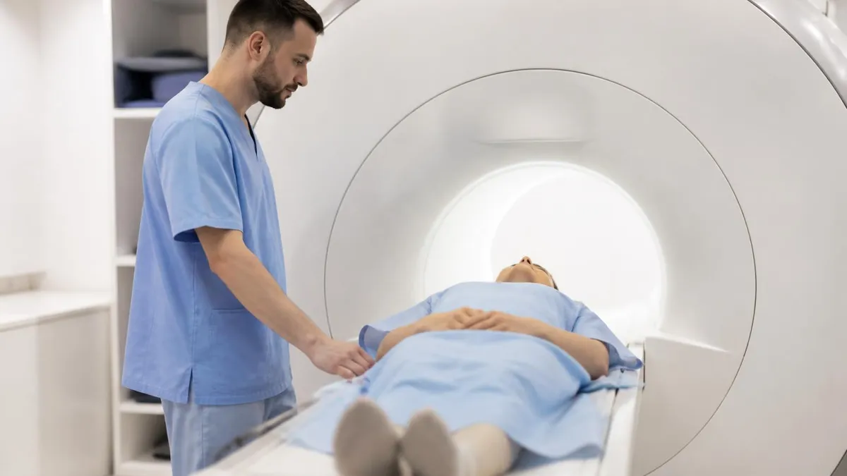
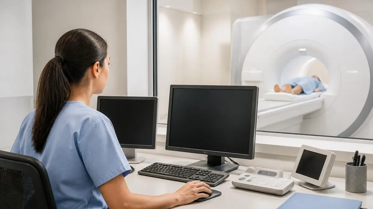
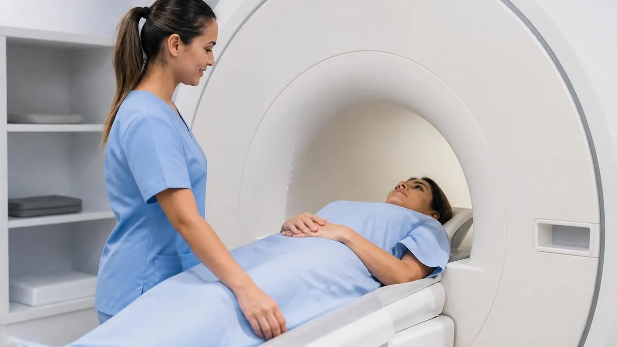
Then there's the MRI technologist. The tech runs the scanner, positions you, and chooses sequences from a protocol the radiologist designed. Techs hold ARRT certification with an MRI sub-specialty. They don't write the report, but they do flag urgent findings to the radiologist on the spot — like a brain bleed picked up on a routine headache scan. If you're heading toward this career, our MRI tech school guide walks through the training path, and the MRI tech salary breakdown shows what techs actually earn by state.
One more role: the referring clinician — your orthopedist, neurologist, or primary care doctor. They read the radiologist's report and translate it for you. Sometimes they spot something the radiologist didn't flag (clinical context matters). Sometimes they miss what the radiologist did flag. That's why getting your own copy of the report — not just the doctor's summary — is worth the effort.
Who Reads MRI Scans: At a Glance
- Training: 4-yr residency + fellowship
- Boards: ABR Core + Certifying Exam
- Role: Reads scan, writes report
- Sub-specialty: Brain, spine, head/neck
- Fellowship: 1-2 years post-residency
- Reads: Brain MRI, spine MRI
- Sub-specialty: Joints, bone, soft tissue
- Fellowship: 1 year
- Reads: Knee, shoulder, hip MRI
- Training: Associate + ARRT-MRI cert
- Role: Runs scanner, picks protocol
- Reports: No — flags urgent findings
The 6-Step Interpretation Framework
Every radiologist follows roughly the same workflow when a study lands in their queue. Different attendings have personal habits, but the bones are universal. Here's the framework most use, in order. Speed varies. A clean brain MRI might take 8 minutes to read. A complex multitrauma whole-body study can eat 45.
1. Identify the Sequences
Before looking for anything pathological, the radiologist confirms what they're looking at. T1, T2, STIR, FLAIR, PD, DWI — each shows tissue differently. Misread a STIR as T2 and you'll misinterpret edema. The first 30 seconds of a read are usually scrolling through each series and labeling them mentally.
2. Check for Contrast
With or without gadolinium? Gadolinium-enhanced sequences highlight blood-brain barrier breakdown, active inflammation, and most tumors. A scan without contrast is a different study with different limitations. The protocol header tells the reader. Our magnetic resonance imaging overview goes deeper on contrast agents and when they're needed.
3. Survey the Anatomy
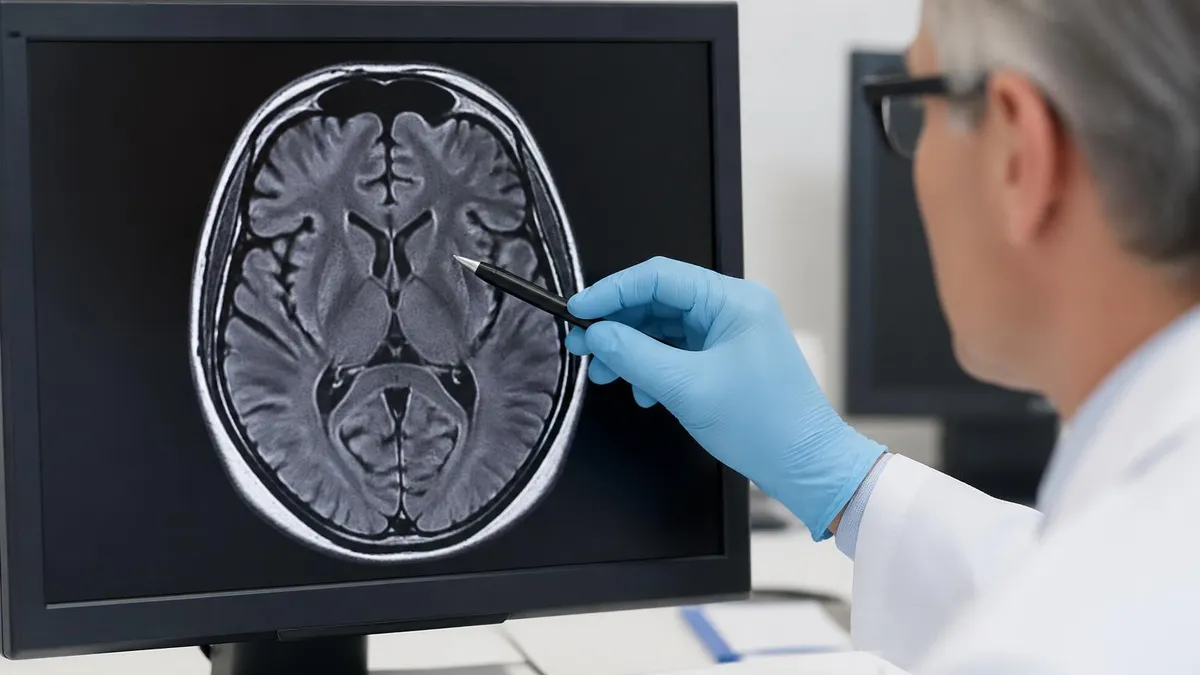
Quick scroll through every slice. Look for the obvious — is the brain symmetric? Is the spinal cord centered? Are the joints intact? This is pattern recognition built over thousands of normal scans. Anything that looks off gets flagged for closer inspection. The radiologist's eye is trained to spot asymmetry first, then focal abnormality, then subtle signal changes.
4. Identify Pathology
Now the slow, deliberate read. Each anatomical region gets a focused look. Brain: ventricles, gray-white differentiation, cortex, cerebellum, brainstem. Spine: alignment, discs, cord signal, neural foramina. Joint: cartilage, ligaments, menisci, marrow. Findings get measured, characterized, and noted. Every measurement matters — a 12mm lesion next time was 10mm today, that's growth.
5. Compare to Prior Studies
If there are old scans on file, the radiologist pulls them up side by side. Is the lesion bigger, smaller, or unchanged? That single comparison drives most clinical decisions. A 5mm brain tumor that's stable for three years means something completely different from a 5mm tumor that wasn't there six months ago. Comparison is also where many misses get caught — a finding overlooked on the prior read might be obvious now.
6. Write the Impression
The impression is the bottom line — usually 2-5 bullet points the referring doc actually reads. Everything above it (the findings section) is technical detail. The impression answers the clinical question: is there a tumor? Is there a fracture? Is the herniated disc still there? Reports follow ACR (American College of Radiology) templates so the structure is predictable. The best impressions are short, specific, and end with a recommendation when appropriate — "recommend short interval follow-up MRI in 6 months" beats vague suggestions every time.
MRI Sequences Decoded
T1-weighted imaging. Fat is bright. Water is dark. CSF in ventricles appears black. Best for anatomy — gray-white differentiation in the brain, marrow signal in bone. With gadolinium added, T1 post-contrast shows enhancement of tumors, infection, and breakdown of the blood-brain barrier.
Quick rule: if you want to see structure, scroll the T1.
Reading Blood on MRI: Aging Hemorrhage
One of the trickiest parts of MRI interpretation is dating a hemorrhage. Blood changes its signal characteristics as hemoglobin breaks down — and the changes are sequence-dependent. A bleed that's bright on T1 today was dark on T1 a week ago. Get this wrong on a board exam and you'll fail neuroradiology. Get it wrong in real life and you'll mis-date a stroke.
The science: hemoglobin moves through five chemical states. Each state has different magnetic properties, which means different MRI signal. Knowing where you are in that timeline tells the radiologist roughly when the bleed happened. The reverse is also true — if the clinical history says "3 days ago" and the imaging looks like a 3-week-old bleed, something doesn't add up. That kind of discrepancy can change a diagnosis from "recent stroke" to "chronic bleed with new event," which changes management completely.
Here's the simplified rule set most residents memorize for the boards. Real bleeds don't always fit neatly into these boxes — patient factors, oxygen levels, and field strength all shift the timeline. But the framework is the backbone of every neuroradiology read. The dating game also matters for trauma cases. Distinguishing an acute hemorrhagic contusion from a chronic hemosiderin scar tells the ER whether you're looking at a fresh injury or an old one.
Worth noting: the brain MRI protocol almost always includes a susceptibility-weighted sequence (SWI or GRE) specifically to catch tiny old bleeds that other sequences miss. Microhemorrhages from amyloid angiopathy or hypertension show up as black dots on SWI long after they're invisible on T1 or T2. In dementia workups, the SWI scan is sometimes the single most important sequence — cerebral amyloid angiopathy can be diagnosed almost entirely on SWI.
One practical tip: when you see a bleed on a scan, look for surrounding edema. Acute bleeds usually have minimal edema. Subacute bleeds often have significant surrounding T2 hyperintensity. Chronic bleeds rarely show edema unless something new has happened. The edema pattern is a useful tiebreaker when the signal characteristics are ambiguous.
Aging of Blood on MRI
- Hemoglobin: Oxyhemoglobin
- T1: Isointense
- T2: Slightly hyperintense
- Hemoglobin: Deoxyhemoglobin
- T1: Isointense
- T2: Hypointense (dark)
- Hemoglobin: Intracellular methemoglobin
- T1: Hyperintense (bright)
- T2: Hypointense (dark)
- Hemoglobin: Extracellular methemoglobin
- T1: Hyperintense (bright)
- T2: Hyperintense (bright)
- Hemoglobin: Hemosiderin
- T1: Isointense to hypointense
- T2: Hypointense (dark rim)
Nerve Root Compression on MRI
Lumbar back pain that radiates down the leg. Neck pain that shoots into the arm. Both point to nerve root compression — and both are read off MRI. This is one of the highest-volume reads any spine radiologist does. It's also where misinterpretation costs patients real time, surgery, or both.
How the Read Works
The radiologist scrolls the sagittal T2 first. Sagittal shows the entire spine length in one image — disc heights, alignment, cord signal. Bright CSF makes the spinal cord and nerve roots stand out as darker structures. Anything pushing into that bright CSF column gets noted: disc bulge, herniation, osteophyte, ligament thickening.
Then comes axial T2 — slice-by-slice cross-sections at each disc level. The axial confirms what the sagittal suggested. A disc that looked herniated in profile might actually be a broad bulge in cross-section. A foraminal narrowing might be obvious on axial but barely visible on sagittal. You need both planes.
Grading the Compression
Standard nomenclature (Fardon/Milette grading) splits findings into:
- Disc bulge — symmetric extension, no focal herniation
- Protrusion — focal extension, base wider than apex
- Extrusion — base narrower than apex, may migrate up/down
- Sequestration — fragment separated from parent disc
For each finding the radiologist describes location (central, paracentral, foraminal, far lateral), the level (L4-5, C5-6), and whether it contacts, displaces, or compresses the nerve root. "Contacts" is mild. "Displaces" is moderate. "Compresses" or "impinges" is severe and usually correlates with the patient's symptoms.
Clinical correlation matters more on spine MRI than almost anywhere else. About 30-40% of asymptomatic adults have disc bulges on MRI. The finding only matters if it explains the symptoms. That's why a good report ends with something like "L5-S1 right paracentral protrusion contacting the right S1 nerve root, correlate clinically with right S1 radiculopathy." The radiologist is matching imaging to story.
MRI vs CT for Nerve Root Compression
- +Soft tissue contrast — sees discs, ligaments, nerve roots clearly
- +No radiation exposure
- +Multiple planes (sagittal, axial, coronal) from one acquisition
- +Sensitive to early changes — edema, inflammation
- +Detects cord signal abnormalities that CT misses
- −Longer scan time (30-45 min vs 5 min for CT)
- −Claustrophobia limits some patients
- −Metal implants and pacemakers may exclude MRI
- −More expensive — often $1,000-$3,000 without insurance
- −Motion artifact ruins images — patient must hold still
Neurosyphilis on MRI
Syphilis is back. CDC data show case counts climbing every year since 2017, and neurosyphilis follows. Most general radiologists go months without seeing a confirmed case, then it shows up unexpectedly — usually in a workup for cognitive decline, stroke in a young patient, or unexplained cranial neuropathy. Knowing what to look for matters.
Imaging Patterns
Neurosyphilis isn't one disease. It's several presentations grouped together, and each has a distinct MRI signature:
- Meningovascular syphilis — the most common modern presentation. Small vessel arteritis causes T2 hyperintense infarcts, often in the middle cerebral artery distribution. Looks like a stroke in someone too young or healthy for typical stroke.
- General paresis — chronic parenchymal involvement. Cortical atrophy with frontal and temporal predominance. T2 hyperintensity in mesiotemporal lobes — easy to confuse with herpes simplex encephalitis or limbic encephalitis on imaging alone.
- Tabes dorsalis — late-stage spinal cord involvement. T2 hyperintensity in posterior columns, cord atrophy.
- Gummas — rare focal mass lesions, enhancing on post-contrast T1. Mimics tumor or abscess.
The mesiotemporal lobe involvement is the high-yield finding for board exams. Bilateral T2 hyperintensity in the mesiotemporal lobes in a patient with progressive cognitive decline — think HSV first, but neurosyphilis is on the differential. Serology (RPR, FTA-ABS) and CSF analysis confirm. Without lab work, imaging alone can't make the call — the radiologist's job is to raise the possibility on the report.
Why It Matters
Treatable. That's the big reason this matters. Neurosyphilis responds to high-dose IV penicillin. If you mis-call it as Alzheimer's or HSV, the patient stays sick. If you raise it as a differential, an LP gets ordered and treatment starts within days. A few cases per year per radiologist — and the right call changes lives. The CDC reported nearly 8,000 cases of neurosyphilis in 2022, up from under 4,000 a decade earlier.
For students preparing for the registry, our MRI meaning guide covers the basics, and the MRI safety primer is worth reviewing before any patient with a vascular stent or recent implant.
MRI Indications: When It's the Right Test
- ✓Suspected stroke (acute or subacute)
- ✓Brain tumor — diagnosis, staging, surveillance
- ✓Multiple sclerosis — diagnosis and treatment response
- ✓Spine pain with neurological signs (weakness, numbness)
- ✓Joint injury — ligament, cartilage, meniscus tears
- ✓Soft tissue mass characterization
- ✓Infection — abscess, osteomyelitis, encephalitis
- ✓Pre-surgical planning for tumors or vascular lesions
- ✓Cardiac function and viability assessment
- ✓Hepatic, renal, or pelvic mass characterization
What MRI Can't Do (Honestly)
MRI is powerful. It's not magic. Every modality has weak spots and MRI has more than most people realize. Knowing the limits matters as much as knowing the indications — for patients, for techs running protocols, and for radiologists fielding 2 AM phone calls from ER docs.
Motion Kills Images
A 45-minute brain MRI requires the patient to hold reasonably still for 45 minutes. Most adults manage. Kids under 6, intoxicated patients, anyone with tremor or pain — they move. Even small motion blurs the image enough to hide pathology or create artifacts that mimic pathology. Repeat sequences eat time. Sedation eats safety margin. Sometimes the scan just gets aborted.
Then the radiologist has to caveat the report — "limited evaluation due to motion artifact, consider repeat under sedation if clinically indicated" — which is a polite way of saying "this read isn't reliable, don't rely on it." Repeat MRIs cost time and money. Sedation adds risk. The chain reaction from one moving patient can ripple through a week of follow-up appointments before anyone gets a usable image.
Claustrophobia Is Real
Roughly 5-10% of patients can't complete a closed MRI. The bore is narrow, the magnet is loud, and the scan time is long. Open MRIs help but often have weaker magnets and lower image quality. Our open MRI guide covers the trade-offs — when it's worth it and when you really need closed bore. For overall scan timing expectations, see how long does an MRI take broken down by body part.
Metal Is a Hard Stop
Pacemakers (older models), cochlear implants, certain aneurysm clips, some inner ear implants — all incompatible with MRI. Modern devices are increasingly MR-conditional, meaning they work in specific field strengths under specific scanning conditions. The screening process is rigorous because mistakes are catastrophic. A retained metal fragment in the eye can move during scanning and cause blindness.
It's Slow and Expensive
A CT scan takes 5 minutes and costs a few hundred dollars. An MRI takes 30-60 minutes and costs $1,000-$3,000 without insurance. For trauma, stroke, or any time-sensitive situation, CT wins on speed even when MRI would give better information. The MRI vs CT scan comparison breaks down when each modality is the right call. And if you're preparing for the registry exam itself, try the MRI practice test PDF or full MRI scan overview.
Reports Follow ACR Templates — Mostly
The American College of Radiology publishes structured report templates for most exam types. Following them helps consistency, but every reader has personal style. A finding labeled "prominent" by one radiologist might be "borderline enlarged" to another. When a report seems vague, ask. Radiologists generally welcome the call — better to clarify than for the patient to get the wrong follow-up.
MRI by the Numbers
MRI Questions and Answers
Related Articles
About the Author

Medical Laboratory Scientist & Clinical Certification Expert
Johns Hopkins UniversityDr. Sandra Kim holds a PhD in Clinical Laboratory Science from Johns Hopkins University and is certified as a Medical Technologist (MT) and Medical Laboratory Scientist (MLS) through ASCP. With 16 years of clinical laboratory experience spanning hematology, microbiology, and molecular diagnostics, she prepares candidates for ASCP board exams, MLT, MLS, and specialist certification tests.
Join the Discussion
Connect with other students preparing for this exam. Share tips, ask questions, and get advice from people who have been there.
View discussion (8 replies)