Brain MRI Lesions: Types, Causes, Appearances, and What They Mean 2026 June
Learn what brain MRI lesions look like, what causes them, and how radiologists classify white matter, enhancing, and other lesion types. 💯
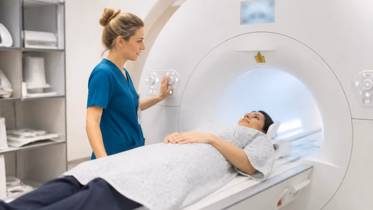
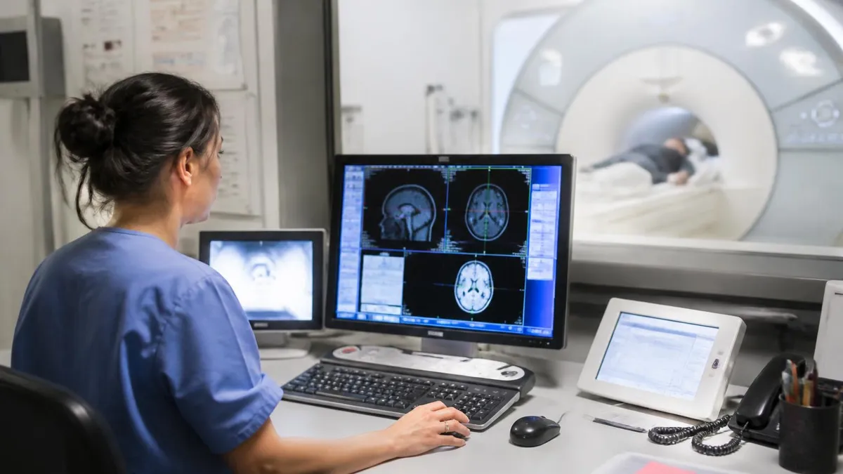
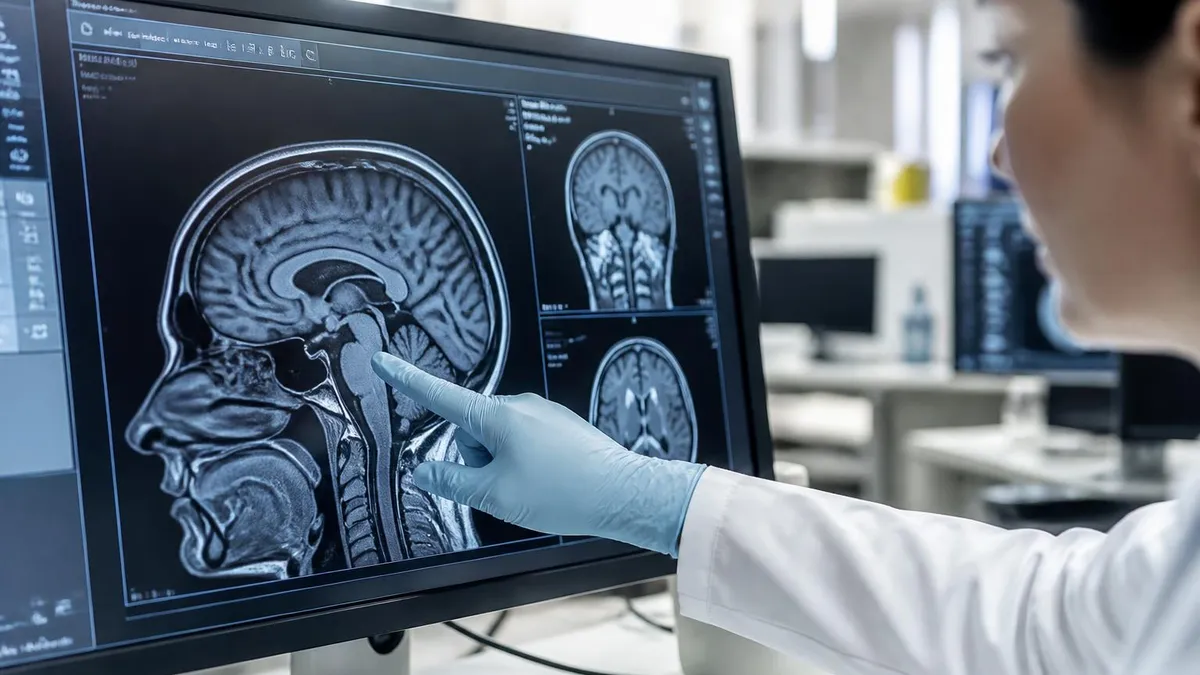
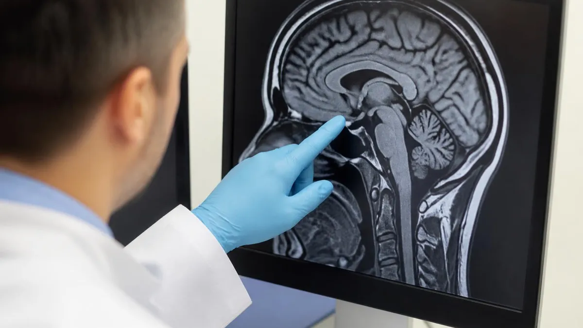
Brain MRI lesions are areas of abnormal signal intensity detected on magnetic resonance imaging that differ from the surrounding normal brain tissue. When a radiologist reads a brain MRI, they are looking for focal or diffuse regions where the signal on T1-weighted, T2-weighted, FLAIR, or diffusion-weighted sequences deviates from what is expected in a structurally and functionally intact brain. Understanding brain mri lesions requires a solid grasp of normal neuroanatomy, MRI physics, and the pathophysiological processes that alter tissue water content, myelination, and blood-brain barrier integrity.
The term "lesion" is intentionally broad. It encompasses everything from small, clinically silent white matter hyperintensities in an elderly patient to large, space-occupying tumors or acute ischemic strokes. Not every lesion signals a dangerous condition, but no lesion should be dismissed without context. The radiologist's job is to characterize each finding by its location, signal characteristics, size, shape, enhancement pattern, and surrounding effects — and then correlate those features with the patient's age, clinical history, and laboratory data to generate a meaningful differential diagnosis.
On T2-weighted and FLAIR sequences, most brain lesions appear bright (hyperintense) because pathological tissue accumulates more water than normal brain parenchyma. This includes demyelinating plaques, infarcts, tumors, infections, and inflammatory conditions. On T1-weighted images, the same lesions often appear dark (hypointense), sometimes called "black holes" in the context of multiple sclerosis. However, some lesions are T1 hyperintense, including subacute hemorrhage, melanoma metastases, and fat-containing lesions, which immediately narrows the differential.
Diffusion-weighted imaging (DWI) adds another crucial layer of characterization. Acute ischemic strokes restrict diffusion because the failure of the sodium-potassium ATPase pump in ischemic neurons causes cytotoxic edema, trapping water molecules intracellularly. Abscesses also restrict diffusion due to the viscous, purulent contents of the cavity. In contrast, cysts, necrotic tumors, and most other lesions do not restrict diffusion, making DWI a powerful tool for narrowing the differential within the first minutes of image interpretation.
Gadolinium contrast enhancement reveals disruptions in the blood-brain barrier. Under normal conditions, tight junctions between endothelial cells prevent gadolinium from entering the brain parenchyma. When pathology — whether inflammatory, neoplastic, or infectious — breaks down those tight junctions, gadolinium leaks into the interstitium and produces enhancement on T1 post-contrast images. Active multiple sclerosis plaques, high-grade tumors, abscesses, and leptomeningeal metastases all enhance in characteristic patterns that help the radiologist distinguish between them.
The clinical significance of a brain MRI lesion depends heavily on patient demographics. A 70-year-old with hypertension, diabetes, and multiple small deep white matter hyperintensities almost certainly has small vessel ischemic disease. The same pattern in a 28-year-old woman with relapsing neurological symptoms should raise immediate concern for multiple sclerosis and prompt additional workup including spine MRI, visual evoked potentials, and cerebrospinal fluid analysis. Age, cardiovascular risk factors, ethnicity, family history, and presenting symptoms all modulate how aggressively a given finding must be pursued.
For MRI technologists, radiologists, and allied health students preparing for board examinations, brain lesion characterization is one of the highest-yield topics. The ARRT and ARMRIT registry examinations test knowledge of lesion signal characteristics, enhancement patterns, and clinical correlations across a wide range of pathologies. Mastering this content not only improves exam performance but directly translates into better patient care by ensuring that imaging findings are accurately interpreted and appropriately communicated to the clinical team.
Brain MRI Lesions by the Numbers
Major Types of Brain MRI Lesions
T2/FLAIR bright foci in the cerebral white matter, ranging from punctate periventricular spots in aging to confluent plaques in demyelinating disease. Prevalence increases with age and vascular risk factors. May be clinically silent or associated with cognitive decline.
Areas that take up gadolinium contrast on T1 post-contrast images, indicating disruption of the blood-brain barrier. Seen in active MS plaques, high-grade gliomas, brain metastases, abscesses, and active inflammation. Enhancement pattern — ring, nodular, leptomeningeal — guides the differential.
Bright on DWI with corresponding low ADC values, signaling restricted water diffusion. Classic for acute ischemic stroke (cytotoxic edema) and pyogenic abscess (pus viscosity). Epidermoid cysts also restrict diffusion, distinguishing them from arachnoid cysts on imaging.
Blood products produce complex signal depending on age. Hyperacute blood is T2 bright; acute blood is T2 dark; subacute blood becomes T1 bright; chronic hemosiderin appears as T2*/SWI dark rim. Gradient echo and susceptibility-weighted imaging (SWI) are most sensitive.
Space-occupying brain lesions include primary gliomas, lymphoma, and metastases. Key features include surrounding vasogenic edema (T2 bright), mass effect with midline shift, heterogeneous enhancement, and restricted diffusion in high-grade tumors. MR spectroscopy and perfusion help grade tumor aggressiveness.
White matter lesions are the most commonly encountered abnormalities on brain MRI in clinical practice, and their correct interpretation can be the difference between reassuring a patient and initiating a life-changing diagnostic workup. The white matter of the brain consists primarily of myelinated axons connecting cortical and subcortical structures. Any process that damages myelin, the axons themselves, or the supporting glial framework will alter the tissue's T1 and T2 relaxation times, producing signal abnormalities visible on MRI. The challenge is that many different diseases produce similar-looking white matter changes, making clinical correlation absolutely essential.
Small vessel ischemic disease is the most prevalent cause of white matter hyperintensities in the adult population, particularly in patients over 55 with hypertension, diabetes mellitus, hyperlipidemia, or a history of smoking. These lesions typically appear as punctate to confluent T2/FLAIR hyperintensities in the periventricular and subcortical white matter, often following the distribution of penetrating arterioles. They are usually symmetric, involve the deep white matter more than the juxtacortical regions, and spare the corpus callosum — a pattern that contrasts sharply with multiple sclerosis, where callosal involvement is a hallmark finding visible on sagittal FLAIR sequences.
Multiple sclerosis plaques are among the most studied white matter lesions in neuroimaging. The McDonald criteria, last updated in 2017, use MRI to establish dissemination in space and time, the two fundamental requirements for an MS diagnosis. Lesions must be ovoid or elliptical (Dawson's fingers when aligned perpendicular to the corpus callosum on sagittal FLAIR), and they must involve at least two of four anatomical regions: periventricular, juxtacortical/cortical, infratentorial, and spinal cord. An actively enhancing lesion or a new T2 lesion on follow-up imaging satisfies dissemination in time without requiring a second clinical attack.
Migraine-associated white matter changes represent another common but often over-interpreted finding. Patients with a history of migraine, particularly migraine with aura, have a higher prevalence of small subcortical white matter hyperintensities than age-matched controls. These lesions are generally small, punctate, and located in the frontal and parietal white matter. While statistically associated with migraine, their clinical significance in individual patients remains uncertain, and they should not be confused with MS plaques or vascular lesions unless accompanied by other supporting features. Most neurologists recommend reassurance and cardiovascular risk factor modification rather than aggressive diagnostic workup for isolated migraine-pattern white matter changes.
Posterior reversible encephalopathy syndrome (PRES) produces a distinctive pattern of white matter edema predominantly affecting the posterior cerebral regions — occipital, parietal, and posterior frontal lobes — in the setting of hypertensive emergency, eclampsia, immunosuppressive therapy, or sepsis. The edema in PRES is vasogenic rather than cytotoxic, meaning it does not restrict diffusion on DWI (a key distinguishing feature from posterior circulation ischemic stroke). The lesions are typically T2/FLAIR bright and bilateral, and they usually resolve completely with blood pressure control or removal of the offending agent, hence the term "reversible."
Toxic and metabolic leukoencephalopathies produce white matter changes through direct myelin injury or axonal damage from exogenous substances or systemic metabolic derangements. Chemotherapy agents, particularly methotrexate and whole-brain radiation, cause a characteristic leukoencephalopathy that appears on MRI as diffuse white matter T2 hyperintensity, sometimes with restricted diffusion in the acute phase. Carbon monoxide poisoning characteristically involves the globus pallidus bilaterally along with white matter changes. Osmotic demyelination syndrome (central pontine myelinolysis) follows rapid correction of hyponatremia and produces a classic T2 bright lesion in the central pons with a characteristic "trident" or "bat-wing" shape on axial imaging.
Infectious and inflammatory causes of white matter lesions include progressive multifocal leukoencephalopathy (PML), caused by JC virus reactivation in immunocompromised patients, which produces asymmetric, confluent white matter lesions without enhancement or mass effect in the early phase. ADEM (acute disseminated encephalomyelitis), typically seen in children following viral infection or vaccination, causes multifocal white matter lesions that are often large, bilateral, and associated with deep gray matter involvement — features that help distinguish it from MS. Lyme disease, HIV encephalopathy, and neurosyphilis can also produce white matter changes that require clinical and serological correlation to correctly characterize.
MRI Signal Patterns: Reading Lesions by Sequence
T2-weighted and FLAIR sequences are the primary workhorses for detecting brain lesions because most pathological tissue accumulates excess water, which prolongs T2 relaxation time and produces bright signal. FLAIR (Fluid-Attenuated Inversion Recovery) adds the advantage of suppressing CSF signal, making periventricular and cortical lesions far more conspicuous than on standard T2 images. T1 sequences provide complementary anatomical detail: most lesions appear hypointense (dark) on T1, but T1 hyperintensity specifically suggests subacute hemorrhage, proteinaceous content, melanin, fat, or gadolinium enhancement.
The combination of T1 and T2 signal allows radiologists to characterize lesion acuity and composition. A lesion that is T2 bright but T1 iso- or hypointense is nonspecific but common for most white matter pathologies. A lesion that is T1 bright before contrast is immediately distinctive and warrants consideration of hemorrhagic, melanotic, or lipid-containing etiologies. Chronic MS lesions may appear as T1 "black holes," reflecting irreversible axonal loss and matrix destruction, making T1 hypointensity a potential surrogate marker of disease severity in longitudinal monitoring.
MRI vs. CT for Brain Lesion Detection
- +Superior soft tissue contrast reveals subtle white matter lesions invisible on CT
- +No ionizing radiation, making it safe for repeated follow-up imaging
- +Multiplanar acquisition without reformatting artifacts improves lesion localization
- +DWI detects acute ischemic stroke within minutes, far earlier than CT
- +Gadolinium contrast shows active inflammation without iodine allergy risk
- +Higher field strength (3T) detects millimeter-scale lesions missed at lower fields
- −Scan times of 30–60 minutes make MRI impractical for critically unstable patients
- −Claustrophobia affects 5–10% of patients and may require sedation
- −Metal implants, pacemakers, and certain devices are absolute or conditional contraindications
- −Higher cost ($1,000–$3,000) compared to CT ($300–$600) limits access in some settings
- −Motion artifacts from patient movement degrade image quality significantly
- −Calcifications and acute hemorrhage are better characterized on CT than on standard MRI
Radiologist Checklist for Evaluating Brain MRI Lesions
- ✓Document lesion location: cortical, subcortical, periventricular, deep white matter, gray matter, infratentorial, or brainstem
- ✓Measure lesion size and count total number of lesions visible on T2/FLAIR
- ✓Assess T1 signal: hypointense (most lesions), isointense, or hyperintense (hemorrhage, fat, melanin)
- ✓Assess T2/FLAIR signal: hyperintense (most lesions) or hypointense (calcification, hemorrhage, high protein)
- ✓Evaluate DWI and ADC map for restricted diffusion indicating cytotoxic edema or abscess
- ✓Review post-contrast T1 images for enhancement pattern: ring, nodular, leptomeningeal, or open-ring
- ✓Check for mass effect, midline shift, sulcal effacement, or herniation
- ✓Assess surrounding vasogenic edema extent relative to lesion size
- ✓Evaluate SWI or GRE for blood products, calcifications, or cavernous malformations
- ✓Correlate imaging findings with patient age, clinical history, and laboratory data before finalizing impression
The Open-Ring Sign Is Pathognomonic for Demyelination
When a brain lesion enhances in a ring pattern but the ring is incomplete — open toward the gray matter — this "open-ring" or "incomplete-ring" sign strongly favors a demyelinating etiology such as multiple sclerosis or tumefactive MS over abscess or high-grade glioma. Knowing this single imaging sign can prevent unnecessary biopsy in a patient with an atypical demyelinating plaque that mimics a tumor on first inspection.
The differential diagnosis for brain MRI lesions is one of the broadest in all of medicine, spanning vascular, inflammatory, infectious, neoplastic, metabolic, and traumatic etiologies. Approaching this differential systematically requires considering the patient's age and clinical presentation before even viewing the images, then using the location, signal characteristics, and enhancement pattern to progressively narrow the possibilities. A useful mnemonic taught in radiology residency is VITAMIN CD: Vascular, Infectious, Traumatic, Autoimmune/inflammatory, Metabolic, Idiopathic, Neoplastic, Congenital, and Degenerative. Applying this framework ensures that no major category is overlooked.
Vascular lesions encompass ischemic stroke, hemorrhage, cavernous malformations, and small vessel disease. Acute ischemic strokes are among the most time-sensitive diagnoses in all of emergency medicine, and MRI with DWI has transformed stroke care by enabling precise lesion localization within minutes of symptom onset.
Hemorrhagic transformation of ischemic stroke — visible as areas of T1 hyperintensity or susceptibility artifact within a previously ischemic territory — alters anticoagulation management and must be recognized promptly. Cavernous malformations appear on T2* or SWI sequences as a characteristic "popcorn ball" lesion with a hemosiderin rim, often with a mixed-signal core reflecting repeated microhemorrhages at different stages of evolution.
Inflammatory and autoimmune lesions represent a rapidly expanding diagnostic category. Beyond classic multiple sclerosis, the neuroradiologist must now consider neuromyelitis optica spectrum disorder (NMOSD), which preferentially involves the optic nerves and spinal cord but can produce large, tumefactive brain lesions near the aquaporin-4–rich periependymal regions around the third and fourth ventricles. MOG antibody-associated disease (MOGAD) produces large cortical and juxtacortical lesions with leptomeningeal enhancement, often in a relapsing-remitting pattern. Anti-NMDA receptor encephalitis typically shows T2 signal abnormalities in the medial temporal lobes resembling herpes encephalitis, underscoring the importance of serological antibody panels in the workup of atypical limbic encephalitis.
Infectious lesions of the brain produce a wide variety of MRI appearances depending on the causative organism, host immune status, and stage of infection. Bacterial cerebritis in its early phase appears as an ill-defined area of T2 hyperintensity without organized rim enhancement; as it evolves into a mature abscess over days to weeks, a T2 dark, T1 bright, restricting rim develops around a central cavity that restricts diffusion.
Herpes simplex encephalitis preferentially involves the medial temporal lobes, insula, and cingulate gyrus in an asymmetric pattern; restricted diffusion and cortical swelling in these regions in a febrile encephalopathic patient should prompt immediate IV acyclovir pending CSF PCR results.
Neoplastic lesions of the brain include primary tumors — glioblastoma, WHO grade 2–4 astrocytomas, oligodendrogliomas, meningiomas, and lymphoma — as well as metastatic disease, which is actually more common than primary brain tumors in the adult population. Glioblastoma (GBM) is the most aggressive primary brain tumor and typically presents as a heterogeneous mass with thick, irregular ring enhancement, central necrosis, and extensive surrounding vasogenic edema.
Primary CNS lymphoma in immunocompetent patients appears as a homogeneously enhancing periventricular mass that often touches the ependymal surface; it is uniquely sensitive to corticosteroids, which can cause dramatic lesion shrinkage within days and paradoxically obscure the diagnosis if steroids are given before biopsy.
Brain metastases most commonly originate from lung, breast, melanoma, renal cell carcinoma, and colorectal cancers. They are typically found at the gray-white matter junction, where microemboli preferentially lodge, and they are often multiple — though up to 30% of patients present with a single brain metastasis. Melanoma metastases are distinguished by their frequent T1 hyperintensity before contrast (due to melanin's paramagnetic properties) and their propensity for spontaneous hemorrhage. Leptomeningeal metastases produce diffuse pachymeningeal or leptomeningeal enhancement on post-contrast FLAIR or high-dose contrast T1 sequences, which are more sensitive than standard post-contrast T1 for detecting early carcinomatous meningitis.
Traumatic brain injury produces a spectrum of MRI findings depending on injury severity and mechanism. Diffuse axonal injury (DAI), caused by rotational acceleration-deceleration forces that shear white matter tracts at the gray-white junction, corpus callosum, and brainstem, is best detected on SWI sequences as multiple small hypointense foci representing petechial hemorrhages along sheared axons.
Standard CT is frequently normal in DAI patients despite severe neurological deficits, making MRI the preferred modality for prognostication and rehabilitation planning. Cortical contusions appear as gyral signal abnormalities at the brain surface, typically in the frontal and temporal poles as a result of contact with the bony skull base during impact.
Certain MRI lesion features require urgent clinical escalation rather than routine follow-up: ring-enhancing lesions with significant mass effect, lesions causing obstructive hydrocephalus, acute diffusion restriction in a stroke distribution in a patient presenting within the thrombolysis window, and lesions with rapid growth on serial imaging. Always communicate these findings directly to the referring clinician rather than relying solely on the written report when the clinical situation is time-sensitive.
Clinical interpretation of brain MRI lesions requires integrating imaging findings with a rich clinical context that the radiologist may only partially possess. The referring clinician — whether a neurologist, emergency physician, or primary care provider — brings crucial information about symptom onset, duration, character, and associated findings that fundamentally change the significance of a given MRI abnormality.
A single periventricular white matter lesion in a 45-year-old with a first episode of optic neuritis carries an entirely different weight than the same lesion in an asymptomatic 72-year-old undergoing imaging for dizziness. Effective communication between imager and clinician is not optional; it is a clinical imperative that directly affects patient outcomes.
The concept of incidental brain lesions — abnormalities found on MRI performed for an unrelated indication — has gained increasing attention as brain MRI utilization expands. Incidental white matter hyperintensities, small enhancing lesions, and even small meningiomas are detected with increasing frequency, creating clinical dilemmas about how aggressively to pursue further workup. Guidelines from major neuroradiology societies recommend a structured reporting approach that characterizes the lesion's most likely etiology, flags features that increase concern for a serious diagnosis, and provides explicit follow-up recommendations rather than leaving clinicians to interpret ambiguous language like "cannot exclude neoplasia."
Quantitative MRI techniques are transforming how radiologists and neurologists track lesion burden over time in conditions like multiple sclerosis, cerebral small vessel disease, and post-treatment tumor response. Automated lesion segmentation software can count and measure lesions across serial MRI studies with precision that exceeds manual counting, reducing inter-reader variability and detecting subtle changes in lesion volume that would escape visual inspection. These tools are particularly valuable in MS clinical trials, where lesion count and volume are primary endpoints, and in neuro-oncology follow-up, where the RANO (Response Assessment in Neuro-Oncology) criteria use precise enhancement measurements to categorize treatment response.
Perfusion MRI adds hemodynamic information about brain lesions that conventional structural sequences cannot provide. Dynamic susceptibility contrast (DSC) perfusion measures cerebral blood volume (CBV), cerebral blood flow (CBF), and mean transit time (MTT) across the brain. High-grade tumors typically show elevated CBV relative to normal white matter, reflecting their pathologically angiogenic vasculature.
In contrast, radiation necrosis — a late complication of brain tumor treatment that can mimic tumor recurrence on conventional contrast-enhanced MRI — typically shows low to intermediate CBV. This distinction is clinically critical because radiation necrosis is managed with steroids and observation, while true tumor progression requires treatment escalation.
MR spectroscopy (MRS) provides a non-invasive metabolic profile of brain lesions by measuring concentrations of key metabolites: N-acetylaspartate (NAA) as a marker of neuronal integrity, choline as a marker of membrane turnover and cellular proliferation, creatine as a stable reference, lactate as an indicator of anaerobic metabolism, and lipids as markers of necrosis.
High-grade tumors characteristically show elevated choline and depressed NAA, reflecting active proliferation and neuronal replacement. Abscesses show a distinctive succinate and acetate peak not seen in tumors. Demyelinating lesions show mildly elevated choline and reduced NAA during active inflammation, which partially normalizes during remission, reflecting the partial reversibility of axonal injury in early MS.
Functional connectivity and advanced diffusion tensor imaging (DTI) are extending brain MRI beyond lesion detection toward understanding how lesions disrupt functional brain networks. DTI tractography maps white matter fiber tracts and can demonstrate disruption of corticospinal, corticothalamic, and callosal connections by space-occupying lesions, guiding surgical planning to minimize postoperative motor or language deficits.
Functional MRI (fMRI) language mapping before tumor resection or epilepsy surgery identifies eloquent cortex adjacent to lesions, allowing neurosurgeons to define safe resection margins. These advanced techniques are standard of care at major academic medical centers and are increasingly available at community hospitals as MRI technology continues to advance.
For students and technologists preparing for MRI registry examinations, understanding not just what lesions look like but why they look the way they do — the underlying physics and pathophysiology — is the key to answering novel questions that fall outside rote memorization.
A thorough grasp of T1 and T2 relaxation, diffusion physics, gadolinium pharmacokinetics, and the pathological basis of signal changes transforms lesion interpretation from pattern matching into mechanistic reasoning. This deeper understanding is exactly what distinguishes top-performing candidates on the ARRT MRI board examination and what makes the difference in clinical practice when an atypical lesion presentation demands careful, first-principles thinking.
Preparing effectively for the MRI registry examination requires a targeted approach to brain lesion content that mirrors the actual exam blueprint. The ARRT MRI examination allocates significant weight to anatomy and pathology, including the recognition of common and clinically important brain lesions across the full spectrum of T1, T2, FLAIR, DWI, and post-contrast imaging. Rather than trying to memorize every possible brain lesion, successful candidates focus on understanding the underlying principles that determine signal behavior, then apply those principles systematically to any lesion they encounter — whether or not they have seen that specific pathology before.
Active recall practice is more effective than passive review for this type of content. Rather than re-reading a textbook description of MS plaques, practice looking at unlabeled MRI images and generating your own differential diagnosis before checking the answer. This approach mirrors the actual examination format and builds the pattern recognition circuitry that allows rapid, accurate lesion characterization. Tools like targeted practice tests, image-based question banks, and flashcard systems specifically designed for MRI pathology outperform general study guides for board preparation.
Understanding the McDonald criteria for MS diagnosis is non-negotiable for any MRI technologist or radiologist working in a neuroimaging environment. These criteria were updated in 2017 to allow a single MRI study to satisfy both dissemination in space and dissemination in time criteria — specifically, if both enhancing and non-enhancing lesions are present simultaneously in characteristic locations.
This means that a single brain MRI in a patient with a first clinical demyelinating event can now be sufficient to diagnose MS, without waiting for a second attack or a follow-up scan showing new lesions. This change has major implications for early treatment initiation and disease-modifying therapy selection.
Stroke protocols deserve special attention because time-critical decision-making depends entirely on rapid and accurate MRI interpretation. The standard acute stroke MRI protocol includes DWI, ADC, T2/FLAIR, GRE or SWI (for hemorrhage exclusion), and MR angiography.
In the first 4.5 hours, the DWI-FLAIR mismatch concept is particularly important: if a lesion is bright on DWI but not yet visible on FLAIR, it suggests the stroke is less than 4.5 hours old, which within the correct clinical framework may support thrombolysis eligibility in patients with an unknown time of onset (so-called "wake-up strokes"). This imaging-based paradigm has extended the treatment window for thousands of patients who would otherwise not qualify for thrombolysis.
Brain tumor follow-up imaging requires familiarity with pseudoprogression — a treatment-related inflammatory response that mimics tumor growth on conventional MRI within the first 3–6 months after concurrent chemoradiation for glioblastoma. Pseudoprogression shows increased T1 enhancement and surrounding edema that may be indistinguishable from true progression on standard sequences. Advanced techniques including perfusion MRI, MR spectroscopy, and amino acid PET can help differentiate these entities, though the distinction remains challenging and often requires multidisciplinary tumor board discussion integrating clinical, imaging, and molecular data.
Pediatric brain lesions present unique diagnostic challenges because the normal developing brain has different signal characteristics than the adult brain, and the differential diagnosis shifts substantially toward congenital, metabolic, and infectious etiologies. The normal myelination pattern progresses in a predictable sequence from the posterior fossa and central white matter outward to the peripheral subcortical white matter, reaching adult-equivalent appearance by approximately 18–24 months of age. Delayed myelination, periventricular leukomalacia (a common finding in premature infants), and leukodystrophies all produce white matter signal abnormalities that must be interpreted against age-appropriate normal standards rather than adult reference patterns.
Incidental findings on brain MRI — the so-called "incidentaloma" problem — represent a growing challenge in clinical neuroradiology. As MRI becomes more accessible and is ordered for an ever-wider range of indications, radiologists increasingly encounter unexpected findings that require clear communication to avoid patient anxiety while ensuring appropriate follow-up.
Small, asymptomatic meningiomas are found in approximately 1% of brain MRIs performed for unrelated reasons. The current consensus recommendation for meningiomas under 3 cm without edema or mass effect in patients over 70 is watchful waiting with follow-up imaging at 1 year, then every 2–3 years if stable — a pragmatic approach that avoids unnecessary surgery while maintaining appropriate surveillance.
MRI Questions and Answers
About the Author
Medical Laboratory Scientist & Clinical Certification Expert
Johns Hopkins UniversityDr. Sandra Kim holds a PhD in Clinical Laboratory Science from Johns Hopkins University and is certified as a Medical Technologist (MT) and Medical Laboratory Scientist (MLS) through ASCP. With 16 years of clinical laboratory experience spanning hematology, microbiology, and molecular diagnostics, she prepares candidates for ASCP board exams, MLT, MLS, and specialist certification tests.
Join the Discussion
Connect with other students preparing for this exam. Share tips, ask questions, and get advice from people who have been there.
View discussion (4 replies)