MRI Report: How to Read, Understand, and Interpret Your Results 2026 June
Learn how to read an MRI report, understand radiology terms, and interpret your results. Complete patient guide with examples. 🔎
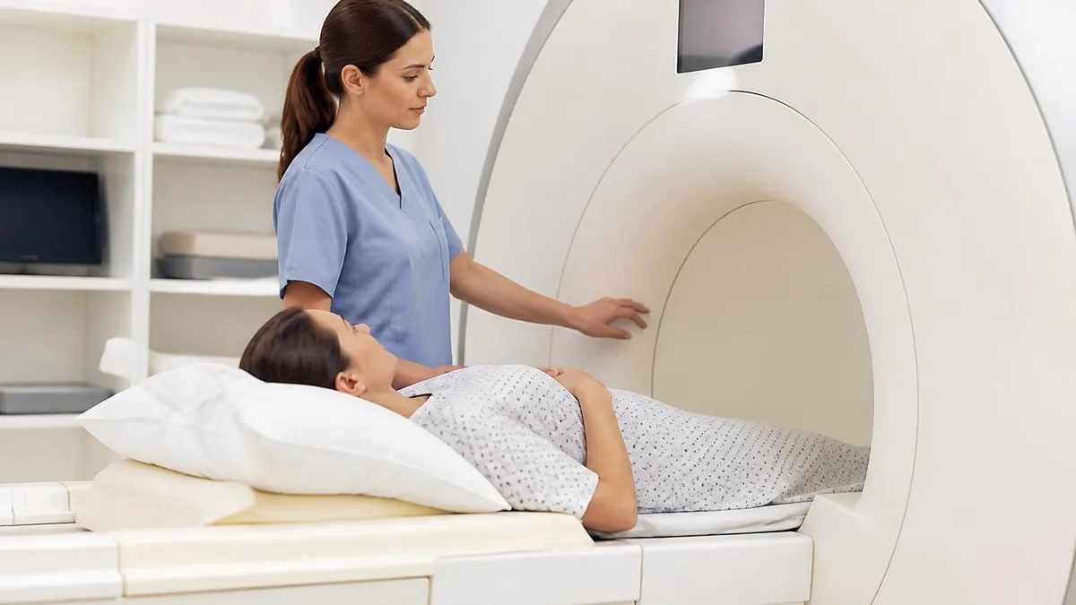
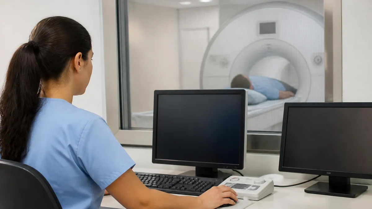
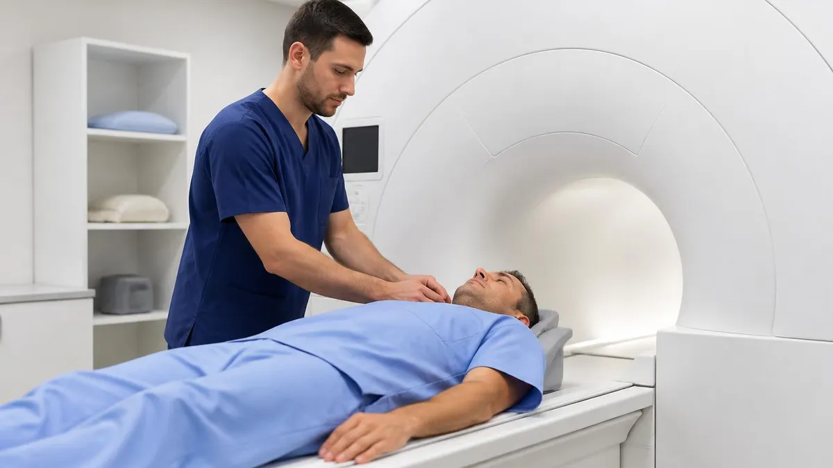
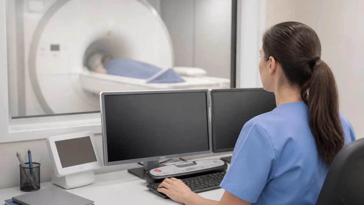
An MRI report is the formal written document a radiologist produces after reviewing your magnetic resonance imaging scans. When your doctor orders an MRI, the images are analyzed by a board-certified radiologist who translates complex visual data into structured clinical language. This report then travels back to your referring physician, who uses it to guide diagnosis, treatment planning, and follow-up care. Understanding what goes into that document — and what the terminology means — can help you have more informed conversations with your healthcare team.
Most patients receive their MRI report within 24 to 72 hours after the scan, though urgent cases are often read within hours. The report is not designed to be read in isolation; it is a communication tool between medical professionals. Nevertheless, patients increasingly have access to their own records through online portals like MyChart, and many want to understand what the findings mean before their next appointment. That curiosity is healthy and worth supporting with clear, accurate information.
A standard MRI report follows a predictable structure, even though the exact wording varies by institution and radiologist. You will typically see sections covering the clinical indication, the imaging technique used, a detailed description of findings, and a final impression or conclusion. Each section serves a specific purpose, and knowing that structure helps you navigate the language more confidently. For example, the "impression" section is arguably the most important: it is the radiologist's bottom-line interpretation of everything observed.
Radiology reports use specialized vocabulary that can feel intimidating at first glance. Terms like "hyperintense signal," "T2 FLAIR," or "mild degenerative changes" mean specific things to a radiologist but can seem alarming or confusing to a patient reading without context. This article breaks down those terms systematically, explaining what they describe and how they relate to common clinical findings. You will also learn which phrases typically signal normal variation and which warrant prompt follow-up with your physician.
It is also worth understanding that a radiologist who writes your report has never met you. They are interpreting images without direct knowledge of your symptoms, history, or physical exam findings. This is why the "clinical indication" section exists — your doctor provides brief context so the radiologist can focus their interpretation appropriately. A finding that looks alarming on imaging may be entirely expected given your clinical history, or it may be an incidental discovery that needs separate evaluation.
For those studying for radiology or MRI credentialing exams, understanding how reports are structured is not just clinically useful — it is directly tested knowledge. Exam questions frequently present a scenario and ask what the correct impression or recommendation should be. Familiarity with report format, standard terminology, and the principles of radiological communication will strengthen your performance on those assessments. This article provides a foundation that serves both patients and students effectively.
Throughout this guide, you will find practical explanations of every major section of the MRI report, the most commonly misunderstood terms, what radiologists look for in different body regions, and how to approach your follow-up appointment armed with meaningful questions. Whether you just received your first mri report or are preparing for a registry exam, the information here will give you a clearer picture of what this critical document contains and why it matters for your care.
MRI Reporting by the Numbers
The Five Sections of Every MRI Report
The reason the scan was ordered, supplied by the referring physician. This section gives the radiologist context — for example, 'right knee pain after sports injury' or 'headache with visual disturbance.' It shapes the entire interpretive focus of the report.
Describes the specific imaging sequences used, the body part scanned, the field strength of the MRI machine (e.g., 1.5T or 3T), and whether contrast agent was administered. This section ensures the study can be reproduced or compared to future scans.
The longest and most detailed section. The radiologist systematically describes what is visible in each anatomical structure — both normal and abnormal. Radiologists document both positive findings and explicitly note normal structures to show the scan was reviewed comprehensively.
The summary conclusion — the most important section for most readers. It translates the detailed findings into a prioritized clinical interpretation and lists specific diagnoses or differential diagnoses ranked by likelihood. Your doctor focuses here first.
Not all reports include this section, but many do. It may suggest additional imaging, clinical correlation, or follow-up timing. For example, a finding may prompt a recommendation for repeat MRI in six months or referral to a specific specialist.
Understanding the language of an MRI report starts with signal intensity — the fundamental building block of MRI interpretation. On MRI, tissues appear bright or dark depending on how their hydrogen atoms respond to radiofrequency pulses. Radiologists describe these appearances using the terms "hyperintense" (bright), "hypointense" (dark), and "isointense" (the same intensity as surrounding tissue). These descriptions are always relative — a structure is described as bright or dark compared to a reference tissue nearby.
The most commonly referenced imaging sequences are T1-weighted and T2-weighted images. On T1-weighted scans, fat appears bright and fluid appears dark. On T2-weighted scans, the reverse is often true — fluid becomes bright and fat can appear intermediate. This matters because abnormalities like edema, inflammation, tumors, and cysts often contain fluid or altered water content, making them particularly visible on T2 sequences. When a report says "T2 hyperintense lesion," it means the abnormal area appears bright on T2-weighted images — consistent with fluid or inflammation.
FLAIR stands for Fluid-Attenuated Inversion Recovery — a specialized T2 sequence that suppresses the signal from cerebrospinal fluid (CSF). This makes it easier to see abnormalities in brain tissue that borders fluid-filled spaces. FLAIR is particularly useful for detecting multiple sclerosis plaques, small strokes, and white matter changes. When you see "FLAIR hyperintensity" in a brain MRI report, it typically indicates an area of tissue damage or pathology that was made visible by suppressing the normally bright CSF signal.
Diffusion-weighted imaging (DWI) and apparent diffusion coefficient (ADC) maps are sequences that measure how water molecules move through tissue. In acute stroke, water movement becomes restricted because cells swell and die. On DWI, restricted diffusion appears bright; on the ADC map, the same area appears dark. This pattern — bright on DWI, dark on ADC — is the hallmark of acute ischemic stroke and is critically important in emergency radiology. Abscess cavities and certain tumors also show restricted diffusion, making DWI a versatile diagnostic tool.
Enhancement refers to brightening that occurs after injection of a gadolinium-based contrast agent. Gadolinium shortens T1 relaxation times, making tissue appear brighter on post-contrast T1-weighted images. Enhancement indicates increased blood flow or a disrupted blood-brain barrier — findings associated with active inflammation, infection, tumors, and other pathologies. A report noting "avid enhancement" signals robust uptake of contrast, which can indicate aggressive pathology. "Rim enhancement" describes a ring-like pattern seen in abscesses and some tumors.
Common terms that confuse patients include "artifact," "incidental finding," and "degenerative changes." An artifact is a technical imperfection in the image caused by patient motion, metallic implants, or equipment — it is not a real anatomical finding. An incidental finding is something discovered that was not the purpose of the scan and may or may not require action. Degenerative changes describe age-related wear in joints, discs, or other structures — they are extremely common and often do not correspond directly to symptoms.
For MRI technologists and radiology students, mastering report terminology is essential because it connects imaging to clinical diagnosis. Exam questions often test whether candidates can correctly identify what a described signal pattern represents or what sequence would best demonstrate a given pathology. Building a working vocabulary of these terms — and understanding the physical principles behind them — is a core competency for anyone pursuing MRI certification or board examination. Reviewing sample reports alongside practice questions reinforces this knowledge in context.
Reading MRI Reports by Body Region
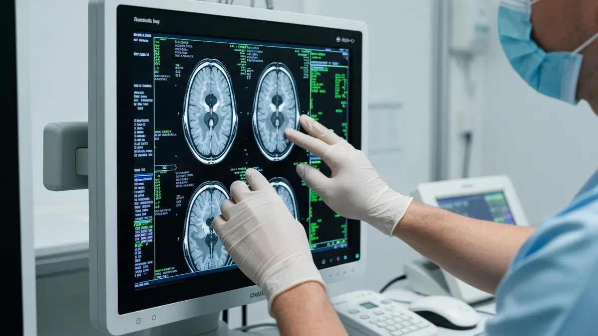
A brain MRI report systematically reviews the cerebral cortex, white matter, basal ganglia, brainstem, cerebellum, and ventricles. Radiologists assess for asymmetry, signal abnormalities, mass effect, and midline shift. The white matter is carefully evaluated for T2/FLAIR hyperintensities, which can represent small vessel disease, demyelination, or prior injury. The ventricles are assessed for size — enlarged ventricles may indicate hydrocephalus or atrophy. The corpus callosum, internal capsule, and posterior fossa are given specific attention depending on the clinical question.
Common phrases in brain MRI reports include "age-appropriate white matter changes," which refers to small areas of T2 signal that are normal in older adults. "No acute intracranial abnormality" is a reassuring statement meaning no stroke, bleeding, or mass was identified. Incidental findings like arachnoid cysts, prominent perivascular spaces, or pineal cysts are extremely common and almost always benign. When contrast is used, the radiologist specifically evaluates for abnormal enhancement that could indicate tumor, infection, or active inflammation — findings that change clinical management significantly.
Accessing Your MRI Report Directly: Benefits and Drawbacks
- +Empowers patients to understand their diagnosis before seeing the doctor
- +Enables more productive, focused conversations with your physician
- +Allows you to catch administrative errors in patient demographics or clinical indication
- +Helps you research your findings from credible medical sources in advance
- +Provides a permanent record you can share with specialists or second-opinion providers
- +Reduces anxiety for some patients who prefer knowing results promptly rather than waiting
- −Medical terminology can be misinterpreted without clinical training or context
- −Incidental findings may cause unnecessary alarm before a doctor explains their significance
- −Reports describe imaging only — they cannot account for your physical exam or full history
- −Radiologists hedge language (e.g., 'cannot exclude') that can feel alarming but is standard practice
- −Patients may fixate on minor findings that clinicians consider unimportant
- −Reading your own report without guidance can lead to incorrect self-diagnosis or unnecessary worry
Patient Checklist: What to Do After Receiving Your MRI Report
- ✓Read the Impression section first — it contains the radiologist's bottom-line conclusion.
- ✓Write down every medical term you do not understand before your follow-up appointment.
- ✓Look up unfamiliar terms in a reputable medical dictionary or ask your physician to explain them.
- ✓Note whether the report mentions any recommendations for follow-up imaging or specialist referral.
- ✓Check that your name, date of birth, and scan date are correct — errors do occur.
- ✓Compare the clinical indication in the report to the reason your doctor said they were ordering the scan.
- ✓Ask your doctor whether any incidental findings require separate evaluation or monitoring.
- ✓Request a copy of the actual MRI images (on CD or via portal) if you plan to seek a second opinion.
- ✓Avoid searching your diagnosis on unvetted websites — stick to academic medical center resources.
- ✓Bring your printed or digital report to every specialist appointment so they have the full text.
The 'Impression' Section Is What Your Doctor Reads First
In a busy clinical setting, physicians often go straight to the Impression section of an MRI report, which distills the radiologist's interpretation into a concise, prioritized conclusion. For patients reviewing their own records, starting here — rather than trying to decode the detailed Findings section line by line — provides the clearest picture of what the scan showed and what action, if any, is recommended. The full Findings section adds depth but the Impression is the clinical bottom line.
Among the most frequently misunderstood findings in MRI reports are those involving the brain's white matter. White matter hyperintensities — bright spots on T2 and FLAIR sequences — are reported in a significant percentage of adults over 50 and increase in frequency with age. They are associated with cardiovascular risk factors including hypertension, diabetes, and smoking. In most cases, small, scattered white matter hyperintensities are considered a normal aging phenomenon and do not require specific treatment, though they may prompt your doctor to optimize cardiovascular risk management.
Disc pathology is the most commonly reported finding in lumbar and cervical spine MRI. It is important to understand that imaging findings often do not correlate directly with symptoms. Studies have consistently shown that disc herniations, bulges, and degenerative changes are present in large percentages of asymptomatic adults — particularly over age 40.
A 2015 systematic review published in the American Journal of Neuroradiology found that more than 50% of adults with no back pain had disc bulges on imaging. This is why radiologists and clinicians emphasize "clinical correlation" — the imaging must be interpreted in the context of what the patient actually feels.
Meniscal tears are among the most common musculoskeletal findings on knee MRI. Radiologists grade meniscal signal using a three-grade system. Grade 1 and 2 signal changes represent intrameniscal degeneration that does not communicate with the articular surface — these are often asymptomatic and do not require surgery. Grade 3 signal reaches the meniscal surface and represents a true tear. Even among grade 3 tears, many middle-aged patients improve with physical therapy and conservative management, making the clinical context essential for treatment decisions.
Brain incidentalomas are common and can cause significant patient anxiety. An incidentaloma is any finding discovered unexpectedly — unrelated to the reason for the scan. Common benign incidentalomas include arachnoid cysts (fluid-filled sacs within the arachnoid membrane), Rathke cleft cysts (near the pituitary gland), pineal cysts, and choroid plexus cysts. These are almost universally benign and require no treatment. Your radiologist will typically include language in the impression noting that such findings are incidental and likely benign, sometimes recommending follow-up imaging to confirm stability over time.
When contrast enhancement is present, the clinical significance depends heavily on the pattern and location. Solitary ring-enhancing lesions in the brain have a well-known differential diagnosis including brain abscess, high-grade glioma, and metastasis. Radiologists will typically offer this differential in the impression and recommend additional workup. Multiple enhancing lesions may suggest metastatic disease or demyelinating conditions like multiple sclerosis. The pattern of enhancement in conjunction with the patient's clinical history — age, cancer history, immune status, fever — narrows the differential significantly.
Vascular findings are increasingly detected on MRI, particularly with dedicated MRA (magnetic resonance angiography) sequences. Small unruptured intracranial aneurysms are found in approximately 3% of the general population. When discovered incidentally, management depends on size, shape, location, and patient risk factors. Most small aneurysms are managed conservatively with surveillance imaging. Larger aneurysms or those with concerning morphology may prompt neurosurgical consultation. Arteriovenous malformations (AVMs) and cavernous malformations are other vascular findings that may be identified on brain MRI, each carrying specific management considerations.
For MRI students and registry exam candidates, understanding these common findings and their clinical significance is directly tested knowledge. Board exams present clinical vignettes describing imaging findings and ask candidates to identify the most likely diagnosis, the appropriate next step, or the imaging sequence best suited to further evaluate the finding. The ability to think systematically about signal patterns, anatomical relationships, and clinical context — exactly the skills that make a radiologist effective — is also what separates high scorers from average performers on standardized exams.
If your MRI report mentions findings such as acute stroke, intracranial hemorrhage, spinal cord compression, or any mass described as "suspicious for malignancy," do not wait for your regularly scheduled follow-up appointment. Contact your referring physician's office immediately to discuss the report. Urgent radiological findings should generate a direct call from the radiologist to the ordering provider — but confirming that communication occurred is reasonable and appropriate patient self-advocacy.
Preparing effectively for your follow-up appointment after receiving an MRI report can transform a potentially stressful interaction into a productive clinical conversation. The most useful thing you can do before that appointment is write down your specific questions in priority order — start with the most important and work down the list. Physicians often have limited time per appointment, so having organized questions ensures you get the most critical information first. Bring the printed report or have it available on your phone so you can reference specific language the radiologist used.
Ask your physician to explain the impression in plain language. Even if you have already researched the terminology, hearing your doctor's interpretation — in the context of your full clinical picture — is invaluable. Phrases like "cannot exclude" or "differential diagnosis includes" are hedging language that radiologists use to communicate uncertainty; your physician can tell you which option on that differential list is most consistent with your specific symptoms and history. This context dramatically changes how you should interpret the language in the report.
If additional imaging is recommended in the report, ask your doctor specifically when it should be scheduled and what you should watch for in the interim. Some follow-up recommendations are time-sensitive — a "repeat MRI in 3 months" recommendation for a growing lesion carries different urgency than a "repeat in 2 years" recommendation for a stable-appearing cyst. Understanding the purpose and timeline of follow-up studies helps you be an active participant in your own care rather than passively waiting for the next appointment.
Second opinions are a legitimate and often valuable option after receiving a significant MRI report finding. Radiology is largely a matter of experienced judgment, and inter-reader variability exists even among expert radiologists. Academic medical centers, subspecialty radiology practices, and teleradiology services all offer formal second-opinion reads. If you are facing a major diagnosis — particularly a suspected tumor, unusual neurological finding, or rare pathology — requesting that your images be reviewed by a subspecialist radiologist with expertise in that body region or disease category is entirely reasonable and appropriate.
For patients who are managing chronic conditions with serial MRI scans — multiple sclerosis, low back pain, brain tumor surveillance — comparing your current report to prior reports is essential. The most clinically meaningful information is often not the absolute finding but the change over time. Reports will typically reference prior studies explicitly, noting whether lesions are "stable," "increased," or "decreased" compared to the previous scan. If you have had multiple scans at different institutions, bringing prior reports and images to each new facility ensures the radiologist has that comparative context.
Understanding your MRI report also matters if you are involved in your own health records management for insurance, disability, or legal purposes. Reports must be accurate and complete for these contexts. If you notice an error — a wrong body part, incorrect date, or description of findings inconsistent with what you were told verbally — you have the right to request a correction through the imaging facility's medical records department. Radiologists can issue addenda to existing reports when a correction or additional clinical context changes the interpretation.
Technology is changing how MRI reports are written and delivered. AI-assisted radiology tools are increasingly used to flag findings, measure lesion volumes, and draft preliminary reports that radiologists then review and finalize. Some health systems now offer automated patient-facing summaries alongside the formal report — plain-language explanations generated from the technical text. While these tools are still evolving in accuracy and reliability, they represent a meaningful step toward making MRI report content more accessible to the patients whose health it describes. Always confirm AI-generated summaries with your physician, as these tools are not infallible.
For MRI technologists preparing for the ARRT Registry Examination in Magnetic Resonance Imaging, understanding how reports are generated and used is part of the foundational knowledge tested on the exam. The registry exam covers patient care, safety, data acquisition, and image production — all of which directly influence the quality of information a radiologist can extract from the images you produce. Poor image quality from inadequate patient preparation, motion artifact, or incorrect sequence selection leads to suboptimal reports that may miss clinically important findings.
One of the most impactful things a technologist does for report quality is patient positioning and preparation. Proper positioning ensures the anatomy of interest is centered in the field of view with optimal coil placement. Patient briefing — explaining what to expect, how long the scan will take, and the importance of staying still — dramatically reduces motion artifact. A report that notes "study limited by patient motion" is a report that has degraded diagnostic value, potentially requiring a repeat scan and additional patient exposure to the scanner environment.
Sequence selection and protocol optimization are also within the technologist's sphere of influence. Most facilities use standardized protocols, but technologists must recognize when protocol modifications are needed — such as adding dedicated sequences for a suspected pathology identified during the scout or adjusting parameters for implanted devices. A technologist who understands why certain sequences are used (and what findings they are designed to detect) is better equipped to adapt appropriately and to communicate with the radiologist when an unexpected finding appears on the scout images.
Image quality factors that directly affect report accuracy include signal-to-noise ratio (SNR), contrast-to-noise ratio (CNR), spatial resolution, and temporal resolution. These parameters involve trade-offs: increasing resolution typically increases scan time; increasing field of view reduces resolution. Technologists must balance these factors based on the clinical question and patient tolerance. Understanding these trade-offs is tested on registry exams through questions about why specific acquisition parameters are chosen and how changing one parameter affects image quality and scan time.
Safety is inseparable from image quality in MRI reporting. A scan that must be interrupted due to a safety screening failure, unexpected patient response to contrast, or equipment malfunction cannot produce a complete report. Technologists bear primary responsibility for the pre-scan screening process — verifying implant compatibility, identifying contraindications to gadolinium contrast, ensuring no ferromagnetic objects enter the scanner room. Registry exam questions on safety are practical and scenario-based, testing decision-making in real-world situations rather than simple recall of rules.
Continuing education requirements for MRI certification reflect the rapidly evolving nature of the field. New sequences, higher field strengths (7 Tesla systems are entering clinical use), AI-assisted reconstruction algorithms, and novel contrast agents all change what radiologists can see — and therefore what they can report. Staying current with these advances is not just a professional obligation but a clinical responsibility. The registry exam is one checkpoint in a career-long learning process that ensures MRI professionals can produce the high-quality images that accurate, meaningful reports depend on.
Ultimately, whether you are a patient trying to understand your own health or a student preparing for an MRI credentialing exam, engaging seriously with MRI report content pays real dividends. For patients, that engagement leads to better conversations with physicians, more informed treatment decisions, and stronger health advocacy. For students and technologists, it builds the clinical reasoning skills that distinguish excellent practitioners from technically competent ones. The MRI report is more than a document — it is a communication bridge between advanced imaging technology and the clinical decisions that affect real people's lives every day.
MRI Questions and Answers
DWI MRI: Diffusion-Weighted Imaging Explained (2026 Guide)
Side Effects of Brain MRI: Understanding Risks, Safety, and What Every Patient Should Know
MRI Lumbar Spine Without Contrast: Complete Guide to the Scan, What It Shows, and How to Prepare
MRI History: From Nuclear Resonance to Modern Scanners
MRI of Cervical Spine: Complete Guide to Imaging, Anatomy, Pathology, and Patient Preparation
About the Author
Medical Laboratory Scientist & Clinical Certification Expert
Johns Hopkins UniversityDr. Sandra Kim holds a PhD in Clinical Laboratory Science from Johns Hopkins University and is certified as a Medical Technologist (MT) and Medical Laboratory Scientist (MLS) through ASCP. With 16 years of clinical laboratory experience spanning hematology, microbiology, and molecular diagnostics, she prepares candidates for ASCP board exams, MLT, MLS, and specialist certification tests.
Join the Discussion
Connect with other students preparing for this exam. Share tips, ask questions, and get advice from people who have been there.
View discussion (4 replies)