Male Pelvis MRI: Complete Guide to Anatomy, Pathology, Protocols, and Patient Preparation
Complete guide to male pelvis MRI 🎯 — anatomy, pathology, scan protocols, and patient prep tips for technologists and students.
A male pelvis mri is one of the most diagnostically powerful tools in modern radiology, offering soft-tissue contrast that far exceeds computed tomography or ultrasound for evaluating the prostate gland, seminal vesicles, bladder, rectum, and surrounding pelvic musculature. Unlike ionizing-radiation-based modalities, MRI relies on radiofrequency pulses and magnetic fields to generate images, making it the preferred imaging method when repeated follow-up scans are anticipated or when fine anatomical detail is critical for surgical planning.
The clinical indications for pelvic MRI in male patients are broad and growing. Prostate cancer staging remains the single most common reason a urologist orders this exam, but it is far from the only one. Bladder cancer local staging, rectal cancer preoperative planning, penile or scrotal pathology evaluation, and assessment of cryptorchidism in adult patients all fall within the scope of a comprehensive male pelvic MRI protocol. Trauma patients presenting with pelvic fractures also benefit from MRI when ligamentous or neurovascular injury is suspected.
Prostate MRI has been transformed in recent years by the adoption of the PI-RADS scoring system — Prostate Imaging Reporting and Data System — now in its version 2.1 iteration. This standardized lexicon allows radiologists across different institutions to communicate findings consistently, enabling urologists to make evidence-based biopsy decisions. A PI-RADS 4 or 5 lesion on a multiparametric prostate MRI carries a clinically significant cancer probability exceeding 70 percent in most published series, fundamentally changing how urologic oncology is practiced.
Multiparametric MRI, often abbreviated mpMRI, combines at minimum three distinct pulse sequence families: T2-weighted imaging for anatomy, diffusion-weighted imaging for cellularity, and dynamic contrast-enhanced imaging for vascularity. Each sequence interrogates different tissue properties and together they dramatically increase sensitivity and specificity compared to any single sequence alone. Understanding how each sequence contributes to the final diagnostic impression is foundational knowledge for MRI technologists, radiologists in training, and students preparing for board certification examinations.
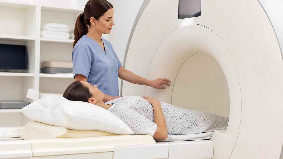
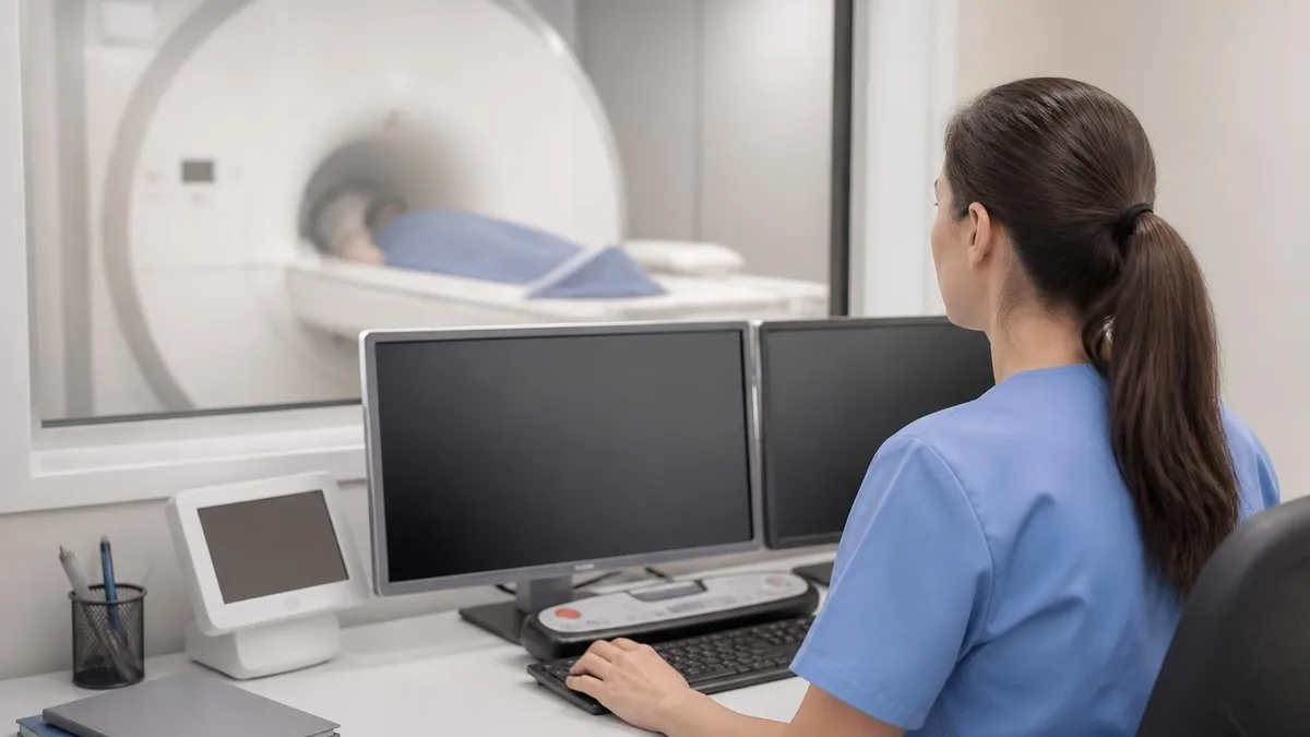
Patient preparation is non-trivial for male pelvis MRI and directly affects image quality. Bowel preparation with antispasmodic agents such as glucagon or hyoscine reduces peristaltic motion artifact that would otherwise degrade T2-weighted sequences. Bladder filling protocols vary by institution but a moderately full bladder — achieved by voiding two hours before the exam and drinking approximately 500 milliliters of water — improves delineation of the bladder wall and perivesical fat planes. Endorectal coils, though less commonly used since the widespread adoption of 3-Tesla scanners, may still be employed at some centers to boost signal-to-noise ratio for prostate imaging.
From a physics standpoint, the pelvis presents specific challenges. The large field of view required to capture all relevant anatomy from the iliac crests to the perineum must be balanced against the need for thin slices and high in-plane resolution for small lesion detection. Phase-encoding direction selection is critical to avoid wrap-around artifacts from the arms or contralateral pelvic sidewall. Parallel imaging acceleration factors of two to three are routinely applied to reduce scan time while maintaining adequate signal, and modern scanners increasingly use compressed sensing reconstruction to further shorten acquisition windows without resolution penalty.
For students and technologists preparing for MRI certification exams, the male pelvis represents a high-yield anatomical region that consistently appears on the ARRT MRI examination and the ARMRIT credentialing process. Understanding not only the normal anatomy but also the characteristic MRI appearance of common pathologies — clinically significant prostate cancer, benign prostatic hyperplasia, seminal vesicle invasion, lymph node metastasis, and bladder tumors — is essential for passing these high-stakes assessments. The sections that follow provide a structured, evidence-based review of everything you need to know.
Male Pelvis MRI by the Numbers
Key Anatomical Structures Visualized on Male Pelvis MRI
Divided into peripheral zone, transition zone, central zone, and anterior fibromuscular stroma. The peripheral zone comprises about 70% of glandular tissue and is where most prostate cancers arise. On T2-weighted images it appears uniformly high signal in healthy tissue.
Paired lobulated structures posterior to the bladder base that appear as feathery high-signal structures on T2W images. Loss of normal architecture or T2 hypointensity within the seminal vesicles raises concern for tumor invasion, upstaging prostate cancer to at least T3b disease.
The bladder wall is best assessed with moderate distension. Muscle-invasive bladder cancer appears as a focal area of T2 hypointensity interrupting the normally bright detrusor muscle layer. DWI with ADC mapping helps distinguish high-grade urothelial carcinoma from benign inflammation.
Critical in rectal cancer staging. The circumferential resection margin is measured from tumor to the mesorectal fascia on high-resolution T2W axial images. Threatened or involved margins guide neoadjuvant chemoradiation decisions and surgical approach selection before total mesorectal excision.
Normal pelvic nodes are oval, less than 8mm short-axis diameter, and show fatty hila. Malignant nodes may be enlarged, rounded, or demonstrate restricted diffusion on DWI. Size criteria alone are imperfect — small nodes can harbor micrometastases while reactive nodes can be enlarged.
A comprehensive male pelvis MRI protocol is built around sequences that each contribute unique and complementary information. T2-weighted fast spin-echo sequences form the anatomical backbone of the examination. Acquired in at least two planes — typically axial and coronal, with sagittal added for rectal or bladder pathology — T2W images provide exquisite depiction of zonal anatomy within the prostate, the fascial planes surrounding the gland, and the relationship between tumor and adjacent structures such as the neurovascular bundles, which are essential for nerve-sparing prostatectomy planning.
Diffusion-weighted imaging has emerged as arguably the most powerful single sequence for prostate cancer detection. DWI exploits the fact that malignant tissue has a higher cellular density than benign tissue, which restricts the random Brownian motion of water molecules. On DWI, cancer appears as a focus of bright signal at high b-values, typically b-1400 or b-2000 when using computed high-b images, with corresponding signal dropout on the apparent diffusion coefficient map. Quantitative ADC values below approximately 0.80 to 0.90 × 10⁻³ mm²/s are associated with clinically significant prostate cancer in most published thresholds.
Dynamic contrast-enhanced MRI adds a temporal dimension by capturing the arrival, peak, and washout of gadolinium-based contrast agent within the prostate parenchyma over a series of rapidly acquired gradient-echo volumes obtained every few seconds over approximately five minutes. Malignant tissue tends to enhance early and wash out quickly — a pattern reflecting tumor neovascularity. In the PI-RADS 2.1 framework, DCE plays a more limited adjunctive role for peripheral zone lesions than it did in earlier versions, but it remains diagnostically important for transition zone lesions and for assessing extraprostatic extension at the prostatic apex.
Magnetic resonance spectroscopic imaging was historically used in some centers to quantify choline-to-citrate ratios within prostate voxels, with elevated ratios indicating malignancy. While technically interesting, MRS is time-consuming, technically demanding, and has largely been supplanted by DWI and DCE in current clinical practice. Most major academic centers no longer include MRS in their routine prostate MRI protocols. Its mention in board examination contexts, however, remains relevant, and candidates should understand its underlying biochemical rationale.
For rectal cancer staging, the protocol departs meaningfully from the prostate-focused approach. High-resolution T2-weighted sequences using a small field of view and thin slices — typically 3mm without interslice gap — oriented perpendicular to the tumor axis allow precise measurement of the mesorectal fascia involvement, the relationship to the peritoneal reflection, and tumor height above the anal verge. These measurements directly inform the surgeon about whether an anterior resection or an abdominoperineal resection is technically feasible while achieving clear margins.
Bladder cancer staging protocols emphasize the importance of adequate bladder distension and motion suppression. T2-weighted images in multiple planes remain the primary staging tool, but DWI adds sensitivity for detecting muscle invasion and small satellite lesions that might be overlooked on T2W alone. Dynamic contrast enhancement helps differentiate viable tumor from treatment-related fibrosis in post-therapy restaging examinations, a clinically critical distinction when deciding whether additional treatment cycles are warranted versus proceeding directly to cystectomy.
Field strength selection substantially influences male pelvic MRI quality. At 1.5 Tesla, the signal-to-noise ratio often necessitates use of an endorectal coil for adequate prostate imaging. At 3 Tesla, the doubled SNR compared to 1.5T allows high-quality prostate imaging with surface phased-array coils alone, improving patient comfort and reducing the rectal discomfort and gas artifact that endorectal coils can introduce. Emerging 7-Tesla whole-body systems show remarkable resolution but remain experimental for pelvic indications and are not yet FDA-cleared for routine clinical use in this body region.
Prostate Cancer Staging and PI-RADS Scoring on Male Pelvis MRI
PI-RADS version 2.1 assigns a score from 1 to 5 to each suspicious prostate lesion, where 1 indicates very low probability of clinically significant cancer and 5 indicates very high probability. The dominant sequence driving the score differs by zone: DWI is the dominant sequence for peripheral zone lesions, while T2-weighted imaging dominates for transition zone lesions. DCE can upgrade a peripheral zone lesion from PI-RADS 3 to PI-RADS 4 when early focal enhancement is present, but it cannot downgrade lesions.
In clinical practice, urologists typically recommend MRI-fusion targeted biopsy for PI-RADS 3 or higher lesions, with systematic biopsy of the remaining gland performed concurrently. PI-RADS 1 and 2 lesions are generally considered benign — with a clinically significant cancer risk below 10 percent in most studies — and surveillance without immediate biopsy is a reasonable management strategy, particularly in patients with low baseline PSA and no prior positive biopsy history. Accurate PI-RADS scoring requires structured training and reader experience at a minimum of 50 to 100 cases.
Male Pelvis MRI vs. Other Imaging Modalities: Advantages and Limitations
- +Superior soft-tissue contrast compared to CT — especially for prostate zonal anatomy and tumor margin assessment
- +No ionizing radiation — safe for repeated follow-up imaging in patients on active surveillance protocols
- +Multiparametric capability — T2W, DWI, and DCE together provide functional and anatomical information in a single session
- +High sensitivity for extraprostatic extension and seminal vesicle invasion critical for surgical planning
- +Enables MRI-fusion biopsy targeting — reducing the number of cores needed while improving clinically significant cancer detection
- +Excellent assessment of the mesorectal fascia margin in rectal cancer staging — directly guides neoadjuvant therapy decisions
- −Longer scan time than CT — typically 45 to 60 minutes, increasing motion artifact risk and patient discomfort
- −Higher cost — male pelvis mpMRI is significantly more expensive than transrectal ultrasound or CT pelvis
- −Contraindicated in patients with certain metallic implants — cardiac pacemakers, cochlear implants, and some aneurysm clips preclude scanning
- −Reader variability — PI-RADS scoring shows moderate inter-reader agreement even among experienced radiologists without formal structured training
- −Limited sensitivity for lymph node micrometastases — morphologic MRI criteria alone miss a large proportion of histologically positive nodes
- −Post-biopsy hemorrhage artifact — scanning within six to eight weeks of prostate biopsy degrades image quality and can obscure small cancers
Patient Preparation Checklist for Male Pelvis MRI
- ✓Screen the patient for MRI contraindications including pacemakers, cochlear implants, metallic orbital foreign bodies, and non-MRI-compatible aneurysm clips before scheduling.
- ✓Instruct the patient to void approximately two hours before the scheduled scan time, then drink 500 mL of water to achieve moderate bladder filling on arrival.
- ✓Administer an antispasmodic agent such as glucagon 1 mg IV or hyoscine butylbromide 20 mg IV immediately before scanning to reduce bowel peristalsis artifact.
- ✓Confirm whether gadolinium-based contrast agent is required and obtain eGFR within the past 12 weeks for patients with known or suspected renal disease.
- ✓Advise the patient to avoid eating a large meal in the two hours preceding the exam to minimize bowel gas accumulation in the pelvis.
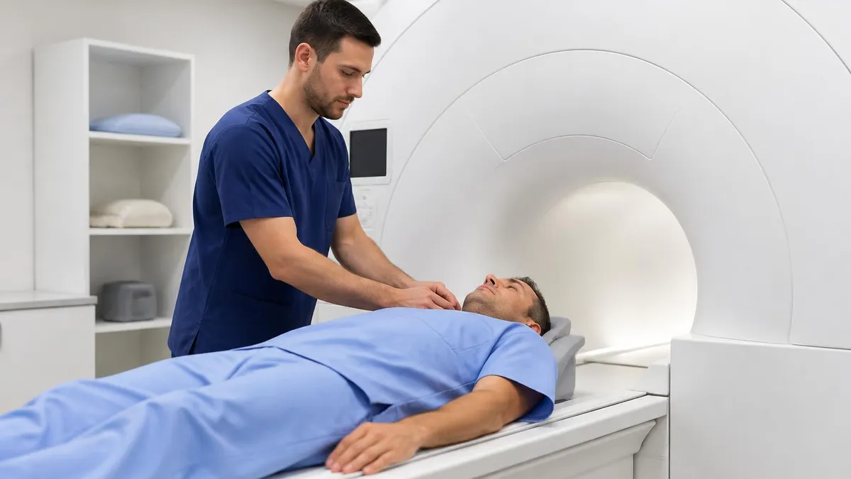
- ✓Position the patient supine feet-first with a surface phased-array pelvic coil centered over the pubic symphysis; confirm coil connectivity and signal before the patient enters the bore.
- ✓Perform a localizer sequence and confirm the field of view covers from the aortic bifurcation superiorly to the perineum inferiorly for adequate lymph node assessment.
- ✓Select axial oblique plane for prostate sequences aligned perpendicular to the long axis of the prostate — not to the patient's body axis — to optimize zonal anatomy depiction.
- ✓Acquire high-b-value DWI images at b-values of 50, 400, and 800, with computed b-1400 or b-2000 images generated automatically by the scanner software.
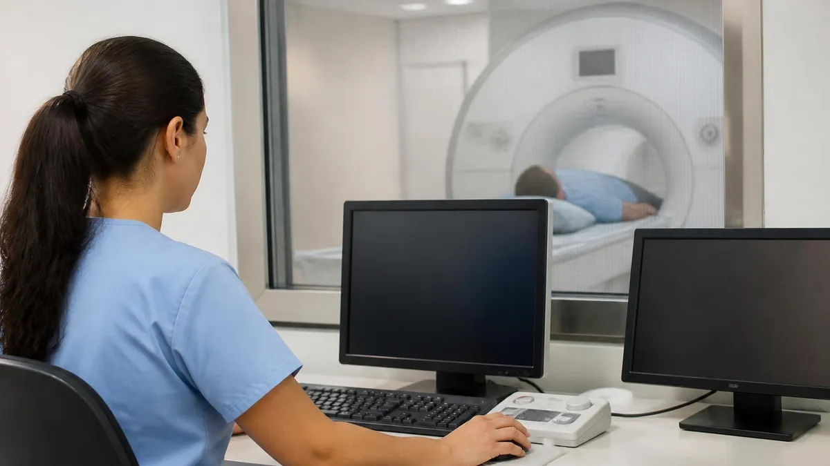
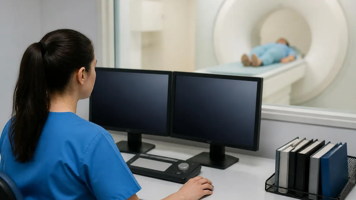
- ✓Review all sequences at the scanner console before releasing the patient — check for motion artifact, adequate bladder filling, and field-of-view coverage before the patient is discharged.
Pre-Biopsy MRI Is Always Preferred Over Post-Biopsy MRI
Post-biopsy hemorrhage from transrectal ultrasound-guided prostate biopsy causes T1 hyperintensity within the gland that can persist for six to eight weeks and significantly degrades T2-weighted and DWI image quality. Whenever clinically feasible, prostate MRI should be performed before biopsy — not after — to obtain the most diagnostically reliable images and to avoid the need for repeat scanning once hemorrhage resolves.
Artifacts are a persistent challenge in male pelvis MRI and technologists must be skilled at both recognizing them and implementing strategies to minimize their impact on diagnostic quality. Motion artifact is the most common degradation encountered in pelvic imaging, arising from two sources: involuntary bowel peristalsis and patient movement during the lengthy acquisition windows required for high-resolution T2-weighted sequences. Antispasmodic premedication addresses the first source, while patient coaching — explaining that even shallow breathing during sensitive sequences can blur images — reduces the second.
Susceptibility artifact from rectal gas is a significant problem, particularly for DWI sequences, which use echo-planar imaging acquisition and are inherently sensitive to field inhomogeneity near air-tissue interfaces. Asking the patient to defecate before the exam or administering a small-volume enema one hour prior to scanning substantially reduces this artifact. Some institutions place a small amount of ultrasound gel in the rectum via a small catheter to displace rectal air with a diamagnetic substance, which dramatically improves DWI quality in the posterior peripheral zone of the prostate adjacent to the rectal wall.
Chemical shift artifact at fat-water interfaces is visible on gradient-echo sequences used for DCE imaging and can occasionally mimic pathology near the prostatic capsule or seminal vesicle walls. Using a higher receiver bandwidth reduces the magnitude of chemical shift artifact in the frequency-encoding direction, as does frequency-selective fat saturation, which is routinely applied for contrast-enhanced sequences. Technologists should verify that fat saturation is homogeneous across the field of view — inhomogeneous suppression creates bright unsuppressed fat signal that can be mistaken for enhancing tissue.
Gibbs ringing artifact, also known as truncation artifact, appears as parallel lines of alternating bright and dark signal near high-contrast interfaces such as the boundary between the low-signal prostatic pseudocapsule and the high-signal peripheral zone. Increasing the matrix size or applying a line-averaging algorithm reduces Gibbs artifact but at the cost of either longer scan time or reduced signal-to-noise ratio. Understanding this trade-off is important for technologists who adjust protocol parameters to compensate for patient-specific challenges encountered on the day of the scan.
Phase-encoding direction selection in the pelvis requires careful consideration. Placing the phase-encoding direction in the anterior-posterior orientation causes wraparound artifact from the patient's arms and abdominal wall into the field of view if the field of view is too narrow. Enlarging the field of view or applying a no-phase-wrap (oversampling) technique prevents this wraparound. For axial oblique prostate sequences, placing phase in the right-left direction moves motion ghosts from bowel away from the central prostate, which is generally the preferred approach in experienced centers.
Endorectal coil artifacts deserve specific mention. When endorectal coils are used — still common at 1.5-Tesla centers — they produce a signal intensity gradient that makes the perirectal fat and posterior peripheral zone extremely bright while signal falls off rapidly toward the anterior gland and bladder base.
Compensation algorithms applied during reconstruction partially correct this gradient, but radiologists must be aware of residual signal non-uniformity when assessing transition zone lesions, where the signal falloff can reduce apparent conspicuity of anterior tumors and lead to false-negative reads. Surface coil arrays at 3 Tesla largely circumvent this problem by providing more uniform sensitivity across the entire gland.
Implant-related susceptibility artifact has become increasingly relevant as more patients present with orthopedic hardware from prior pelvic fracture repair, hip arthroplasty, or sacral neuromodulator devices. Titanium hardware produces relatively minor susceptibility artifact, while stainless steel implants cause extensive signal voids and geometric distortion that can render pelvic imaging non-diagnostic. When implant type is uncertain, consulting the manufacturer's MRI conditional labeling and, when possible, obtaining a metal artifact reduction sequence such as MARS or VAT, is the appropriate technologist response before beginning the examination.
Nephrogenic systemic fibrosis, a rare but serious condition associated with gadolinium-based contrast agents, has been reported almost exclusively in patients with severe renal impairment (eGFR below 30 mL/min/1.73m²). Group II (macrocyclic) agents carry substantially lower NSF risk than Group I (linear) agents and are preferred in patients with reduced renal function. Always obtain a current eGFR before administering gadolinium to any patient with known chronic kidney disease, diabetes, or age over 60 years.
Preparing for MRI certification examinations requires a systematic approach that maps the depth of knowledge expected to the specific domains tested by each credentialing body. The American Registry of Radiologic Technologists MRI examination covers patient care, safety, image production, and procedures in roughly equal proportion, with procedures including the clinical indications, anatomical landmarks, protocol parameters, and artifact recognition skills applicable to all major body regions including the male pelvis. The ARMRIT examination places comparatively greater emphasis on physics and safety but similarly tests clinical competency in pelvic imaging.
When studying male pelvic MRI for board examinations, candidates should build their knowledge around three core frameworks. First, understand the zonal anatomy of the prostate thoroughly — peripheral zone, transition zone, central zone, and anterior fibromuscular stroma — because nearly every prostate MRI question anchors to this organizational scheme.
Second, master the dominant sequence rule in PI-RADS 2.1 because examiners frequently present a clinical scenario with imaging findings and ask which sequence is most important for scoring a given lesion. Third, know the MRI findings of extraprostatic extension cold, because distinguishing organ-confined from locally advanced prostate cancer is the single most clinically important determination made on these studies and appears repeatedly on practice examinations.
Beyond prostate-specific content, board candidates should be comfortable recognizing the MRI appearances of bladder cancer staging, the key measurements required for rectal cancer staging reports, and the normal MRI signal characteristics of the seminal vesicles, vas deferens, and neurovascular bundles. These structures appear in clinical vignette questions where the test-taker must identify which structure is involved by pathology based on its location, signal characteristics, and relationship to the prostate or bladder. Systematic review of labeled MRI images in a dedicated atlas or high-quality online resource accelerates recognition skills more efficiently than reading text alone.
Physics content relevant to male pelvis MRI on certification exams includes understanding why echo-planar imaging is used for DWI and why it is susceptible to distortion near air-tissue interfaces. Examiners frequently ask about the relationship between b-value and diffusion sensitivity, the definition of the apparent diffusion coefficient, and the clinical interpretation of ADC maps. Candidates should understand that a low ADC value indicates restricted diffusion — as seen in hypercellular tumors — and that this appears dark on ADC maps and bright on high-b-value DWI images, a combination described as restricted diffusion.
Dynamic contrast-enhanced MRI physics questions on board exams often focus on the temporal resolution requirements for meaningful pharmacokinetic analysis. DCE sequences must acquire an entire volume of the prostate in approximately five to ten seconds per frame to capture the arterial input function and early tumor enhancement.
This requires a rapid gradient-echo sequence with very short repetition time, usually three to five milliseconds, and a flip angle optimized for T1 contrast — typically between 12 and 15 degrees. Candidates who understand why these parameters are chosen, rather than simply memorizing them, perform better on scenario-based examination questions that present slightly modified conditions.
Practice questions that simulate the format and difficulty level of the actual MRI board examination are among the most effective study tools available. Working through large banks of questions under timed conditions trains not only content recall but also the metacognitive skills needed to identify which answer choices are distractors and which correctly apply the underlying principle being tested.
After answering each question, reviewing the full rationale — including why the incorrect choices are wrong, not just why the correct one is right — deepens conceptual understanding in a way that passive reading cannot replicate. This approach is particularly effective for high-yield topics like male pelvis MRI where a small set of core concepts generates a disproportionately large number of examination questions.
Simulation-based learning, including clinical rotations in centers with active mpMRI programs, provides exposure to real-world protocol decision-making that no textbook fully reproduces. Watching an experienced MRI technologist adjust slice orientation to the prostate axis, troubleshoot a bowel motion artifact on-the-fly by repositioning phase encoding, or recognize a susceptibility artifact from rectal air and request that the patient attempt defecation before restarting the sequence — these practical competencies are acquired through supervised clinical experience.
Students preparing for certification should actively seek case review sessions with supervising radiologists to build the pattern recognition skills that close the gap between textbook knowledge and examination performance.
Emerging applications of male pelvis MRI continue to expand the clinical utility of this modality beyond its established roles. Whole-gland treatment planning for focal therapy of prostate cancer — including high-intensity focused ultrasound, cryoablation, and photodynamic therapy — relies heavily on mpMRI to define the target lesion and its margins with sufficient precision to guide energy delivery while sparing the neurovascular bundles and external urinary sphincter. Pre-treatment MRI, intraoperative fusion guidance, and post-treatment follow-up imaging are all part of the focal therapy workflow, making MRI-literate technologists particularly valuable in urologic oncology programs.
Synthetic MRI is an emerging technique that acquires quantitative tissue property maps — T1 relaxation time, T2 relaxation time, and proton density — from a single acquisition and then synthesizes any desired conventional contrast weighting mathematically. In the male pelvis, synthetic T2-weighted images have been shown to correlate well with conventional T2W sequences for prostate zonal anatomy, and synthetic contrast-enhanced images can be generated without gadolinium injection by exploiting endogenous tissue contrast differences.
While not yet standard of care, synthetic MRI may eventually reduce or eliminate the need for exogenous contrast agents in routine prostate MRI, which would benefit patients with renal impairment and streamline workflow by eliminating the intravenous access and contrast administration steps.
Artificial intelligence and machine learning applications for prostate MRI are advancing rapidly. Automated prostate segmentation algorithms can now delineate the peripheral and transition zones with accuracy approaching that of expert radiologists in a matter of seconds, providing consistent volume measurements for PSA density calculation and enabling automated lesion detection pipelines.
Computer-aided detection systems trained on large annotated datasets are being evaluated as second-reader tools that flag potential PI-RADS 3–5 lesions for the radiologist's review, potentially reducing the number of clinically significant cancers missed on initial reads. Understanding the strengths and limitations of these AI tools — particularly their tendency to false-positive on post-biopsy hemorrhage and BPH nodules — prepares technologists and radiologists for the increasingly AI-assisted clinical environments they will practice in throughout their careers.
Quantitative MRI biomarkers for treatment response assessment represent another active research frontier. After radiation therapy, androgen deprivation therapy, or systemic chemotherapy for metastatic prostate cancer, changes in ADC values have been proposed as early indicators of tumor response before conventional size changes are measurable on anatomical sequences.
Increasing ADC within a previously low-ADC lesion suggests cell death and treatment success, while persistently low or declining ADC may indicate treatment resistance. Serial quantitative DWI — performed with identical acquisition parameters across time points to enable reliable ADC comparison — is being evaluated in multicenter trials as a response biomarker that could guide adaptive treatment strategies in the future.
The integration of MRI with PSMA PET imaging, either as separate modalities or as combined PET-MRI on dedicated hybrid scanners, addresses the complementary weaknesses of each modality. PSMA PET provides whole-body sensitivity for lymph node and bone metastasis detection far exceeding MRI morphologic criteria, while MRI provides local staging detail at the primary tumor site that PET resolution cannot match. Simultaneous PET-MRI acquisition reduces registration error between the two datasets and shortens total examination time compared to separate sequential scans, making it particularly attractive for restaging patients with biochemical recurrence after prostatectomy.
For the practicing MRI technologist, staying current with these evolving applications requires active engagement with continuing education resources. SMRT — the Society for Magnetic Resonance Technologists — publishes position papers and educational modules covering emerging MRI applications including pelvic imaging advances.
ISMRM — the International Society for Magnetic Resonance in Medicine — annual meeting proceedings are publicly available and provide early access to research that will shape clinical protocols in coming years. Committing to ongoing professional development beyond initial certification ensures that technologists remain effective practitioners as male pelvis MRI continues to evolve from a diagnostic tool into a fully integrated component of precision oncology workflows.
Board examination candidates who invest time in understanding the clinical context of the sequences they perform — not just the button-press mechanics — consistently outperform those who approach MRI as a purely technical skill. A technologist who understands why the radiologist needs thin axial oblique T2W images through the prostate apex will spontaneously re-acquire that series when motion artifact degrades the first attempt, rather than releasing the patient with a suboptimal dataset. Clinical understanding translates directly into better patient care, more reliable diagnostic datasets, and stronger performance on high-stakes certification examinations that test integrated competency, not isolated memorization.
MRI Questions and Answers
DWI MRI: Diffusion-Weighted Imaging Explained (2026 Guide)
Side Effects of Brain MRI: Understanding Risks, Safety, and What Every Patient Should Know
MRI Lumbar Spine Without Contrast: Complete Guide to the Scan, What It Shows, and How to Prepare
MRI History: From Nuclear Resonance to Modern Scanners
MRI of Cervical Spine: Complete Guide to Imaging, Anatomy, Pathology, and Patient Preparation
About the Author
Medical Laboratory Scientist & Clinical Certification Expert
Johns Hopkins UniversityDr. Sandra Kim holds a PhD in Clinical Laboratory Science from Johns Hopkins University and is certified as a Medical Technologist (MT) and Medical Laboratory Scientist (MLS) through ASCP. With 16 years of clinical laboratory experience spanning hematology, microbiology, and molecular diagnostics, she prepares candidates for ASCP board exams, MLT, MLS, and specialist certification tests.
Join the Discussion
Connect with other students preparing for this exam. Share tips, ask questions, and get advice from people who have been there.
View discussion (6 replies)