Rockledge MRI & PET Center: Complete Guide to Advanced Imaging Services
Rockledge MRI & PET Center explained — services, what to expect, prep tips & how MRI works. ✅ Complete patient guide.
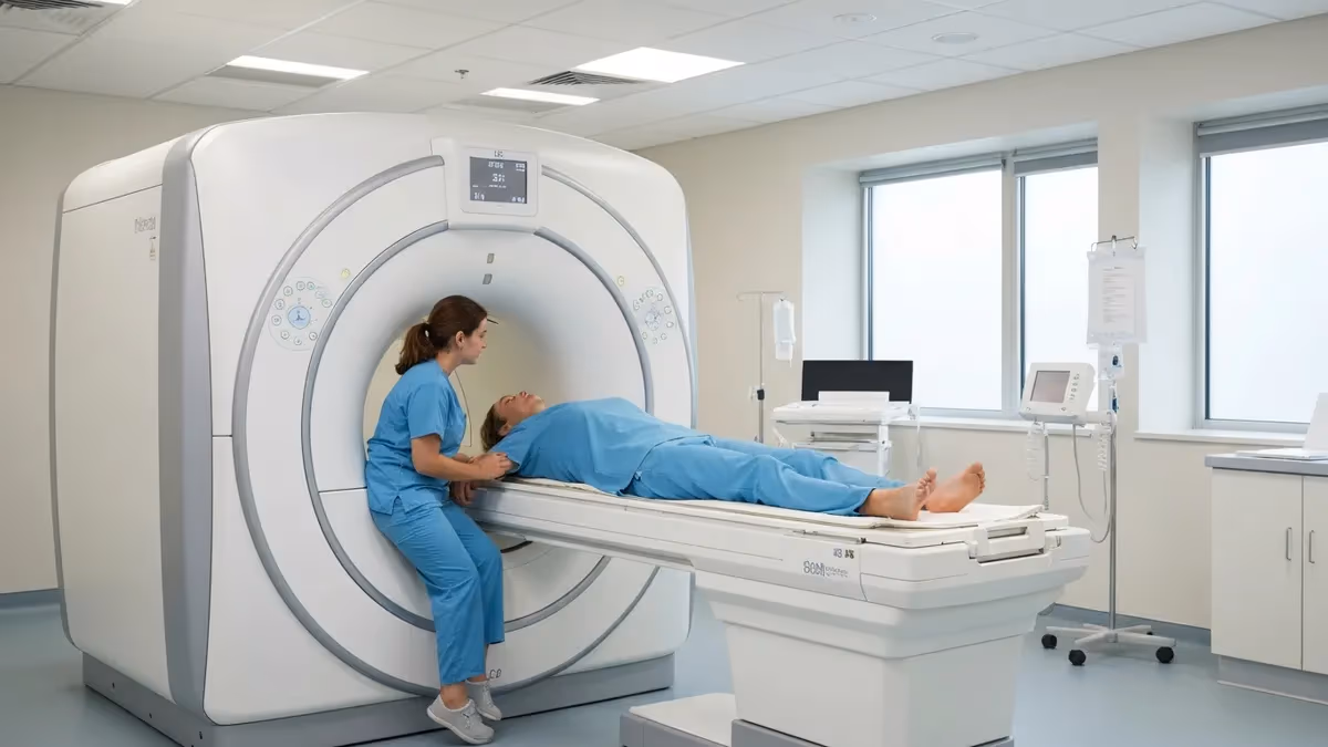
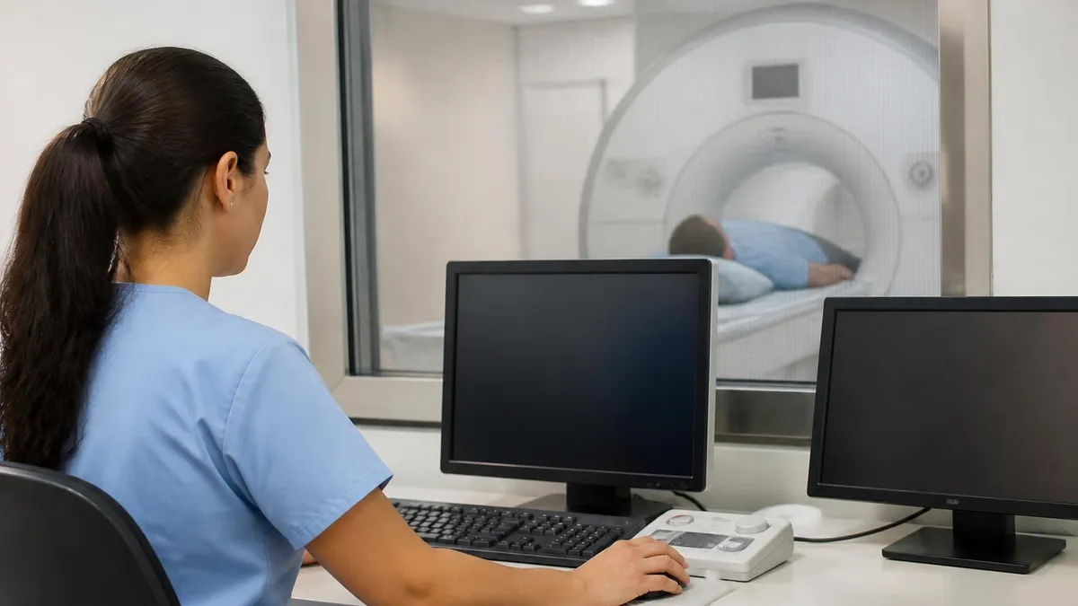
The rockledge mri & pet center is a dedicated outpatient imaging facility located in Rockledge, Florida, offering a comprehensive suite of diagnostic imaging services including magnetic resonance imaging (MRI), positron emission tomography (PET), and combined PET/CT scanning. Patients across Brevard County seek out this center for its advanced technology, board-certified radiologists, and commitment to delivering accurate, timely diagnostic results. Whether you are managing a chronic condition or investigating a new symptom, understanding what this facility provides can help you navigate your care with confidence.
Magnetic resonance imaging has transformed modern medicine by allowing clinicians to visualize soft tissues, organs, joints, and the central nervous system without exposing patients to ionizing radiation. Unlike X-rays or CT scans, MRI uses powerful magnetic fields and radiofrequency pulses to generate detailed, three-dimensional images. At Rockledge MRI & PET Center, scanners are regularly maintained and calibrated to ensure image quality meets radiological standards, which in turn supports accurate physician interpretation and treatment planning for a broad spectrum of conditions.
PET imaging complements MRI by measuring metabolic activity at the cellular level. By injecting a small amount of a radioactive tracer — most commonly fluorodeoxyglucose (FDG) — PET scans reveal how tissues and organs consume energy. This metabolic perspective is invaluable for oncology, cardiology, and neurology, where structural imaging alone may not distinguish between active disease and scar tissue, or between a benign lesion and a malignant tumor. Facilities that combine PET with CT — sometimes called PET/CT — overlay functional and anatomical data into a single, precisely correlated image.
Choosing an outpatient imaging center like Rockledge MRI & PET rather than a hospital-based radiology department often provides several practical advantages. Scheduling flexibility tends to be greater, wait times shorter, and the atmosphere more relaxed. Many patients report feeling less anxious when they arrive at a dedicated imaging center compared to a busy hospital radiology suite. Cost can also differ substantially; outpatient imaging centers frequently have lower facility fees, which matters for patients responsible for a significant share of their diagnostic costs under high-deductible health plans.
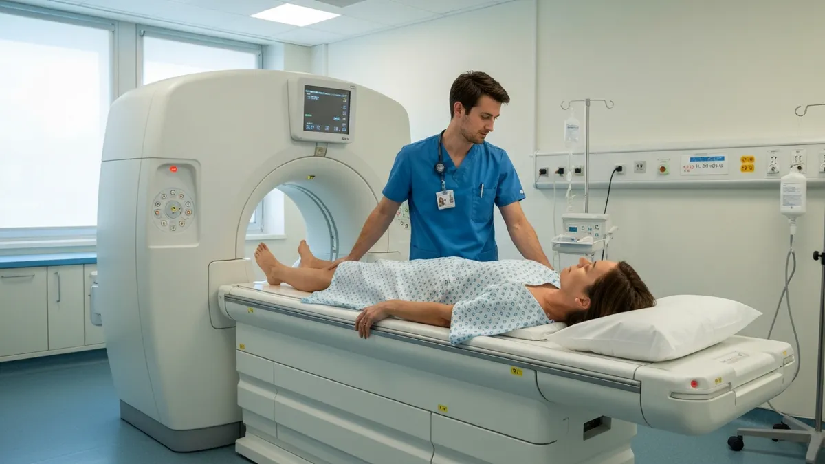
The referral process for advanced imaging typically begins with your primary care physician or specialist ordering a scan based on clinical findings, symptoms, or lab results. Once the order is received, the imaging center coordinates insurance authorization, schedules the appointment, and provides preparation instructions tailored to the specific type of study ordered. Patients who need contrast-enhanced MRI studies will receive additional guidance about fasting requirements and any medications that may need to be adjusted before the scan, particularly for those with kidney conditions that affect contrast clearance.
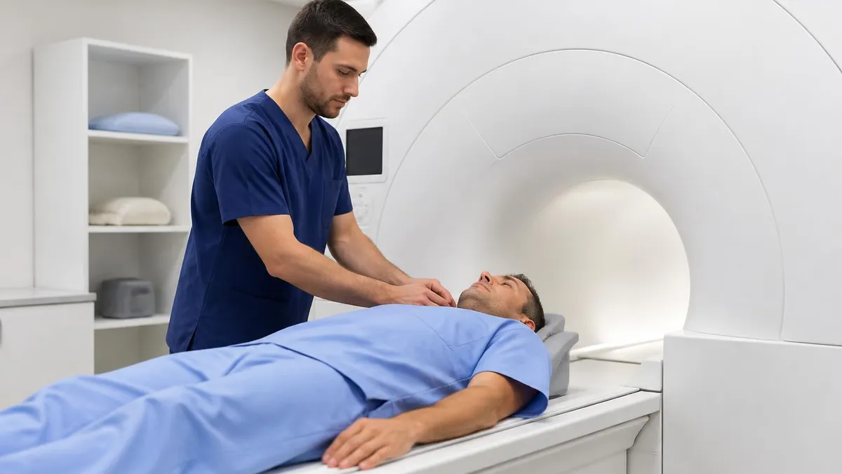
Technologists at accredited MRI and PET centers hold certification from organizations such as the American Registry of Radiologic Technologists (ARRT) or the American Registry for Diagnostic Medical Sonography (ARDMS). These credentials ensure that the individuals positioning patients, selecting imaging protocols, and monitoring scan quality have completed rigorous educational programs and passed standardized examinations. Continuing education requirements keep technologists current with evolving imaging techniques, safety protocols, and equipment capabilities, which directly benefits patient outcomes and diagnostic accuracy.
This guide covers the full scope of what patients, students, and healthcare professionals need to know about MRI and PET imaging services at centers like Rockledge MRI & PET Center. From the physics underlying each modality to practical preparation tips and frequently asked questions, the sections below walk through every dimension of the imaging experience. Whether you are preparing for your own scan, studying for a radiology credentialing exam, or simply curious about how these technologies work, you will find detailed, accurate information designed to inform and empower.
MRI & PET Imaging by the Numbers
Core Imaging Services at MRI & PET Centers
High-resolution soft tissue imaging using magnetic fields and radiofrequency pulses. Ideal for brain, spine, joints, and abdominal organs. No radiation exposure. Available with or without gadolinium-based contrast agents depending on clinical indication.
Metabolic imaging using radioactive tracers such as FDG to detect cancer, assess heart viability, and evaluate neurological disorders. Scans reveal biological activity rather than anatomy alone, enabling earlier and more precise diagnoses.
Simultaneous acquisition of metabolic PET data and anatomical CT images. The fusion product precisely locates abnormal metabolic activity within the body's structural context, improving staging accuracy for oncology and cardiology patients.
Intravenous gadolinium contrast improves visualization of tumors, inflammation, vascular structures, and blood-brain barrier disruption. Contrast protocols are selected based on referring physician orders and patient safety screening results.
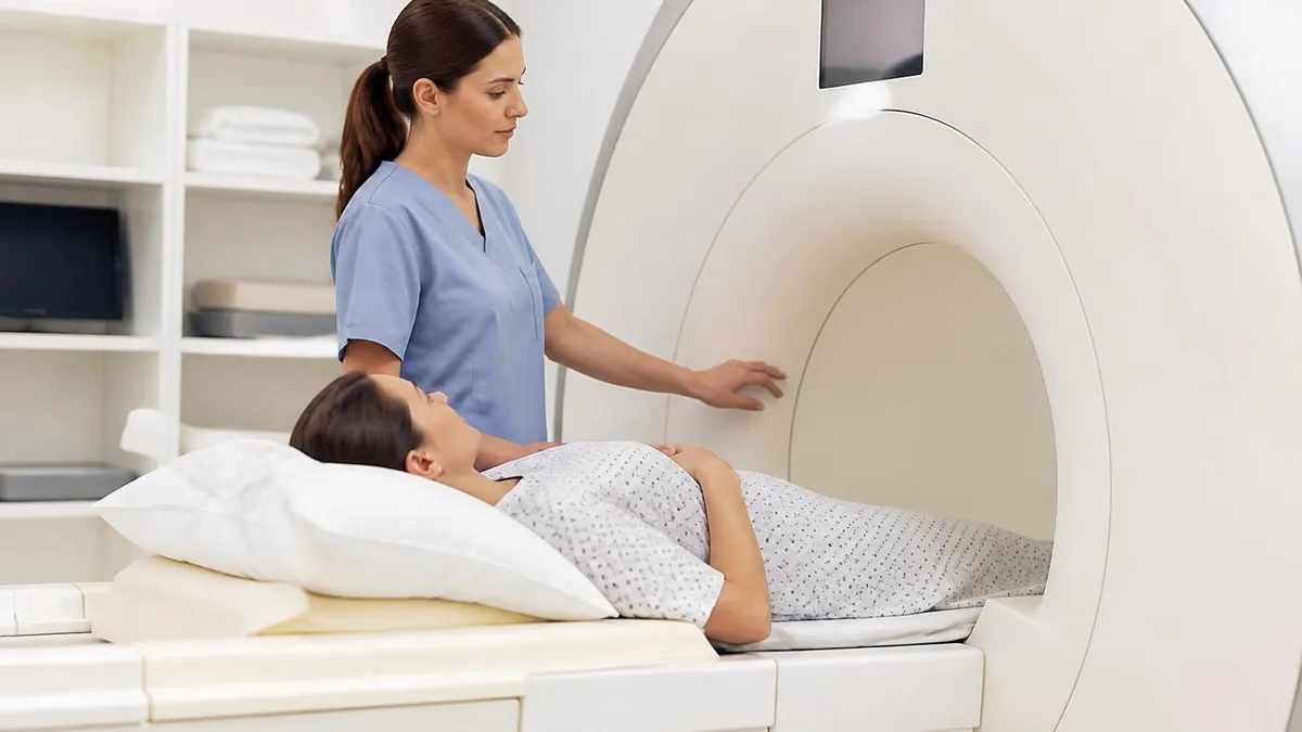
Wide-bore 70 cm scanners reduce claustrophobia and accommodate larger body habitus patients. Open MRI configurations offer even greater patient comfort for those with severe anxiety, though field strength and resolution may differ from closed-bore systems.
Understanding how MRI works at a fundamental level helps patients feel less anxious about the procedure and helps radiology students build the conceptual framework needed for credentialing examinations. MRI exploits the magnetic properties of hydrogen atoms, which are extraordinarily abundant in the human body due to the water and fat content of biological tissues. When a patient enters the bore of an MRI scanner, the powerful static magnetic field — measured in Tesla (T) — causes hydrogen protons to align along the field direction, much like compass needles aligning with Earth's magnetic field.
Once protons are aligned, the scanner delivers precisely tuned radiofrequency (RF) pulses at the Larmor frequency, which is the specific frequency at which hydrogen protons resonate within a given magnetic field strength. These RF pulses knock the protons out of alignment, and as the protons return to their resting state — a process called relaxation — they emit RF energy that the scanner's receiver coils detect. The timing characteristics of this relaxation process, described by the longitudinal relaxation time T1 and the transverse relaxation time T2, differ across tissue types and form the basis for MRI contrast.
T1-weighted images display fat as bright and water as dark, making them excellent for anatomical detail and post-contrast enhancement. T2-weighted images display water as bright and fat as intermediate, which highlights edema, cysts, cerebrospinal fluid, and many pathological processes. Proton density (PD) sequences represent a third fundamental weighting that reflects the concentration of hydrogen protons in a tissue, useful in musculoskeletal imaging of cartilage and fibrocartilaginous structures such as the menisci of the knee and the labrum of the shoulder and hip.
PET imaging operates on entirely different physical principles. A PET radiopharmaceutical — a molecule labeled with a positron-emitting radioisotope — is injected intravenously and distributed throughout the body according to normal metabolic pathways. For oncologic PET, FDG is the dominant tracer; because cancer cells typically have markedly elevated glucose metabolism (the Warburg effect), they accumulate FDG disproportionately compared to surrounding normal tissue. As the radioisotope decays, it emits positrons that travel only a short distance before annihilating with electrons to produce pairs of 511 keV gamma rays traveling in opposite directions.
PET detectors arranged in a ring around the patient detect these coincident gamma ray pairs and use their timing and angular information to reconstruct a three-dimensional map of tracer concentration. Modern PET scanners incorporate time-of-flight (TOF) technology, which uses the tiny difference in arrival times of the two coincident photons to localize the annihilation event more precisely along the line of response, dramatically improving image signal-to-noise ratio and enabling shorter scan times or reduced tracer doses without sacrificing diagnostic quality.
Gradient magnetic fields are an equally important component of MRI scanner hardware. Three sets of gradient coils — X, Y, and Z — allow the scanner to spatially encode the NMR signal by creating slight, predictable variations in magnetic field strength across the imaging volume.
This spatial encoding, combined with Fourier transformation of the raw data (called k-space), allows the reconstruction team to generate images at any desired slice orientation — axial, coronal, sagittal, or oblique — without physically moving the patient. The flexibility of oblique imaging planes is one of MRI's greatest advantages over CT for certain anatomical regions, particularly the knee, wrist, and temporomandibular joint.
Shimming is another technical process central to MRI quality. Because even minor inhomogeneities in the main magnetic field degrade image quality, every modern scanner includes active shimming coils that compensate for field irregularities measured during a brief prescan calibration. Passive shimming using strategically placed iron plates inside the magnet bore provides a coarser level of correction. Together, active and passive shimming ensure that the field uniformity across the imaging volume meets the tight tolerances needed for high-quality diagnostic imaging, which is why properly maintained, well-shimmed scanners produce consistently sharper images that support accurate radiological interpretation.
MRI Scan Types and Clinical Protocols
Brain MRI protocols typically include T1, T2, and FLAIR (Fluid-Attenuated Inversion Recovery) sequences in multiple planes, with diffusion-weighted imaging (DWI) added when acute stroke is suspected. Contrast-enhanced sequences using gadolinium help characterize tumors, abscesses, demyelinating plaques, and meningeal disease. A standard brain MRI without contrast takes approximately 30 to 45 minutes, while a comprehensive contrast protocol may extend to 60 minutes or longer depending on institutional protocols and clinical indication.
Spine MRI follows similar T1/T2 weighting principles but emphasizes sagittal and axial planes through the cervical, thoracic, or lumbar spine as directed by clinical symptoms. Disc herniations, spinal stenosis, cord signal abnormalities, and vertebral marrow lesions are among the most common findings. Fat suppression techniques such as STIR (Short Tau Inversion Recovery) increase sensitivity for marrow edema and inflammatory conditions, making them essential in protocols evaluating spondylitis, metastatic disease, and post-traumatic injury patterns.
Outpatient Imaging Center vs. Hospital Radiology: Key Differences
- +Shorter scheduling wait times — often same-week appointments available at outpatient centers
- +Lower facility fees, reducing out-of-pocket costs under high-deductible health plans
- +Relaxed, patient-centered environment with dedicated MRI and PET staff
- +Convenient parking and streamlined check-in processes typical of outpatient settings
- +Faster turnaround on radiology reports, often within 24 to 48 hours
- +Specialized technologists focused exclusively on advanced imaging rather than general radiology
- −On-site emergency support is limited compared to a full-service hospital campus
- −Some complex multi-modality protocols may require a hospital setting for coordination with clinical teams
- −Not all centers offer intraoperative or interventional MRI procedures
- −Insurance authorization may differ; some plans require hospital-based imaging for specific indications
- −After-hours availability may be restricted compared to 24/7 hospital radiology departments
- −Immediate physician consultation is typically not available if the scan reveals an urgent finding
MRI & PET Scan Preparation Checklist
- ✓Inform your scheduling coordinator of any metal implants, pacemakers, cochlear implants, or surgical hardware before your appointment.
- ✓Complete the MRI safety screening questionnaire honestly and thoroughly — disclose all prior surgeries and occupational metal exposure.
- ✓Follow fasting instructions for your specific scan — contrast MRI and PET studies often require 4-6 hours of fasting beforehand.
- ✓Arrive 15-20 minutes early to allow time for registration, safety screening, and changing into a provided gown.
- ✓Remove all metal objects including jewelry, piercings, hair clips, belts, and underwire bras before entering the MRI suite.
- ✓Leave credit cards, key fobs, and hearing aids outside the magnet room, as the magnetic field can demagnetize and damage these devices.
- ✓Inform the technologist of any kidney disease, diabetes, or allergy to contrast media if a contrast study has been ordered.
- ✓For PET scans, avoid strenuous exercise for 24 hours before your appointment to minimize muscular FDG uptake artifacts.
- ✓Arrange transportation home if you have been prescribed a sedative or anxiolytic medication for claustrophobia management.
- ✓Bring your insurance card, photo ID, and any prior imaging discs or reports that your radiologist should review for comparison.
Always Disclose All Metal Implants — Even Older Ones
Many patients assume older implants are automatically MRI-safe because they passed through airport security without triggering alarms. Airport metal detectors are not sensitive to the same materials that can be hazardous in a magnetic field. Always bring implant documentation to your MRI appointment, and never assume safety based on prior non-MRI screening experiences. Your technologist will verify conditional implant safety using the implantreference.com database or manufacturer documentation before proceeding with the scan.
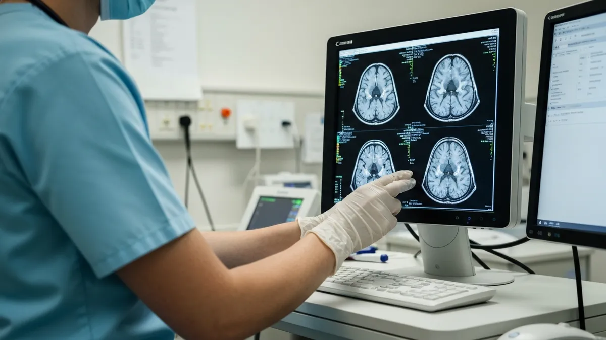
After your scan is complete, the raw image data is transferred to a PACS (Picture Archiving and Communication System) workstation where a board-certified radiologist reviews the images and prepares a written report. At most outpatient imaging centers, preliminary reports are available within a few hours for urgent studies, while routine studies are typically reported within 24 to 48 business hours. Your referring physician receives the report electronically and will contact you to discuss the findings, recommend follow-up, or adjust your treatment plan based on the imaging results.
Understanding the structure of a radiology report can help patients navigate their results more confidently. Most reports begin with a clinical history section that summarizes the indication for the scan, followed by a technique section describing the imaging protocol used. The findings section provides a systematic description of all structures visualized, noting both normal and abnormal observations. The impression section at the end summarizes the most clinically significant findings and may include recommendations for additional imaging, follow-up, or correlation with clinical or laboratory data.
Radiology reports use standardized terminology that can initially seem opaque to patients unfamiliar with medical language. Terms like "hyperintense" mean bright on the particular sequence being described, while "hypointense" means dark. An "incidental finding" refers to something discovered that was not the original target of the study — common examples include small liver cysts, adrenal nodules, or pulmonary nodules seen at the margins of an abdominal scan. Most incidental findings are benign, but some warrant follow-up imaging to establish stability over time, which is why radiologists frequently include management recommendations in their impressions.
For PET scan reports, the standard measure of metabolic activity is the Standardized Uptake Value (SUV), which normalizes tracer uptake relative to the injected dose and patient body weight. An SUV above a threshold — typically around 2.5 for FDG, though context-dependent — suggests hypermetabolic activity consistent with malignancy, infection, or inflammation. Oncologists use SUV measurements both to characterize lesions at initial staging and to assess treatment response on follow-up PET studies, with a meaningful decrease in SUV between baseline and post-treatment scans indicating metabolic response.
Radiological follow-up recommendations, often abbreviated in reports as "clinical correlation recommended" or "short-interval follow-up suggested," are not reasons for alarm — they reflect the radiologist's obligation to acknowledge interpretive uncertainty and suggest a path toward resolution. When a finding is truly indeterminate, additional imaging using a different modality or technique often provides the decisive information needed. For example, a liver lesion that appears indeterminate on a routine abdominal MRI may be definitively characterized as a benign hemangioma on a dedicated liver MRI using hepatobiliary contrast and perfusion sequences.
Patients have the legal right under HIPAA to request copies of their imaging studies and radiology reports. Most imaging centers provide discs or digital downloads of image data upon request, and many now use patient portals through which results can be accessed directly. Having your own copies of prior studies is valuable because comparison imaging is one of the most powerful tools radiologists use — the ability to demonstrate that a finding was present and unchanged years earlier can instantly convert a "suspicious" lesion into a "stable, likely benign" finding, avoiding unnecessary additional workup and patient anxiety.
Second opinions in radiology are entirely appropriate for complex or high-stakes findings and are readily available through academic medical centers, teleradiology services, and subspecialty radiologists. If you receive an unexpected diagnosis — particularly a new malignancy or a structural abnormality requiring surgical intervention — requesting a second radiological opinion before proceeding with treatment is a reasonable and commonly taken step. Many academic radiology departments offer formal second-opinion consultation services, and your imaging center can facilitate transfer of image data to support that process.

Gadolinium-based contrast agents (GBCAs) are generally safe but carry a rare risk of nephrogenic systemic fibrosis (NSF) in patients with severe renal impairment (eGFR below 30 mL/min/1.73m²). Always disclose any history of kidney disease, dialysis, or reduced kidney function before a contrast-enhanced MRI. Your imaging center will screen your renal function and may require a recent creatinine or eGFR lab result before administering contrast, or may choose a gadolinium agent with a lower NSF risk profile.
For MRI technologists, radiologic technologists, and radiology students, the knowledge base required to perform and interpret MRI studies extends well beyond operating the scanner console. The American Registry of Radiologic Technologists (ARRT) offers an MRI post-primary certification examination for eligible candidates who hold primary ARRT certification in radiography, nuclear medicine, or radiation therapy. The exam covers a breadth of content domains including patient care, safety, imaging procedures, and data acquisition and processing — all of which are directly applicable to daily clinical practice at facilities like Rockledge MRI & PET Center.
The ARRT MRI examination consists of 200 multiple-choice questions, of which 175 are scored and 25 are unscored pilot questions distributed randomly throughout the exam. Candidates have three hours and thirty minutes to complete the examination. The content specifications weight patient care and safety at approximately 25%, imaging procedures at 35%, and data acquisition, processing, and quality control at 40%, reflecting the technical complexity of modern MRI practice. Staying current with these content areas through structured study, practice examinations, and continuing education is essential for both initial certification and 10-year renewal cycles.
Beyond the ARRT credential, the American Registry of Magnetic Resonance Imaging Technologists (ARMRIT) offers an alternative MRI-specific credential that some technologists pursue as either a primary or supplemental certification. ARMRIT certification requires completion of an accredited MRI program or equivalent clinical hours, passing a written examination, and demonstrating clinical competency in the modality. Both ARRT and ARMRIT credentials are recognized by employers, though ARRT certification is more universally required as a condition of employment in hospital and large outpatient imaging center settings across the United States.
Nuclear medicine technologists who perform PET imaging typically hold certification from the Nuclear Medicine Technology Certification Board (NMTCB) or the ARRT nuclear medicine post-primary certification. PET-specific competencies include radiopharmaceutical handling and preparation, dosimetry, radiation safety, quality control of PET scanner performance, and patient care protocols for the unique requirements of PET studies. As PET/CT becomes standard at advanced imaging centers, technologists increasingly need cross-training in both nuclear medicine and computed tomography to efficiently operate combined scanners and produce diagnostic-quality fused datasets.
Continuing education (CE) requirements ensure that credentialed imaging professionals maintain and expand their knowledge throughout their careers. The ARRT requires 24 CE credits per biennium, with specific requirements for structured education and professional development. Topics most relevant to MRI technologists pursuing CE include advances in pulse sequence design, new contrast agent applications, MR safety updates, and emerging clinical protocols such as MR-guided focused ultrasound and MR-LINAC radiation therapy — areas where MRI technology is expanding its therapeutic as well as diagnostic role in modern medicine.
Salary data from the Bureau of Labor Statistics (BLS) and professional surveys consistently shows that MRI technologists earn above-average wages relative to the broader allied health workforce. The BLS Occupational Employment and Wage Statistics program reports median annual wages for radiologic and MRI technologists in the range of $64,000 to $80,000 nationally, with significant variation by geography, experience level, specialty certification, and practice setting. Florida wages tend to track slightly below national medians but are competitive given the state's lower cost of living in many regions outside major metropolitan areas.
Career advancement opportunities for MRI technologists include supervisory and lead technologist roles, educational positions in radiologic technology programs, application specialist positions with MRI equipment manufacturers, and clinical research roles supporting imaging trials in academic medical centers. Some experienced MRI technologists transition into MRI safety officer positions, where they lead institutional safety programs, review implant compatibility, train staff, and serve as the clinical expert resource for complex patient safety questions. These senior roles reflect the depth of expertise that accumulates over years of clinical practice in a modality as technically demanding as MRI.
Practical preparation for your MRI or PET appointment at any advanced imaging center involves both logistical and psychological dimensions. Logistically, confirming your insurance authorization status before the appointment day is one of the most important steps patients often overlook. Insurance pre-authorization for advanced imaging studies can take anywhere from 24 hours to several business days, and a scan performed without valid authorization may result in a substantial claim denial. Your imaging center's scheduling team typically manages this process, but confirming authorization is complete — and getting the authorization number — gives you a safety net if billing issues arise later.
Anxiety about confined spaces is among the most common reasons patients delay or cancel MRI appointments. Approximately 5 to 10 percent of patients experience significant claustrophobia during MRI scans, and many more feel some degree of discomfort in the bore.
Practical strategies that reduce MRI-related anxiety without medication include listening to music through MRI-compatible headphones (widely available at most centers), requesting a mirror attachment that allows you to see out of the bore, practicing slow abdominal breathing during the scan, and asking the technologist to explain each sequence before it begins so you know what to expect in terms of noise, duration, and required stillness. For patients with severe claustrophobia, low-dose oral anxiolytics prescribed by your physician can make the difference between a completed diagnostic study and an aborted attempt.
Noise is a distinctive feature of MRI that surprises many first-time patients. The loud banging and knocking sounds produced during scanning arise from rapid switching of gradient magnetic field currents, which cause the gradient coils to expand and contract abruptly. These sounds can reach 100 decibels or more within the bore, which is why all MRI facilities provide hearing protection — either foam earplugs, MRI-compatible headphones, or both.
Accepting and using hearing protection is not optional; sustained exposure to MRI acoustic noise without protection can contribute to cumulative noise-induced hearing threshold shift, particularly in technologists who work in close proximity to active scanners throughout their shifts.
For PET scan appointments, the uptake period — the time between tracer injection and imaging — typically runs 60 minutes for FDG PET studies. During this period, patients are asked to rest quietly, avoid speaking, and minimize muscle activity to reduce non-target FDG uptake in vocal cords and skeletal muscles.
Keeping warm during the uptake period also matters: brown adipose tissue (BAT), concentrated in the neck and supraclavicular regions, is metabolically active in response to cold and can create intense FDG uptake patterns that may mimic or obscure pathological findings. Many PET centers provide blankets and keep uptake rooms comfortably warm for this reason.
Hydration after contrast-enhanced MRI studies is frequently recommended to support renal clearance of gadolinium. While gadolinium chelates are rapidly cleared by the kidneys in patients with normal renal function — with over 90 percent excreted within 24 hours — maintaining good hydration supports this clearance process. Patients with impaired renal function may be advised to undergo additional monitoring or to receive alternative non-contrast protocols when the clinical information sought can be obtained without contrast administration. Your radiologist and the ordering physician together evaluate the risk-benefit balance of contrast use for each individual study.
Following up with your ordering physician promptly after receiving imaging results ensures that findings are acted upon within clinically appropriate timeframes. For urgent findings such as acute stroke, pulmonary embolism, aortic dissection, or high-grade spinal cord compression, most imaging centers have a critical results notification protocol that contacts the ordering physician immediately by telephone rather than waiting for the routine written report. Understanding that this system exists — and that your physician will be called directly for life-threatening findings — can provide reassurance that serious conditions will not go unaddressed during the reporting interval.
Finally, maintaining a personal health record that includes copies of your imaging reports and disc images supports longitudinal care across multiple providers and health systems. When you see a new specialist, arrive at an emergency room, or relocate to another state, having your prior imaging history readily accessible allows radiologists and clinicians to make accurate comparisons without delay. Cloud-based personal health record applications now make it easier than ever to store, organize, and share imaging data securely, and many imaging centers can directly upload your images to patient-accessible portals with a simple request made at the time of your appointment.
MRI Questions and Answers
DWI MRI: Diffusion-Weighted Imaging Explained (2026 Guide)
Side Effects of Brain MRI: Understanding Risks, Safety, and What Every Patient Should Know
MRI Lumbar Spine Without Contrast: Complete Guide to the Scan, What It Shows, and How to Prepare
MRI History: From Nuclear Resonance to Modern Scanners
MRI of Cervical Spine: Complete Guide to Imaging, Anatomy, Pathology, and Patient Preparation
About the Author
Medical Laboratory Scientist & Clinical Certification Expert
Johns Hopkins UniversityDr. Sandra Kim holds a PhD in Clinical Laboratory Science from Johns Hopkins University and is certified as a Medical Technologist (MT) and Medical Laboratory Scientist (MLS) through ASCP. With 16 years of clinical laboratory experience spanning hematology, microbiology, and molecular diagnostics, she prepares candidates for ASCP board exams, MLT, MLS, and specialist certification tests.
Join the Discussion
Connect with other students preparing for this exam. Share tips, ask questions, and get advice from people who have been there.
View discussion (5 replies)