MRI of Prostate: Complete Guide to Prostate MRI Imaging, PI-RADS Scoring, and What to Expect
🟢 MRI of prostate explained: multiparametric protocols, PI-RADS scoring, preparation, results, and what patients and technologists need to know.
An MRI of prostate, formally called multiparametric mri MRI or mpMRI, has become the gold standard imaging examination for detecting, localizing, and characterizing prostate cancer in men with elevated PSA, abnormal digital rectal exams, or prior negative biopsies. Unlike older ultrasound-guided biopsy approaches that sampled tissue blindly, prostate MRI delivers high-resolution anatomical images plus functional information that lets radiologists target suspicious lesions with millimeter precision. For radiologic technologists, urologists, and patients alike, understanding this exam is now essential to modern men's health care in 2026.
The exam combines three core sequences: T2-weighted imaging for zonal anatomy, diffusion-weighted imaging (DWI) with apparent diffusion coefficient (ADC) maps for cellular density, and dynamic contrast mri-enhanced (DCE) imaging for vascularity assessment. When interpreted using the PI-RADS v2.1 scoring system, multiparametric prostate MRI achieves sensitivity of roughly 89% for clinically significant prostate cancer, dramatically outperforming PSA testing alone, which has well-documented false-positive rates exceeding 70% in some screening populations.
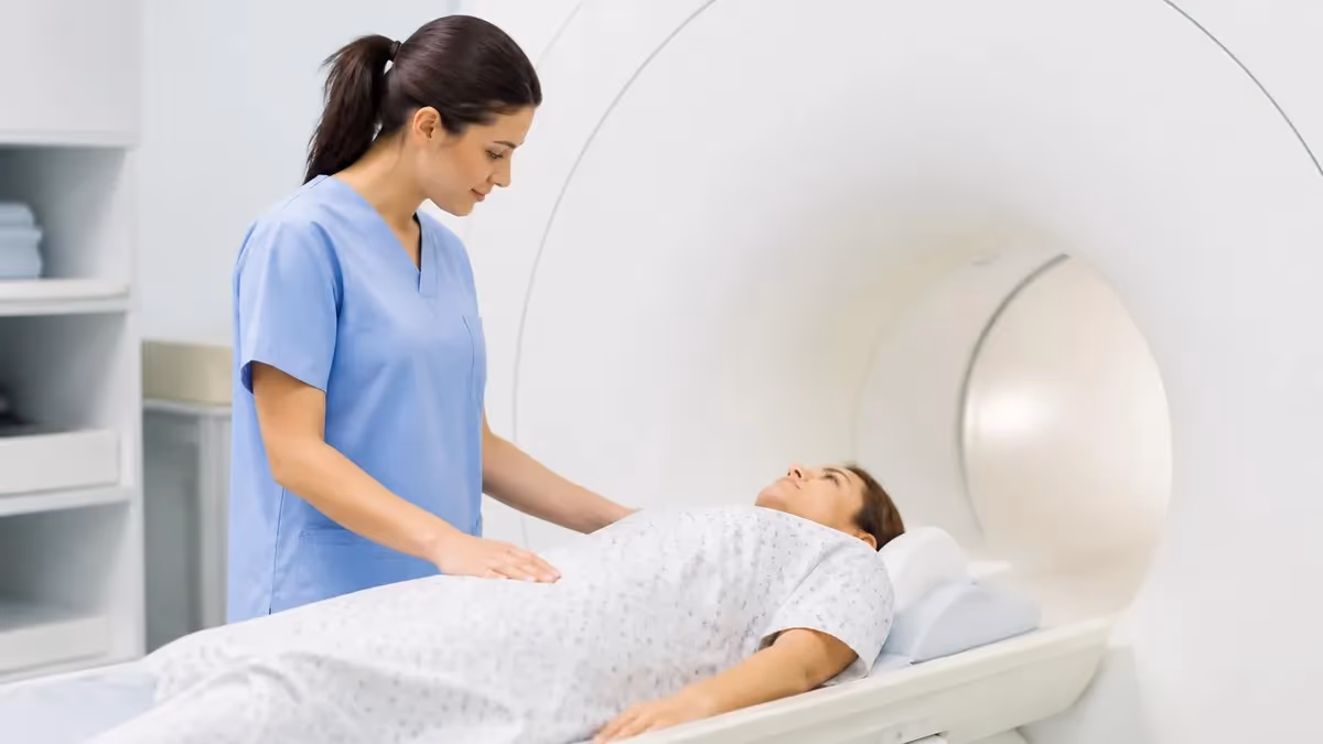
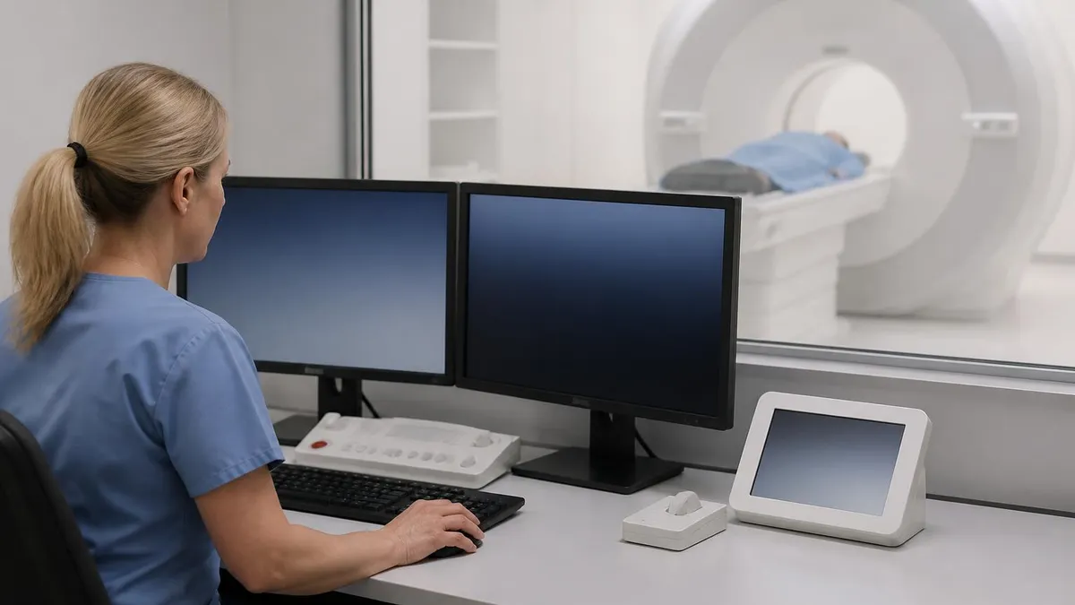
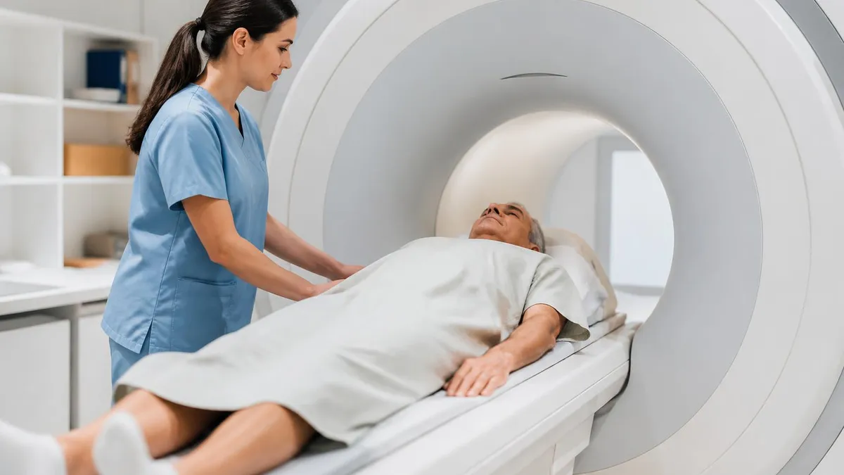
Most prostate MRI exams in the United States are now performed on 3.0 Tesla scanners using a surface phased-array coil placed over the pelvis, replacing the older endorectal coil that many patients found uncomfortable. Modern 3T technology delivers superior signal-to-noise ratio, allowing radiologists to identify lesions as small as 5 millimeters in diameter. Exam times typically range from 30 to 45 minutes, and patients undergo the scan supine with arms above their head, similar to other pelvic MRI protocols.
The clinical impact has been transformative. Before widespread mpMRI adoption around 2013, men with elevated PSA went directly to systematic 12-core transrectal ultrasound biopsy, which missed approximately 30% of clinically significant cancers while finding many indolent ones that would never cause harm. Now, MRI-first pathways using PI-RADS scoring allow up to 27% of men to avoid biopsy entirely when imaging is negative, while improving detection of aggressive Gleason 7 or higher tumors by roughly 18% according to the landmark PRECISION trial.
Preparation for the exam is straightforward but important. Patients typically need a gentle enema two hours before scanning to reduce rectal gas artifacts on diffusion-weighted sequences, must avoid ejaculation for three days before imaging to optimize seminal vesicle distention, and should stop antispasmodic medications only on physician advice. Most facilities require fasting four hours prior if gadolinium contrast will be administered, though many centers now perform biparametric MRI without contrast for screening indications.
For technologists preparing for ARRT MRI certification or working in advanced practice, prostate imaging represents one of the highest-growth subspecialties in MRI. Mastering coil placement, motion-reduction strategies, and protocol optimization for this exam directly impacts diagnostic quality and patient outcomes. This complete guide covers everything from the physics of each sequence to how radiologists assign the final PI-RADS score that drives biopsy decisions.
Throughout this article we will reference comparison topics such as whats difference between mri and ct scan when relevant to prostate imaging choices, and walk through patient experience, image interpretation principles, common artifacts, contraindications, and what an abnormal PI-RADS score actually means for next steps in clinical care.
MRI of Prostate by the Numbers
Prostate MRI Protocol Steps from Arrival to Image Review
Patient Intake & Screening
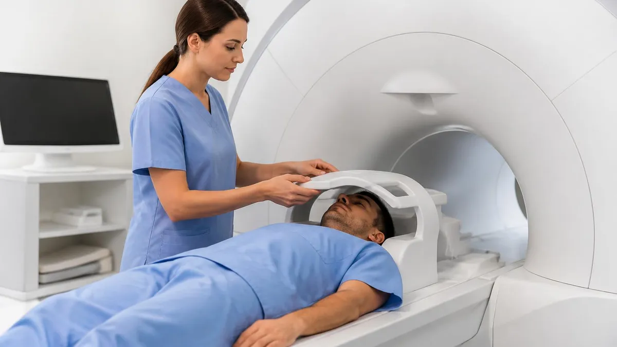
Coil Placement & Positioning
T2 Localizers & High-Res Imaging
Diffusion-Weighted Imaging
Dynamic Contrast-Enhanced
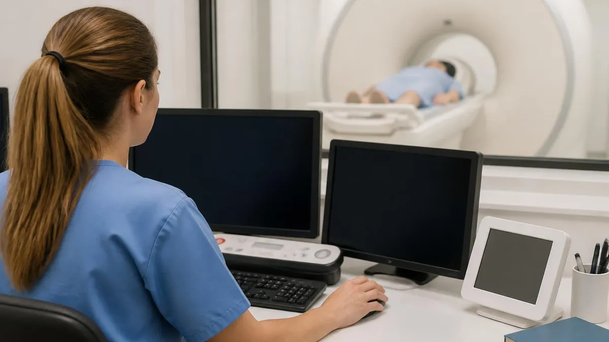
Image Review & Reporting
The diagnostic power of an MRI of prostate flows directly from combining anatomical and functional sequences, each interrogating different tissue properties. Understanding how each sequence contributes to the final interpretation is essential for technologists optimizing protocols and for clinicians counseling patients. The multiparametric approach is what separates modern prostate MRI from older T2-only studies that produced disappointing results in the 1990s and early 2000s.
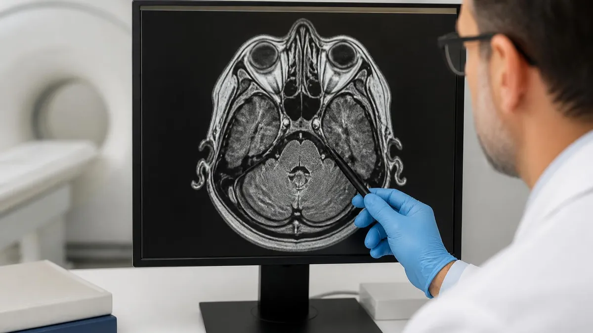
T2-weighted imaging forms the anatomical foundation. Acquired with fast spin echo techniques at 3 millimeter slice thickness and small field of view, T2 images depict the prostate's zonal anatomy with exquisite clarity. The peripheral zone appears uniformly hyperintense, the central and transition zones appear heterogeneous and lower in signal, and the surgical capsule between them is often visible. Cancers in the peripheral zone typically appear as focal hypointense nodules, while transition zone cancers show ill-defined, lentiform low signal that radiologists describe as an erased charcoal appearance.
Diffusion-weighted imaging measures the random Brownian motion of water molecules and is the single most important sequence for detecting clinically significant peripheral zone cancer. Malignant tissue has densely packed cells that restrict water diffusion, appearing bright on high b-value images (typically b=1400 to 2000 s/mm²) and correspondingly dark on the apparent diffusion coefficient or ADC map. Quantitative ADC values below 750 to 900 mm²/s strongly suggest aggressive disease, though absolute thresholds vary by scanner and vendor.
Dynamic contrast-enhanced imaging captures the wash-in and wash-out of gadolinium through prostate tissue. Cancers, being highly vascular with leaky neovasculature, typically show rapid early enhancement followed by rapid washout, while normal tissue and benign prostatic hyperplasia nodules show slower, persistent enhancement curves. DCE is most useful for upgrading equivocal PI-RADS 3 peripheral zone lesions to PI-RADS 4 when focal early enhancement is seen, though it plays a smaller role in transition zone scoring.
Knowing how an MRI works at the physics level helps technologists troubleshoot artifacts that frequently affect prostate imaging. Rectal gas creates susceptibility artifacts on echo-planar diffusion sequences, motion from peristalsis blurs T2 images, and patient movement during the 3-minute DCE acquisition can ruin the entire perfusion analysis. Mitigation strategies include pre-exam enemas, antiperistaltic agents like glucagon, and careful patient coaching about breathing and stillness.
Recently, biparametric prostate MRI (bpMRI) omitting the DCE sequence has gained traction for screening populations because it shortens exam time, eliminates contrast risks, and reduces cost. Multiple studies including the PRIME trial published in 2024 showed non-inferior cancer detection rates for bpMRI versus mpMRI in average-risk men, though most centers still default to multiparametric for biopsy-naive patients and equivocal cases.
Vendor-specific sequences like Siemens RESOLVE diffusion, GE FOCUS, and Philips ZOOM-DWI provide reduced field-of-view diffusion imaging with less geometric distortion, particularly valuable at 3T where echo-planar artifacts can be severe. Technologists should know which advanced sequences their scanner offers and when to apply them, especially in patients with hip prostheses, rectal gas, or large body habitus where standard EPI-DWI fails.
MRI Practice Test Questions
Prepare for the MRI - Magnetic Resonance Imaging exam with our free practice test modules. Each quiz covers key topics to help you pass on your first try.
MRI Knowledge
MRI Exam Questions covering Knowledge. Master MRI Test concepts for certification prep.
MRI Physics
Free MRI Practice Test featuring Physics. Improve your MRI Exam score with mock test prep.
MRI Anatomy and Pathology
MRI Test Prep for MRI Anatomy and Pathology. Practice MRI Quiz questions and boost your score.
MRI Anatomy and Positioning
MRI Questions and Answers on MRI Anatomy and Positioning. Free MRI practice for exam readiness.
MRI Contrast Agents
Free MRI Quiz on MRI Contrast Agents. MRI Exam prep questions with detailed explanations.
MRI Patient Care and Positioning
MRI Practice Questions for MRI Patient Care and Positioning. Build confidence for your MRI certification exam.
PI-RADS v2.1 Scoring for MRI of Prostate
PI-RADS 1 means clinically significant cancer is highly unlikely, with a normal-appearing prostate on all sequences and no focal abnormalities. PI-RADS 2 indicates unlikely significant cancer, where minor findings such as linear or wedge-shaped low signal on T2 may be present but without restricted diffusion or focal enhancement. These scores typically do not warrant biopsy unless PSA velocity or clinical suspicion is unusually high.
Patients receiving PI-RADS 1 or 2 reports are usually managed with continued PSA surveillance every 6 to 12 months rather than immediate biopsy. The negative predictive value for clinically significant cancer in this group exceeds 95% in large meta-analyses, providing meaningful reassurance to men anxious about elevated PSA results. Repeat MRI is generally recommended only if PSA continues rising.
Multiparametric MRI of Prostate vs Traditional TRUS Biopsy First
- +No radiation exposure to the pelvis or reproductive organs
- +Identifies clinically significant cancer 18% more often than systematic biopsy alone
- +Allows up to 27% of men with negative MRI to safely avoid biopsy entirely
- +Targets MRI-visible lesions precisely via fusion biopsy, improving sampling accuracy
- +Provides local staging information including extraprostatic extension and seminal vesicle invasion
- +Detects fewer indolent low-grade cancers, reducing overdiagnosis and overtreatment
- −Higher cost compared to ultrasound, typically $1,200-$2,500 in the United States
- −Requires patient cooperation and stillness for 30-45 minutes inside a noisy bore
- −Contrast-enhanced exams cannot be performed in patients with severe renal impairment
- −Susceptibility artifacts from hip prostheses or rectal gas can degrade diagnostic quality
- −Interpretation accuracy is highly reader-dependent and requires specialty training
- −Claustrophobic patients may need sedation or open-bore scanner accommodations
Patient Preparation Checklist for MRI of Prostate
- ✓Avoid ejaculation for at least 3 days before exam to maximize seminal vesicle distention
- ✓Perform a Fleet enema or equivalent 2 hours prior to scan to clear rectal contents
- ✓Avoid solid food for 4 hours before contrast-enhanced exams to reduce nausea risk
- ✓Continue regular medications with water unless explicitly told otherwise by the radiologist
- ✓Verify recent serum creatinine and eGFR if gadolinium contrast is planned
- ✓Remove all metallic objects including jewelry, hearing aids, dentures, and body piercings
- ✓Complete the MRI safety screening form documenting all surgeries and implants
- ✓Inform staff of any claustrophobia in advance so sedation can be arranged
- ✓Arrive 30 minutes early to allow for IV placement and antispasmodic administration
- ✓Bring previous prostate imaging and biopsy reports for radiologist comparison
MRI-First Pathways Reduce Unnecessary Biopsies by 27%
Multiple randomized trials including PRECISION (2018) and the more recent PRIME and PROMIS studies confirm that performing an MRI of prostate before biopsy in biopsy-naive men allows safe avoidance of biopsy when imaging is normal. This approach simultaneously increases detection of aggressive Gleason 7 or higher tumors while reducing diagnosis of clinically insignificant Gleason 6 disease that would never cause harm if left untreated.
Receiving the results of an MRI of prostate is often a moment of significant anxiety for patients, and clinicians who can clearly explain what each PI-RADS score means provide enormous value. The report will typically include prostate volume measurement, zonal description, PSA density calculation, lesion location using a 41-sector map, individual lesion measurements, sequence-specific scores, and a final overall PI-RADS assessment. Modern structured reporting templates ensure consistency across institutions.
For a PI-RADS 1 or 2 result, the negative predictive value for clinically significant cancer exceeds 95%. Patients with these scores generally do not need biopsy and can return to PSA surveillance with their urologist or primary care provider. However, men with very high PSA densities above 0.20 ng/mL/cc, strong family histories of aggressive prostate cancer, or known germline mutations in BRCA2 or HOXB13 may still warrant biopsy despite negative imaging because the residual risk, though low, is not zero.
PI-RADS 3 is the most clinically challenging category, often described as the diagnostic equipoise zone. Approximately one in five PI-RADS 3 lesions ultimately proves to be clinically significant cancer on targeted biopsy. Modern management combines PSA density thresholds, patient age and life expectancy, family history, and shared decision-making. Some institutions perform short-interval repeat MRI at 6 to 12 months to assess for lesion progression rather than immediate biopsy in low-risk men.
PI-RADS 4 and 5 findings should always trigger urology referral and discussion of MRI-targeted fusion biopsy. In fusion biopsy, the diagnostic MRI is co-registered with real-time transrectal or transperineal ultrasound, allowing the urologist to direct biopsy needles into the MRI-visible lesion with millimeter accuracy. This approach detects clinically significant cancer in 60 to 85% of PI-RADS 4-5 lesions, dramatically outperforming systematic biopsy.
The MRI report also provides local staging information vital for treatment planning. Extraprostatic extension, seminal vesicle invasion, neurovascular bundle involvement, and adjacent lymph node enlargement all influence whether a man is a candidate for nerve-sparing prostatectomy, external beam radiation, brachytherapy, or focal therapies such as HIFU and cryoablation. For locally advanced disease, additional staging with PSMA-PET CT or bone scan is typically recommended before treatment decisions.
Active surveillance has emerged as a major management option for low-risk Gleason 6 disease, and prostate MRI plays a central role in following these patients longitudinally. Most active surveillance protocols now incorporate annual or biennial MRI to detect progression, replacing the older practice of routine systematic re-biopsy every 1-3 years. Stability of MRI-visible lesions over time provides reassurance, while growing or new lesions trigger confirmatory biopsy.
For prostate cancer survivors, MRI also plays a critical role in detecting biochemical recurrence after definitive treatment. When PSA rises after prostatectomy or radiation, dynamic contrast-enhanced MRI of the prostate bed can identify focal recurrence amenable to salvage radiation or focal therapy. Combined with PSMA-PET, modern imaging has revolutionized our ability to detect and treat recurrent disease at much earlier stages than was previously possible.
Gadolinium-based contrast agents used in dynamic contrast-enhanced prostate MRI are generally safe but carry rare risks of nephrogenic systemic fibrosis in patients with severe renal impairment (eGFR below 30 mL/min/1.73m²). All patients receiving contrast should have recent renal function testing, and macrocyclic agents such as gadobutrol or gadoterate meglumine are preferred over older linear agents. Discuss any prior contrast reactions, asthma, or pregnancy with the technologist before injection.
Safety considerations for an MRI of prostate begin long before the patient enters the magnet room. The MRI safety screening must identify all implants, devices, and surgical hardware that could pose risks in the 3 Tesla static field. While most modern joint replacements, surgical clips, and stents are MR-conditional, older devices, cardiac pacemakers without MR-conditional labeling, cochlear implants, certain neurostimulators, and metallic fragments near the eyes remain absolute or relative contraindications requiring careful risk-benefit analysis.
Hip prostheses deserve special attention in prostate imaging because they sit immediately adjacent to the prostate gland and produce severe susceptibility artifacts on echo-planar diffusion sequences. For patients with bilateral hip replacements, dedicated metal artifact reduction sequences such as MARS and reduced field-of-view diffusion techniques can salvage diagnostic image quality. Reviewing MRI MARS protocol fundamentals is valuable for technologists encountering these challenging cases.
Claustrophobia affects approximately 5 to 10% of patients undergoing prostate MRI and can be addressed through several strategies. Wide-bore 70 cm scanners, prone positioning when feasible, prism glasses allowing the patient to see outside the bore, headphones with music, and oral or IV anxiolytics like lorazepam can all help. In severe cases, open-bore 1.5T systems can perform multiparametric prostate MRI, though diagnostic performance is slightly inferior to 3T closed-bore systems.
Contrast safety is another important domain. Modern macrocyclic gadolinium agents such as gadobutrol, gadoterate meglumine, and gadoteridol have excellent safety profiles, with severe adverse reaction rates well below 0.01%. However, patients with severe renal impairment, prior allergic-like contrast reactions, or active pregnancy require careful evaluation. Biparametric MRI without contrast is increasingly used as a safe alternative in these populations.
Limitations of prostate MRI include reader dependence, lesion size threshold, and false positives from prostatitis. Diagnostic accuracy varies significantly between expert academic radiologists and general radiologists, with some studies showing PI-RADS 3 lesion detection accuracy differing by 20% or more between readers. Lesions smaller than 5 mm are often invisible even on optimal 3T imaging. Acute and chronic prostatitis can mimic cancer with restricted diffusion and early enhancement, leading to false-positive PI-RADS 4 calls.
Cost and accessibility remain practical barriers in many parts of the United States. Out-of-pocket costs for self-pay prostate MRI range from $1,200 to $2,500, and even insured patients face significant co-pays. Geographic disparities in MRI availability mean rural patients may travel hours to reach a center with prostate-specific expertise. Advocacy for broader Medicare and commercial coverage continues, with most major insurers now covering MRI before biopsy for men with elevated PSA.
Quality assurance is critical to reliable prostate imaging. The American College of Radiology offers prostate MRI accreditation, and the recently created PI-QUAL scoring system allows radiologists and technologists to objectively rate the technical quality of each exam on a 1 to 5 scale. PI-QUAL scores of 4 or 5 ensure that the diagnostic conclusions are well-supported by adequate image quality across all sequences, while scores of 1 or 2 prompt repeat imaging.
For patients scheduled for an MRI of prostate, several practical tips can dramatically improve both the experience and the diagnostic quality of the exam. First, take the ejaculation abstinence requirement seriously. Recent ejaculation collapses the seminal vesicles and can mask invasion by aggressive cancers. Three full days of abstinence is the minimum, and some centers recommend five days for optimal seminal vesicle distention and accurate staging assessment.
Second, complete the pre-exam enema thoroughly. Rectal gas and stool produce devastating susceptibility artifacts on the diffusion-weighted sequences that drive PI-RADS scoring. A Fleet enema 2 hours before the scan, followed by sitting on the toilet to fully evacuate, dramatically improves image quality. If the technologist sees significant gas on the localizer, they may ask the patient to use the restroom again before scanning resumes.
Third, communicate honestly about claustrophobia, anxiety, and physical limitations. The prostate MRI exam requires lying still in a noisy enclosed space for 30 to 45 minutes. Patients who think they may struggle should request a tour of the scanner before exam day, ask about oral lorazepam premedication, or inquire whether the facility has a wide-bore scanner. Hiding anxiety until the day of often results in incomplete or non-diagnostic exams that must be repeated.
Fourth, technologists should optimize protocols proactively rather than reactively. Reviewing the patient's history for hip prostheses, recent biopsy with post-biopsy hemorrhage, or known prostatitis allows preemptive protocol adjustments. Post-biopsy hemorrhage can mimic cancer on T2 imaging and persists for 6 to 8 weeks, so scheduling MRI at least 6 weeks after biopsy is preferred when possible. Bowel preparation orders should be standardized and confirmed at scheduling.
Fifth, leverage structured reporting and PI-QUAL scoring. Modern PACS and reporting systems offer prostate-specific templates that ensure all required elements appear in every report. The PI-QUAL system rates technical exam quality on a 1-5 scale, providing accountability for image quality and helping identify systemic protocol issues. Radiologists should regularly audit their PI-RADS scoring against biopsy outcomes to refine interpretive accuracy.
Sixth, stay current with evolving guidelines. The PI-RADS scoring system has gone through versions 1, 2, 2.1, and is actively being updated. Biparametric MRI without contrast is gaining acceptance for screening but remains controversial for biopsy-naive men. Artificial intelligence tools for prostate MRI segmentation and lesion detection received FDA clearance starting in 2021 and are increasingly integrated into clinical workflow, though human radiologist oversight remains essential.
Finally, for technologists preparing for the ARRT MRI advanced certification, prostate imaging questions appear frequently on the registry exam. Understanding zonal anatomy, b-value selection for diffusion imaging, dynamic contrast injection protocols, common artifacts and their mitigation, and PI-RADS scoring fundamentals will pay dividends both on the exam and in daily practice. Practice questions specifically targeting prostate MRI, pelvic anatomy, and contrast-enhanced techniques are an efficient way to consolidate knowledge before sitting for certification.
MRI Questions and Answers
MRI MARS Protocol: Complete Guide to Metal Artifact Reduction Sequences for Orthopedic Imaging
How Does an MRI Work: Magnetic Resonance Imaging Explained
MRI With or Without Contrast: What to Expect

The History of MRI: From Discovery to Modern Medicine
What's the Difference Between MRI and CT Scan? A Complete Comparison Guide
About the Author
Medical Laboratory Scientist & Clinical Certification Expert
Johns Hopkins UniversityDr. Sandra Kim holds a PhD in Clinical Laboratory Science from Johns Hopkins University and is certified as a Medical Technologist (MT) and Medical Laboratory Scientist (MLS) through ASCP. With 16 years of clinical laboratory experience spanning hematology, microbiology, and molecular diagnostics, she prepares candidates for ASCP board exams, MLT, MLS, and specialist certification tests.
Join the Discussion
Connect with other students preparing for this exam. Share tips, ask questions, and get advice from people who have been there.
View discussion (6 replies)