What's the Difference Between MRI and CT Scan? A Complete Comparison Guide
What's the difference between MRI and CT scan? Compare imaging technology, costs, scan times, radiation, and which test is right for your diagnosis. ❓
If your doctor has ever ordered imaging and you found yourself wondering what's the difference between MRI and CT scan, you are not alone. These two technologies look similar from the outside — both involve lying still inside a large machine — but they work on completely different physical principles, image different tissues with different clarity, and serve very different clinical purposes. Understanding the distinction can help you ask better questions, prepare correctly, and feel more confident about why a particular scan was chosen for you.
A computed tomography (CT) scan uses ionizing X-ray radiation rotated around the body to build cross-sectional slices, while magnetic resonance imaging (MRI) uses a powerful magnetic field and radio waves to map the behavior of hydrogen protons in soft tissue. That single technical difference cascades into nearly every other distinction you will read about — cost, scan duration, image quality, contraindications, and the kinds of pathology each test is best at finding. Neither is universally better; they are complementary tools.
CT scans tend to be fast, widely available, and excellent for visualizing bone, acute hemorrhage, lung tissue, and trauma. Emergency departments rely on CT for the same reason: a head CT can be completed in under a minute, which matters when a patient may be having a stroke or internal bleeding. MRI scans, by contrast, take 20 to 60 minutes and are much better at distinguishing between soft tissues with similar densities — ligaments, brain matter, spinal cord, cartilage, and tumors that would appear as vague gray blobs on a CT.
Cost is another major divider. In the United States, a CT scan typically runs between $300 and $1,500 depending on the body part and whether contrast is used. An MRI usually costs $1,000 to $5,000 for the same anatomic region, partly because the scanner itself costs millions of dollars and partly because scan times tie up the machine longer. Insurance coverage, prior authorization requirements, and your deductible all influence what you actually pay out of pocket.
Safety profiles also differ in important ways. CT exposes you to ionizing radiation — a chest CT delivers roughly the equivalent of two years of natural background radiation in a single sitting. MRI uses no radiation at all, which is why it is often preferred for pregnant patients, children, and anyone needing repeated imaging. However, MRI has its own contraindications: pacemakers, certain metal implants, cochlear implants, and severe claustrophobia can all rule it out or require modification.
Even the patient experience feels different. CT scanners are open, donut-shaped rings; you slide through quickly and are done. MRI scanners are long tubes that can feel confining, the machine is loud (often 100+ decibels), and you must hold still for extended periods. Some MRIs require contrast injection with gadolinium, while CT contrast uses iodine — both have their own allergy and kidney-function considerations that the imaging team will screen for before your appointment.
This guide breaks down every meaningful difference between MRI and CT, when each one is the right tool, what to expect from preparation through recovery, and how to interpret the recommendation your physician gave you. If you are also weighing whether your scan requires intravenous dye, our deep dive into mri with and without contrast cpt walks through the specifics in detail.
MRI vs CT by the Numbers
How MRI and CT Scanners Actually Work
A CT scanner spins an X-ray tube and detector array around your body, capturing hundreds of cross-sectional images per rotation. A computer reconstructs these into 2D slices or 3D models. Speed is its hallmark — modern scanners image the chest in under five seconds.
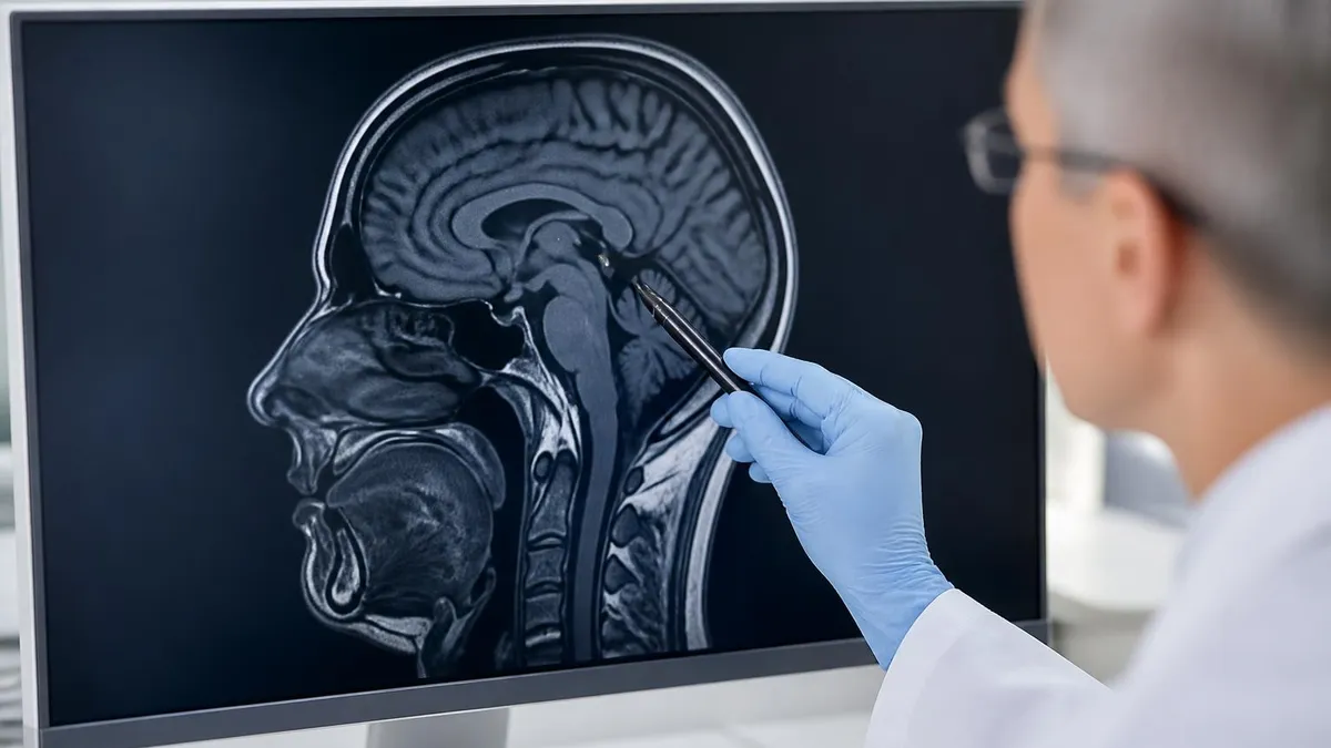
An MRI scanner uses a superconducting magnet to align hydrogen protons in your body, then radio-frequency pulses knock them out of alignment. As the protons relax, they emit signals that the machine translates into stunningly detailed soft-tissue images.
CT uses iodine-based contrast injected intravenously to highlight blood vessels and vascular lesions. MRI typically uses gadolinium-based agents, which behave differently — enhancing certain tumors, inflammation, and breakdown of the blood-brain barrier with remarkable sensitivity.
Both technologies produce digital slices that radiologists can scroll through on high-resolution monitors. CT slices are typically 0.5-5 mm thick; MRI offers multiple sequences (T1, T2, FLAIR, DWI) that highlight different tissue properties in the same anatomical region.
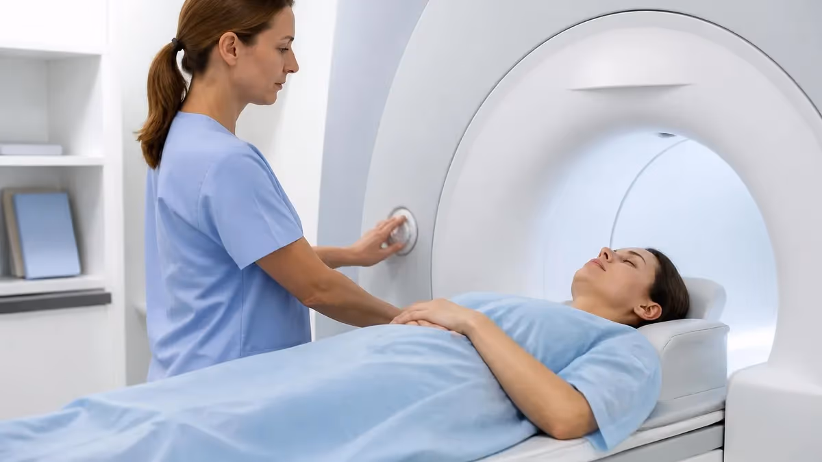
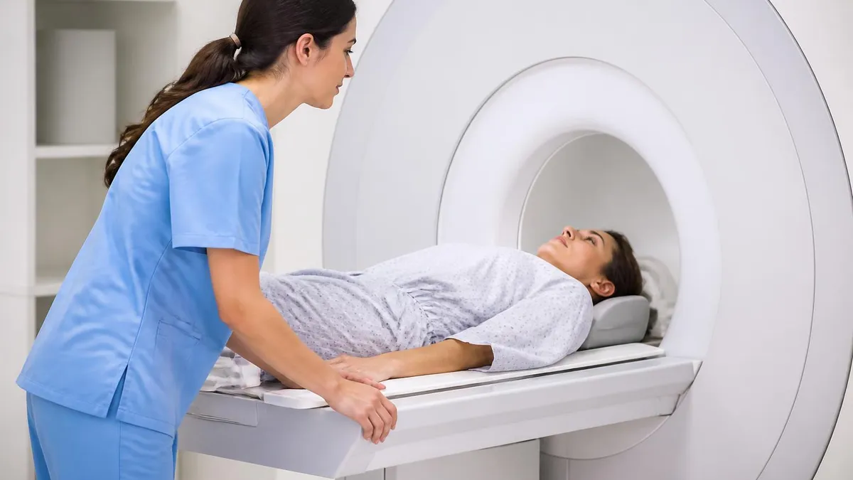
CT scans are quick and the gantry is open and shallow. MRI requires lying inside a longer, narrower bore for extended periods. Open MRI machines exist for claustrophobic patients but generally produce lower-resolution images than closed-bore systems.
When it comes to image quality, each modality has a clear domain of superiority. CT is unmatched for visualizing dense structures — bones, calcifications, kidney stones, and acute bleeding. Because X-rays pass through soft tissue more easily than bone, you get sharp contrast at tissue boundaries that involve density differences. A radiologist looking at a CT of the abdomen can quickly identify a kidney stone the size of a grain of rice or detect free air outside the bowel indicating perforation. CT is the workhorse of trauma medicine for exactly this reason.
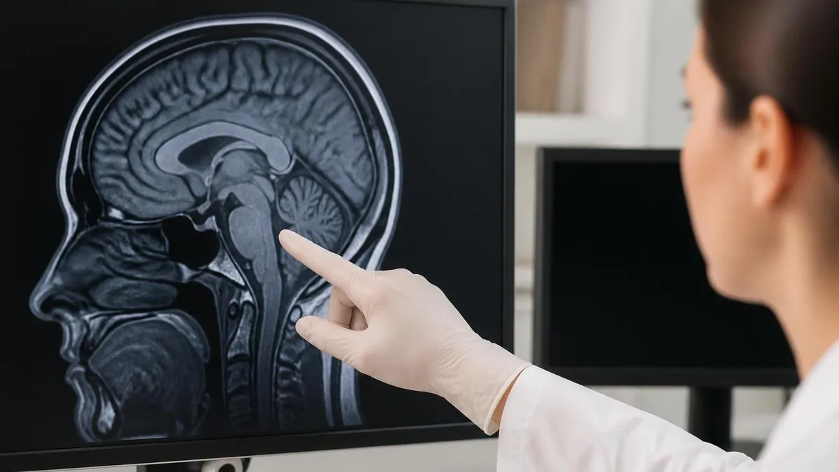
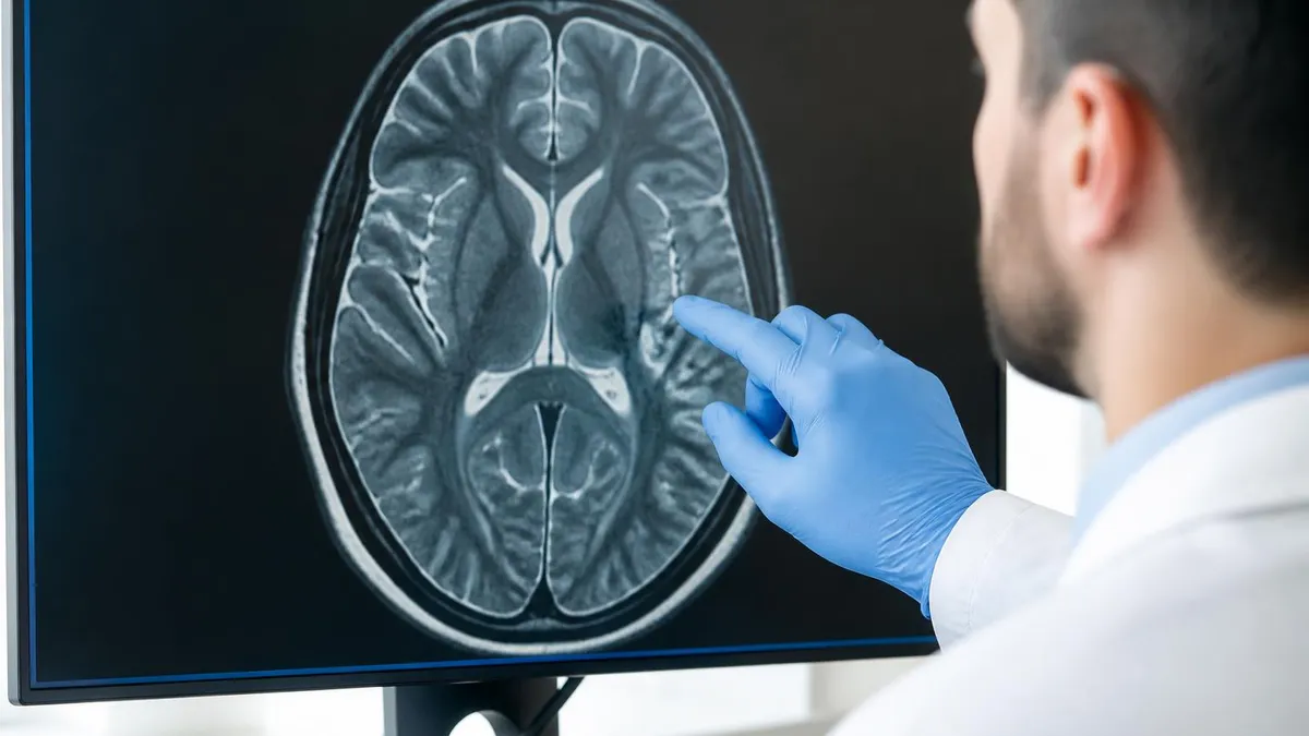
MRI dominates anywhere soft tissue contrast matters. Brain MRI can distinguish between gray matter, white matter, cerebrospinal fluid, blood vessels, and lesions as small as a few millimeters. Spinal MRI shows discs, nerve roots, and the spinal cord itself with clarity no CT can match. Joint MRI reveals ligament tears, cartilage damage, and meniscal injuries — the kinds of findings that send athletes for surgical evaluation. MRI is the gold standard for staging many cancers because it captures tumor margins so precisely.
Specific clinical scenarios tend to dictate which scan you receive. Stroke evaluation usually starts with a non-contrast head CT to rule out hemorrhage in minutes, then progresses to MRI within hours to visualize the ischemic penumbra and confirm the diagnosis. Suspected pulmonary embolism gets a CT angiogram of the chest because the technology images the pulmonary arteries quickly and reliably. Multiple sclerosis, on the other hand, is diagnosed and tracked almost exclusively with MRI because demyelinating lesions show up beautifully on FLAIR and T2 sequences.
Musculoskeletal complaints follow a similar pattern. A patient with acute trauma and possible fracture goes straight to CT or X-ray. The same patient returning weeks later with persistent knee pain after a normal X-ray would be sent for MRI to evaluate ligaments, cartilage, and meniscus. Sports medicine, orthopedic surgery, and physical medicine specialists all rely heavily on MRI to plan interventions. CT may be added later if a complex fracture needs 3D reconstruction for surgical planning.
Cancer imaging combines both modalities depending on the organ involved. Liver, brain, prostate, and breast tumors are typically characterized with MRI. Lung nodules, pancreatic masses, and many lymphomas are followed with CT because of speed and reproducibility. PET-CT and PET-MRI fusion technologies are increasingly common in oncology centers, marrying the metabolic information of positron emission tomography with the anatomical detail of either CT or MRI.
Pediatric imaging strongly favors MRI when feasible because children are more vulnerable to lifetime radiation exposure. Hospitals increasingly avoid CT in kids under age 18 unless the clinical question genuinely requires it. Ultrasound is often the first-line alternative, with MRI as second choice. For chronic conditions requiring repeat imaging — inflammatory bowel disease, congenital heart disease, certain cancers — MRI protocols have been developed specifically to spare young patients cumulative radiation dose.
Pregnant patients face their own considerations. CT involves radiation that can pose risks to a developing fetus, particularly in the first trimester, so MRI is generally preferred when imaging is medically necessary. Gadolinium contrast for MRI is typically avoided in pregnancy unless absolutely essential, while non-contrast MRI is considered safe in all trimesters. For background on how the technology evolved into the diagnostic powerhouse it is today, see the full history of MRI and its key milestones.
MRI Practice Test Questions
Prepare for the MRI - Magnetic Resonance Imaging exam with our free practice test modules. Each quiz covers key topics to help you pass on your first try.
MRI Knowledge
MRI Exam Questions covering Knowledge. Master MRI Test concepts for certification prep.
MRI Physics
Free MRI Practice Test featuring Physics. Improve your MRI Exam score with mock test prep.
MRI Anatomy and Pathology
MRI Test Prep for MRI Anatomy and Pathology. Practice MRI Quiz questions and boost your score.
MRI Anatomy and Positioning
MRI Questions and Answers on MRI Anatomy and Positioning. Free MRI practice for exam readiness.
MRI Contrast Agents
Free MRI Quiz on MRI Contrast Agents. MRI Exam prep questions with detailed explanations.
MRI Patient Care and Positioning
MRI Practice Questions for MRI Patient Care and Positioning. Build confidence for your MRI certification exam.
MRI vs CT Scan Side-by-Side Comparison
CT scans use ionizing X-ray radiation rotated 360 degrees around the patient. Detectors capture how much radiation passes through tissues of different densities, and software reconstructs cross-sectional slices. The whole imaging process for a body region typically takes seconds. CT excels at bone, lung tissue, acute hemorrhage, and dense structures because X-ray attenuation varies sharply with tissue density.
MRI uses no radiation. Instead, a powerful magnet (commonly 1.5 or 3 Tesla) aligns hydrogen protons in your body. Radio-frequency pulses temporarily knock these protons out of alignment, and as they relax, they emit signals decoded into images. Different pulse sequences highlight different tissue properties, giving MRI unmatched soft-tissue contrast for brain, spinal cord, muscles, ligaments, and tumors.
MRI vs CT: When Each One Wins
- +MRI gives superior soft-tissue detail for brain, spine, and joints
- +MRI uses zero ionizing radiation — safer for repeat imaging and pregnancy
- +MRI sequences (T1, T2, FLAIR, DWI) reveal different tissue properties in one session
- +MRI is the gold standard for multiple sclerosis, ligament tears, and many cancers
- +MRI contrast (gadolinium) highlights tumors and inflammation with high sensitivity
- +MRI can image in any plane without repositioning the patient
- −CT is 10-50x faster — critical in trauma, stroke, and emergency settings
- −CT costs about one-third as much as MRI on average
- −CT excels at bone, lung tissue, kidney stones, and acute hemorrhage
- −CT has fewer contraindications — pacemakers and metal implants are not problems
- −CT is more accessible in rural hospitals and smaller imaging centers
- −CT scans are far better tolerated by claustrophobic or anxious patients
Preparation Checklist: What to Do Before Your MRI or CT Scan
- ✓Tell your doctor about all medical implants, including pacemakers, stents, and joint replacements.
- ✓Disclose any history of kidney disease before contrast is administered.
- ✓List all allergies, especially to iodine (CT) or gadolinium (MRI) contrast agents.
- ✓Confirm with your provider whether you should fast — usually 4-6 hours before contrast studies.
- ✓Wear comfortable clothing without metal zippers, buttons, or underwire.
- ✓Remove all jewelry, watches, hairpins, and piercings before MRI scanning.
- ✓Bring a list of current medications and any prior imaging on disc.
- ✓Arrange a ride home if you'll receive sedation for claustrophobia or pediatric scans.
- ✓Tell the technologist if you are or might be pregnant before any imaging begins.
- ✓Arrive 15-30 minutes early to complete screening forms and answer safety questions.
Neither scan is universally better — they are complementary tools
The most common misconception is that MRI is always 'better' than CT because it's more expensive and more detailed. In reality, the right scan is the one that answers the clinical question. Trust your provider's judgment: CT can save a life in minutes during a stroke or trauma, while MRI may be essential for diagnosing a subtle brain lesion or evaluating cartilage damage that no CT could ever detect.
Cost is one of the biggest practical differences between MRI and CT, and understanding what drives the price helps you plan financially and advocate for yourself. The MRI scanner itself can cost a hospital anywhere from $1 million to $3 million, plus annual maintenance contracts in the six-figure range. The cryogen cooling system that keeps the magnet superconducting consumes helium continuously. CT scanners are cheaper to purchase and operate, but they still represent a major capital investment that gets recouped through scan fees over time.
For patients in the United States, the sticker price of imaging varies wildly by facility, region, and insurance contract. The same MRI of the lumbar spine might cost $400 at an independent imaging center and $3,200 at a hospital outpatient department — for the exact same study. CT pricing shows similar variation but at a lower absolute range. Asking for the negotiated cash price, comparing facilities, and using transparency tools available through your insurer can save thousands of dollars on a single elective study.
Insurance coverage adds another layer of complexity. MRI scans almost always require prior authorization for elective indications, meaning your physician's office must submit clinical documentation justifying the test before insurance will pay. CT scans face less stringent authorization requirements because they are often ordered emergently or for clearer indications. If your prior authorization is denied, you have appeal rights — and many denials are overturned when additional clinical context is provided.
Access varies geographically as well. Almost every hospital with an emergency department has a CT scanner running 24/7. MRI availability is more concentrated; rural areas may have one machine serving an entire region, with appointment waits of weeks for non-urgent scans. Mobile MRI units that rotate among smaller hospitals help bridge this gap but typically operate only on scheduled days. Finding a convenient nearby facility for your study is worth the effort, especially when follow-up imaging is anticipated.
Scan duration also affects access and scheduling. Because MRI ties up a multimillion-dollar scanner for 30-60 minutes per patient, facilities run fewer studies per day. CT can image dozens of patients per hour, which is why emergency departments rely on it for high-throughput evaluation. If a same-day appointment matters to you, CT availability is typically easier to find than MRI. For urgent MRI needs, many academic centers maintain after-hours coverage but at premium pricing.
The financial picture also depends on what comes after the scan. CT contrast is iodine-based and inexpensive; gadolinium MRI contrast is more costly. Sedation for claustrophobic patients adds anesthesia fees. Specialized sequences — cardiac MRI, MR enterography, prostate MRI — command higher reimbursement than basic protocols. Always ask the imaging center for an itemized estimate that includes professional radiologist fees, which are usually billed separately from the technical scan fee.
One practical workaround for cost-conscious patients: standalone outpatient imaging centers are typically 40-70% cheaper than hospital-based imaging departments for the identical study. As long as the facility is accredited (ACR accreditation is the gold standard) and your ordering physician accepts their report, you can often save substantial money by choosing an independent center. Tools that help you locate an MRI scan near me often include pricing comparisons and patient reviews to guide the decision.
The MRI magnet is always on — even when no scan is in progress. Never bring ferromagnetic metal into the scan room. Pacemakers, certain aneurysm clips, cochlear implants, and even some tattoo pigments can pose serious risks. Always complete the screening form honestly and disclose every implant, no matter how minor it seems. Failure to do so can result in burns, device malfunction, or fatal projectile injury.
Choosing between MRI and CT is rarely something patients do on their own — your physician will make the recommendation based on what they are trying to diagnose. But understanding the logic behind that decision empowers you to ask informed questions and engage meaningfully in your own care. The first question is always: what are we looking for? Bone fractures, kidney stones, acute bleeding, and lung pathology point toward CT. Soft tissue injuries, neurological symptoms, joint problems, and most tumors point toward MRI.
The urgency of the situation also shapes the decision. In trauma, stroke, or suspected pulmonary embolism, every minute matters and CT wins because of speed. In chronic conditions or surgical planning where detail matters more than time, MRI is typically chosen. There are also clinical scenarios where both scans are performed — a head CT to rule out hemorrhage followed by brain MRI to confirm ischemic stroke, or a chest CT for lung nodules followed by MRI of the adrenal glands found incidentally.
Your individual risk profile also influences which scan is chosen. A 28-year-old woman with abdominal pain who might be pregnant would likely receive ultrasound first, MRI second, and CT only if absolutely necessary. A 75-year-old man with the same complaint would likely go straight to CT because the radiation risk over his remaining lifetime is negligible compared to the diagnostic value. Pediatric protocols are especially conservative with radiation — many children's hospitals have radically reduced CT usage in favor of MRI alternatives.
Contraindications matter too. If you have a non-MRI-compatible pacemaker, severe kidney disease that prohibits gadolinium, or extreme claustrophobia that cannot be managed with sedation, MRI may simply not be an option. CT becomes the default in those cases, sometimes supplemented with ultrasound or nuclear medicine studies. Conversely, if you have a severe iodine contrast allergy and cannot tolerate even premedicated CT contrast, MRI with gadolinium may be the safer alternative for vascular imaging.
Insurance authorization plays a real role in clinical decision-making, frustrating as it is. If your insurer requires you to fail conservative treatment for six weeks before approving MRI for back pain, your physician may need to start with X-ray or CT to justify the eventual escalation. Understanding your plan's imaging policies in advance can save weeks of back-and-forth and reduce surprise bills. Most insurance websites publish their imaging guidelines openly.
Second opinions on imaging are entirely reasonable, especially when surgery, cancer treatment, or chronic disease management hinges on the findings. Sub-specialized radiologists — neuroradiologists, musculoskeletal radiologists, abdominal imagers — often catch subtle findings that general radiologists miss. Many academic centers offer formal second-read services for outside imaging, and the cost is typically modest compared to the stakes of an incorrect diagnosis. Ask your physician whether a second read is appropriate for your situation.
Finally, remember that imaging is one piece of a larger diagnostic puzzle. A normal MRI doesn't mean nothing is wrong, and an abnormal finding doesn't always require treatment. Radiologists describe what they see; clinicians interpret what it means for you specifically. If you want context on how findings are reported and what common terminology means, our overview of common MRI findings covers cpt codes for mri and joint results in plain language.
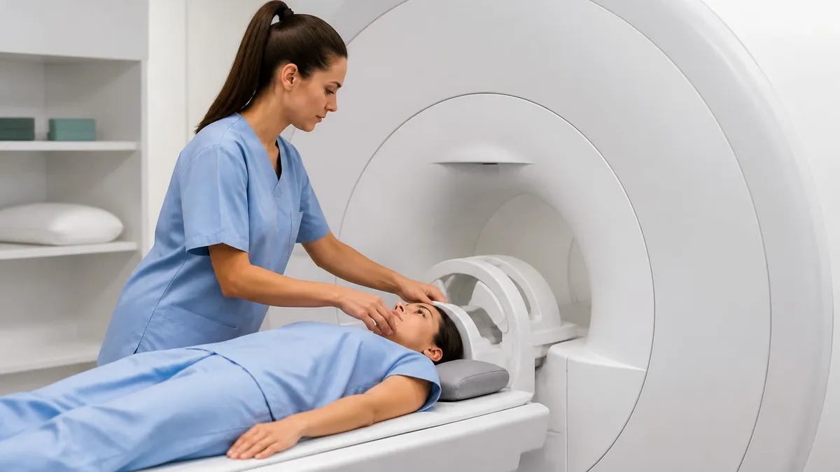
On the day of your imaging study, a little preparation goes a long way. For MRI specifically, plan to arrive 15-30 minutes early to complete the safety questionnaire. The form will ask in detail about any metal in your body, surgical history, prior reactions to contrast, and current medications. Be thorough — even small metal fragments from old shrapnel injuries, dental work, or occupational metalworking can matter. The technologist will use this information to determine whether you can safely enter the scan room and which sequences are appropriate.
Wear comfortable, loose-fitting clothing free of metal. Many facilities provide gowns, but if you wear your own clothes, choose items without zippers, snaps, underwire bras, or metallic threads. Leave jewelry, watches, hearing aids, and dentures at home or with a family member. Even small magnetic objects can become projectiles in the scan room or cause image artifacts that obscure the area being studied. Hair products containing metallic pigments can occasionally cause issues — when in doubt, skip them on scan day.
For CT scans, preparation is usually simpler. If contrast is being used, you may be asked to fast for four to six hours beforehand to reduce the small risk of nausea. Drinking water is usually encouraged unless instructed otherwise. Some abdominal CT studies require oral contrast — a flavored barium or iodine drink consumed an hour before the scan to outline the digestive tract. The taste varies from pleasant to chalky, but it goes down with cold water and a quick chaser.
During the MRI scan, expect noise. The machine produces loud knocking, buzzing, and humming as the magnetic field gradients switch on and off. You'll be given earplugs, headphones, or both, and many centers offer music or movies to make the time pass. You'll have a squeeze ball to alert the technologist if you need to stop. Try to stay still — even small movements blur images and may require sequences to be repeated. Breathing instructions are minimal for most studies but critical for cardiac or abdominal imaging.
During CT, the experience is briefer and quieter. You'll lie on a table that slides through a donut-shaped opening. Breath-hold instructions are common — the technologist will tell you when to hold your breath for 5-10 seconds during image acquisition. If contrast is being injected, you may feel a warm flushing sensation throughout your body and a metallic taste in your mouth. Both are normal and resolve within seconds. Tell the technologist immediately if you feel itching, difficulty breathing, or chest tightness.
After the scan, you can usually resume normal activities immediately. If you received intravenous contrast, drink extra water for the next 24 hours to help your kidneys clear the dye. Watch for delayed allergic reactions — rare but possible up to 24-48 hours later. Symptoms like hives, swelling, or wheezing warrant immediate medical evaluation. For breastfeeding mothers, gadolinium and iodine contrast are both considered safe to continue nursing, though some lactation consultants suggest pumping and discarding for 24 hours as an abundance of caution.
Results typically take 24-72 hours, though emergency studies are read within minutes. The radiologist's report is sent to your ordering physician, who will discuss findings with you. Don't be alarmed by technical language — terms like 'unremarkable,' 'no acute findings,' or 'within normal limits' are reassuring. Words like 'incidental finding' often refer to harmless variations that don't require treatment. If your report uses unfamiliar terminology, ask your physician to translate, and request a copy of both the report and the images for your personal records.
MRI Questions and Answers


About the Author
Medical Laboratory Scientist & Clinical Certification Expert
Johns Hopkins UniversityDr. Sandra Kim holds a PhD in Clinical Laboratory Science from Johns Hopkins University and is certified as a Medical Technologist (MT) and Medical Laboratory Scientist (MLS) through ASCP. With 16 years of clinical laboratory experience spanning hematology, microbiology, and molecular diagnostics, she prepares candidates for ASCP board exams, MLT, MLS, and specialist certification tests.
Join the Discussion
Connect with other students preparing for this exam. Share tips, ask questions, and get advice from people who have been there.
View discussion (6 replies)