MRI Scan Near Me: How to Find, Schedule, and Prepare for a Local MRI 2026 July
✍🏼 Searching for an MRI scan near me? Compare local imaging centers, costs, wait times, scanner types, and prep tips to book the right scan fast.
If you have typed mri scan near me into a search bar, you are likely juggling a referral, a worried question, and a calendar that will not cooperate. The good news is that magnetic resonance imaging has become widely available across the United States, with hospitals, outpatient imaging centers, mobile units, and specialty clinics all competing for your appointment. The harder news is that price, wait time, scanner strength, and report quality vary enormously between facilities that sit only a few miles apart, which means a quick zip-code search rarely tells the full story.
This guide walks you through everything you need to evaluate when choosing a local MRI provider, from understanding what the scanner actually does to comparing 1.5T versus 3T magnets, open versus wide bore designs, and accredited versus non-accredited facilities. We will also unpack realistic pricing, insurance authorization timelines, and the prep instructions that actually matter on scan day. Whether your referral is for a brain, spine, knee, abdomen, or breast study, the same evaluation framework applies.
Most patients do not realize they have negotiating power when scheduling. A self-pay MRI at a hospital can run $2,500, while the same anatomy at an accredited outpatient center across town might cost $400. Insurance contracts, equipment age, radiologist subspecialty, and even the time of day you book all influence the experience and the bill. Understanding these variables turns a frustrating search into a decision you can defend.
We will also address the questions patients commonly hesitate to ask out loud. Will I fit in the scanner? Can I take my anxiety medication beforehand? How fast can I get results, and who actually reads the images? Are walk-in scans real, or is that just marketing? Each of these has a concrete answer that depends partly on the facility you choose, which is why the search itself matters more than most people assume.
By the end of this guide you will know how to filter local options, what questions to ask when you call, which red flags suggest you should keep looking, and how to prepare physically and mentally so the appointment runs smoothly. You will also see how MRI fits into the broader diagnostic workflow, including when a CT, ultrasound, or follow-up study may be more appropriate than the scan you were originally referred for.
Finally, we built this article with a focus on accuracy over hype. MRI is a remarkable, non-ionizing imaging modality, but it is not magic and not always the right first step. The best outcome from your search is a scan performed at a qualified facility, interpreted by a subspecialty radiologist, delivered to your ordering clinician promptly, and billed at a fair price. Let us help you find exactly that.
If you are studying MRI rather than scheduling one, this article will also give you a patient-facing perspective that complements your clinical training and helps you communicate clearly with the people lying inside the bore.
MRI Access in the U.S. by the Numbers
Types of MRI Facilities You Will Find Near You
Attached to acute-care hospitals, these run 24/7 and handle inpatients, ER cases, and complex protocols. Expect higher prices, longer waits for outpatient slots, but access to advanced sequences and on-site subspecialty radiologists.
Free-standing facilities focused exclusively on imaging. Pricing is typically 40-70 percent lower than hospitals, scheduling is faster, and many run extended evening and Saturday hours. Quality varies, so check ACR accreditation.
Clinics that scan a narrow set of anatomy, often with extremity-only or dedicated neuro scanners. Radiologists subspecialize, producing detailed musculoskeletal or brain reports for surgeons and neurologists.
Tractor-trailer scanners that rotate between rural hospitals and clinics. Convenient for underserved areas, though magnet strength is usually 1.5T and protocols may be standardized rather than tailored.
Facilities marketing claustrophobia-friendly designs. Open MRIs use lower field strengths, while wide-bore 3T scanners now offer a 70 cm opening with full diagnostic image quality for most studies.
Cost is usually the second question after location, and it is the variable with the widest spread. A self-pay lumbar spine MRI without contrast can range from about $380 at an accredited outpatient center in Texas to over $3,200 at a major academic hospital in the Northeast. Same anatomy, same diagnostic value when performed correctly, very different bills. Always ask for the cash price even if you have insurance, because in some cases the cash rate beats your in-network coinsurance after deductible.
Insurance authorization is the second pricing variable. Most commercial plans require prior authorization for non-emergency MRI, a process that typically takes one to seven business days. Your ordering physician submits clinical justification, the insurer reviews against criteria like InterQual or ACR Appropriateness Criteria, and either approves, denies, or requests a peer-to-peer call. Denials are common when conservative therapy has not been documented, particularly for back and shoulder studies.
If you have a high-deductible plan, you are functionally self-pay until the deductible resets, so shop aggressively. Use tools like FAIR Health Consumer, Healthcare Bluebook, or call three local facilities directly and ask for the global rate, which includes both the technical fee and the professional read. Beware quotes that exclude the radiologist interpretation, because that bill arrives separately weeks later and surprises patients who thought they had paid in full.
Medicare reimburses MRI under the Hospital Outpatient Prospective Payment System for hospital-based scans and under the Medicare Physician Fee Schedule for free-standing centers. The free-standing rate is roughly half the hospital rate, which is why Medicare beneficiaries with secondary coverage often pay less at outpatient facilities even before considering convenience.
Contrast adds cost. A gadolinium-enhanced study typically runs $150 to $400 more than a non-abdominal mri cpt code reflecting the cost of the agent, IV placement, and additional sequences. Not every study needs contrast. Routine knee, shoulder, and lumbar spine MRIs are usually performed without it. Brain, breast, abdomen, pelvis, and tumor follow-up studies generally require contrast unless your kidney function is impaired.
Finally, watch for facility fees. Hospital-owned outpatient imaging centers can legally bill a facility fee on top of the technical and professional charges if they meet provider-based criteria, which can add hundreds of dollars. Truly independent imaging centers cannot bill this fee. When you call, ask directly whether the center is hospital-owned and whether a facility fee will appear on your bill.
If you anticipate a need for repeat imaging, such as tumor surveillance or MS monitoring, establishing care with a single high-quality outpatient center pays off. Consistent technique, the same scanner, and the same reading radiologist make subtle interval changes far easier to detect than scattering scans across different facilities.
MRI Practice Test Questions
Prepare for the MRI - Magnetic Resonance Imaging exam with our free practice test modules. Each quiz covers key topics to help you pass on your first try.
MRI Knowledge
MRI Exam Questions covering Knowledge. Master MRI Test concepts for certification prep.
MRI Physics
Free MRI Practice Test featuring Physics. Improve your MRI Exam score with mock test prep.
MRI Anatomy and Pathology
MRI Test Prep for MRI Anatomy and Pathology. Practice MRI Quiz questions and boost your score.
MRI Anatomy and Positioning
MRI Questions and Answers on MRI Anatomy and Positioning. Free MRI practice for exam readiness.
MRI Contrast Agents
Free MRI Quiz on MRI Contrast Agents. MRI Exam prep questions with detailed explanations.
MRI Patient Care and Positioning
MRI Practice Questions for MRI Patient Care and Positioning. Build confidence for your MRI certification exam.
Scanner Types You May See at an MRI Scan Near Me
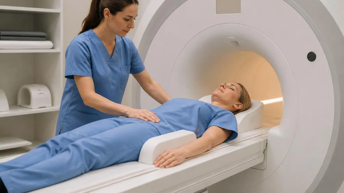
The 1.5 Tesla closed bore scanner is the workhorse of community imaging. It produces excellent diagnostic quality for nearly every clinical indication, from brain and spine to abdomen and musculoskeletal studies. Bore diameter is typically 60 cm, which accommodates most adults but can feel tight for larger patients or those with claustrophobia. Scan times are slightly longer than at 3T but artifacts from metal implants are usually less pronounced.
For most outpatient referrals, 1.5T is not a downgrade. The American College of Radiology accredits both field strengths under the same diagnostic standards, and many subspecialty radiologists prefer 1.5T for cardiac, body, and post-operative imaging where 3T susceptibility artifacts can obscure findings. If a facility quotes a lower price for 1.5T, that is not a quality compromise for routine studies.
Outpatient Imaging Center vs Hospital MRI
- +Significantly lower cost, often 40-70 percent less than hospital pricing
- +Faster scheduling, with same-week and Saturday slots common
- +Free, easy parking and shorter check-in lines
- +Focused environment with techs who perform MRI all day every day
- +No hospital facility fees added to your bill
- +Cash-pay and bundled pricing options frequently available
- +Streamlined workflow for stable outpatient indications
- −Limited after-hours and weekend coverage at some centers
- −Cannot accommodate acutely ill patients needing monitoring
- −Fewer subspecialty radiologists on-site for complex cases
- −Some advanced protocols like cardiac MRI may be unavailable
- −Equipment may be older at lower-cost facilities
- −Contrast reactions managed with less immediate backup
- −Records may not auto-flow to a hospital electronic health record
Pre-Scan Checklist for Your MRI Scan Near Me
- ✓Confirm your insurance prior authorization is approved and the authorization number is on file
- ✓Complete the safety screening form, listing every implant, surgery, and metal exposure
- ✓Bring prior imaging on CD or via image-sharing portal so the radiologist can compare
- ✓Wear or bring metal-free clothing such as sweatpants and a cotton t-shirt without graphics
- ✓Remove jewelry, watches, hearing aids, dentures, hairpins, and body piercings before arrival
- ✓Eat normally unless instructed otherwise, except for abdominal or pelvic studies with prep
- ✓Hydrate well if contrast is planned, which makes IV placement faster and easier
- ✓Arrive 20 to 30 minutes early to complete paperwork and change into a gown
- ✓Discuss anxiety medication with your ordering provider at least 48 hours in advance
- ✓Arrange a driver home if you take sedation, since you cannot drive for several hours after
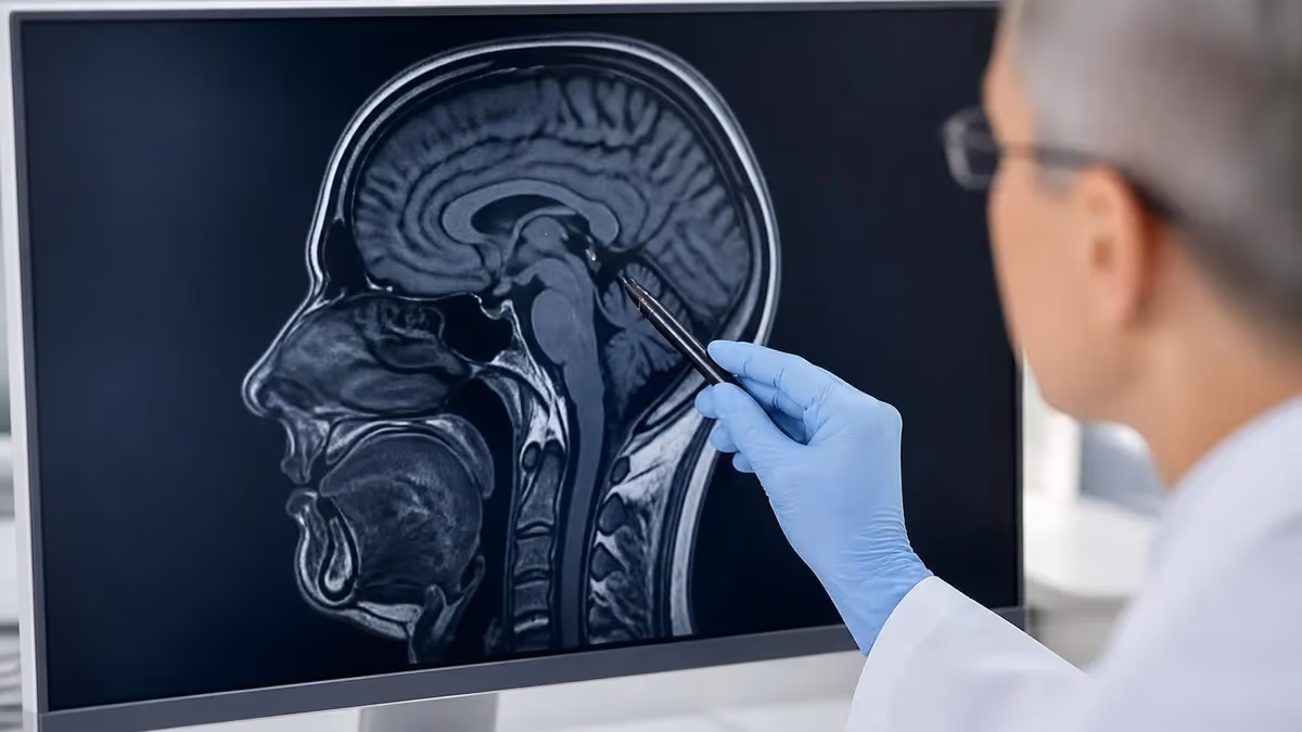
Who is reading my MRI, and what is their subspecialty?
The radiologist who interprets your scan matters as much as the scanner that acquires it. A fellowship-trained musculoskeletal radiologist reading your knee MRI catches details a general radiologist may miss, and the same applies to neuro, body, breast, and cardiac imaging. Ask the facility whether they use subspecialty readers and request the report be sent directly to you in addition to your ordering physician.
Wait times for an MRI scan near you depend heavily on geography, urgency, and facility type. Major metropolitan areas typically offer outpatient appointments within three to seven days, while rural regions or hospital-only markets can stretch to two or three weeks. If your referral is marked urgent or STAT, most facilities will work you in within 24 to 48 hours, sometimes the same day if a slot opens. Always ask the scheduler to put you on a cancellation list, because gaps appear constantly.
Scheduling tip from the inside: late afternoon and early evening slots get cancelled most often, which makes them the easiest to grab last minute. Sunday and Monday morning slots tend to be the most reliable but book furthest out. If a facility offers walk-in MRI for self-pay patients, that means they have unused capacity and you can often negotiate a lower price on top of the convenience.
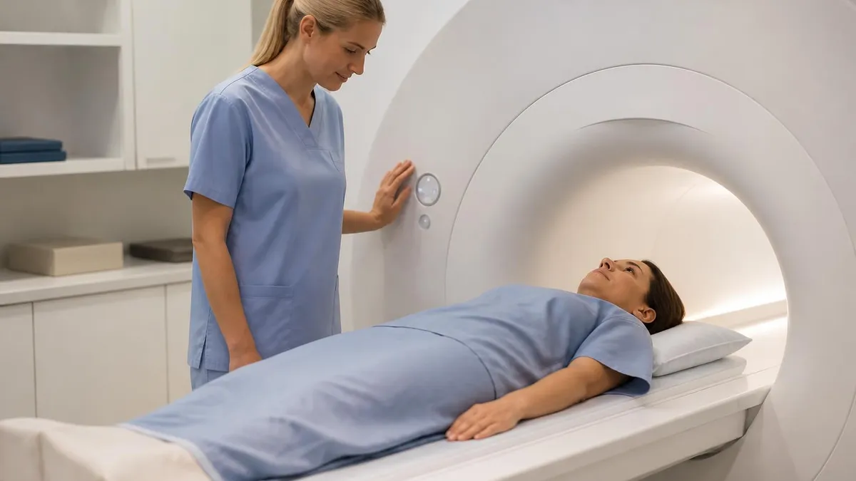
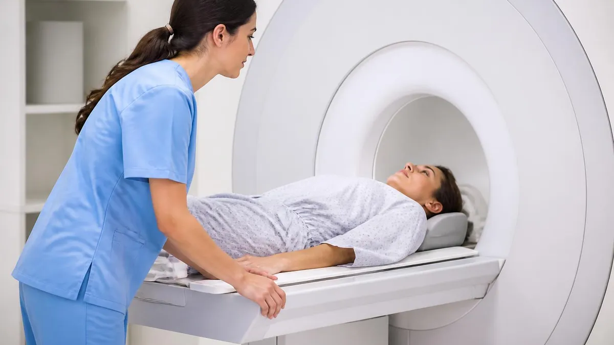
On the day of the scan, plan to be at the facility for 60 to 90 minutes total. Check-in and paperwork take 20 to 30 minutes, the actual scan runs 15 to 60 minutes depending on the body part and whether contrast is used, and post-scan observation if you received gadolinium adds another 15 minutes. Knee and brain scans are usually quickest, while multi-region spine studies, breast MRI, and cardiac studies sit at the longer end.
Inside the bore, expect loud knocking and buzzing noises from the gradient coils switching on and off. Hearing protection is mandatory, usually foam earplugs plus headphones playing music of your choice. Stay as still as possible during each sequence, breathe normally unless instructed to hold your breath, and use the squeeze ball to signal the technologist if anything goes wrong. Most patients describe the experience as boring rather than scary.
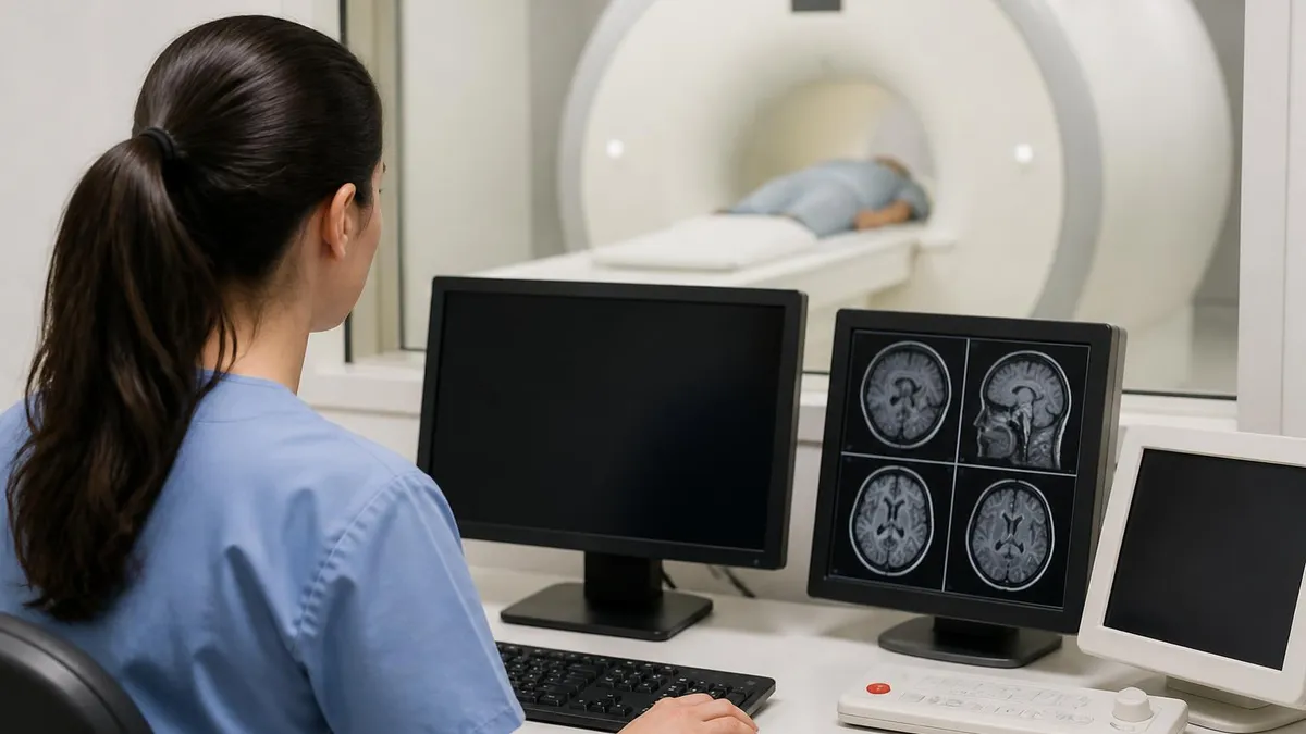
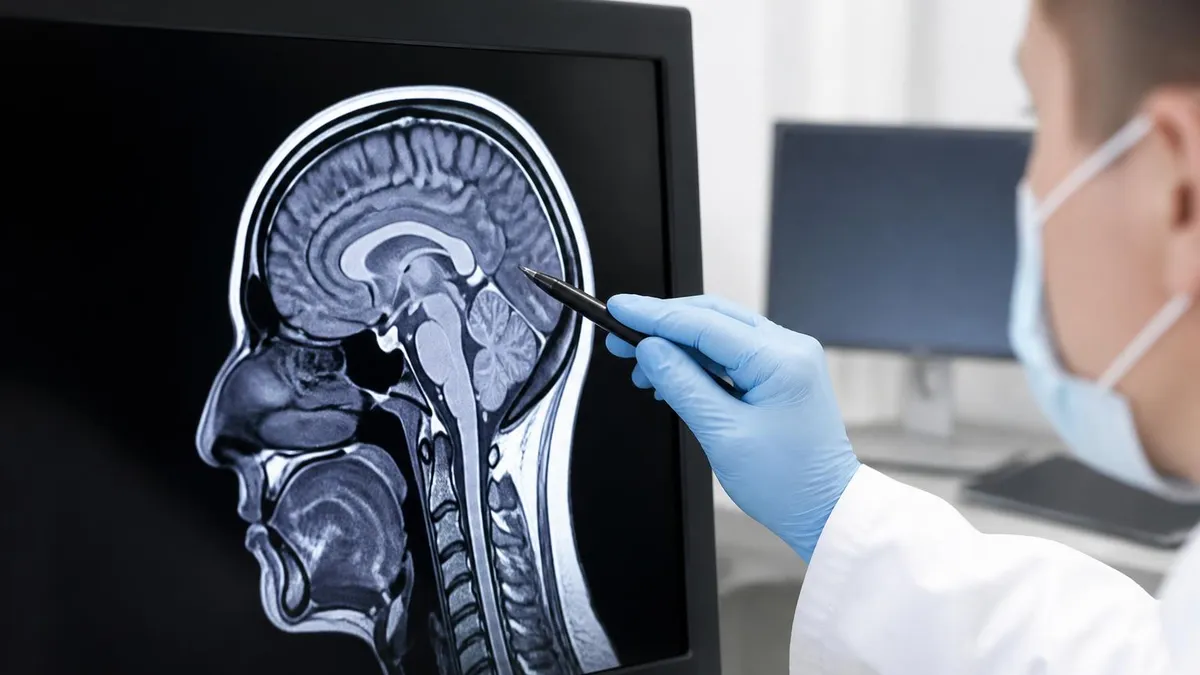
Image acquisition is followed by interpretation, which is where the timeline can stretch. A routine outpatient MRI is typically read within 24 to 48 business hours, while STAT studies on hospital inpatients are read within 30 to 60 minutes. Reports flow electronically to your ordering provider, who is responsible for contacting you with results. Many facilities now offer patient portals that release the report directly to you within a few days.
If you have not heard from your ordering physician within five business days of the scan, call their office. Reports occasionally fail to route correctly, especially between independent imaging centers and large health systems on different electronic record platforms. Asking the imaging center to fax or upload a copy directly to your portal is a reasonable backup. You are entitled to your own report under federal information-blocking rules.
For complex findings, expect a follow-up appointment to discuss results rather than a quick phone call. Imaging findings rarely tell the whole story on their own, and your physician will integrate them with symptoms, exam findings, and other tests before recommending a treatment plan. A good radiology report contains both findings and an impression, but it is your clinician who turns that into a decision.
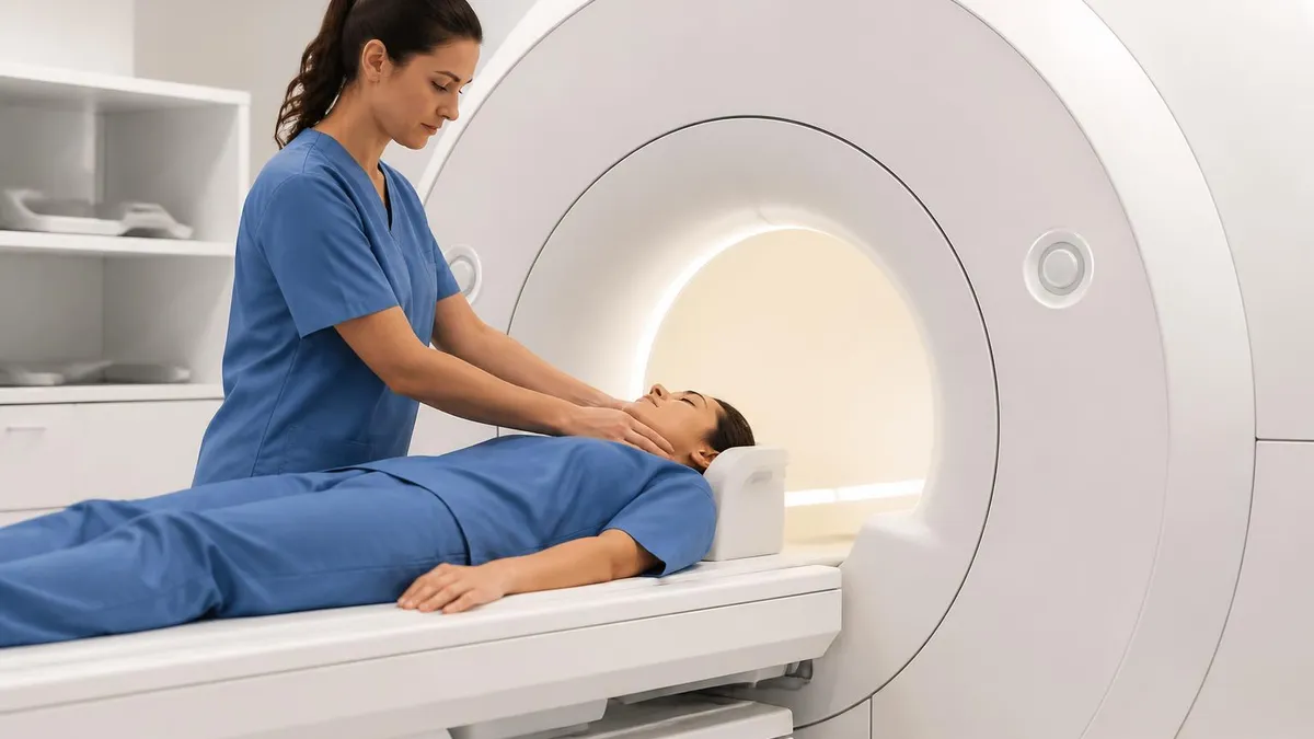
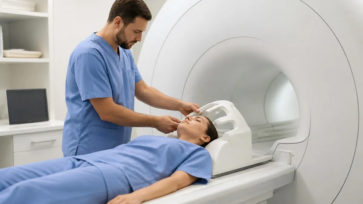
Every patient must complete a safety questionnaire before entering the scan room. Pacemakers, certain aneurysm clips, cochlear implants, metal fragments in the eye, and some neurostimulators can be life-threatening in the magnetic field. Tell the technologist about every surgery, occupational metal exposure, and tattoo, even if it seems unrelated. When in doubt, the facility will look up your implant card or contact the manufacturer before scanning.
Quality matters even more than convenience when choosing a local MRI. The single most useful credential to look for is ACR accreditation, granted by the American College of Radiology after a peer review of equipment performance, image quality, personnel qualifications, and quality control programs. An ACR seal on the website is not a marketing claim, it is an audited standard. The Intersocietal Accreditation Commission offers a similar program, also reputable.
Beyond accreditation, ask whether the radiologists are board certified by the American Board of Radiology and whether subspecialty fellowship training is available for your study type. For a brain MRI looking at multiple sclerosis or stroke, a neuroradiologist is ideal. For a knee or shoulder, a musculoskeletal radiologist. For a prostate or rectal study, an abdominal radiologist with specific prostate MRI experience. Many outpatient centers contract with teleradiology groups that include subspecialists.
Technologist credentials matter too. The American Registry of Radiologic Technologists offers an MRI certification, ARRT (MR), which requires both didactic education and clinical experience. Ask the facility whether their technologists are ARRT-certified in MRI specifically rather than general radiography. A well-trained MRI technologist tailors sequences to your anatomy and tolerance, catches motion artifacts in real time, and communicates findings to the radiologist that improve interpretation.
Equipment age is a fair question. A scanner installed in the last five years will have current pulse sequences, improved coils, and faster reconstruction. Older scanners can still produce diagnostic images for routine indications, but advanced applications like diffusion tensor imaging, MR elastography, or cardiac MRI may not be available. The American Hospital Association reports that the average MRI scanner in the U.S. is about eight years old, with significant variation by market.
Reviews are useful but read them critically. Patients tend to comment on parking, friendliness, and scheduling, which matter for experience but not for diagnostic accuracy. Look for repeated mentions of unexplained billing surprises, lost results, or technologist communication problems, which signal operational issues. Reviews mentioning specific clinical scenarios are rare but valuable when you find them. For broader context on what scanners can detect, see our guide on MRI diagnostic capabilities.
If you have a body habitus consideration, ask specifically about the bore diameter and weight limit. Standard scanners accept up to 350 pounds with a 60 cm bore, wide-bore scanners go to 550 pounds with a 70 cm bore, and dedicated bariatric units exist in some markets. Calling ahead to confirm fit avoids a wasted trip and the awkward moment of being turned away at check-in.
Finally, consider whether the facility offers patient-friendly scheduling tools like online booking, automated reminders, and digital intake forms. These are not luxuries, they reduce errors in the safety screening process and free technologists to focus on patient care rather than paperwork. A facility that has invested in patient experience usually invests in clinical quality too.
Final preparation comes down to a handful of small choices that make a meaningful difference. The night before your scan, get reasonable sleep but skip alcohol if you struggle with claustrophobia, since hangovers amplify anxiety. Eat a normal breakfast unless your study requires fasting, which usually applies only to abdominal MRCP, pelvic MRE, and some cardiac protocols. Take your regular medications with water, including blood pressure and antiseizure medications, unless your ordering physician has said otherwise.
Dress strategically. Soft sweatpants without metal zippers, a plain cotton shirt, and slip-on shoes save you from changing into a gown for shorter studies. Leave jewelry at home rather than in a locker, because facilities are not liable for losses. If you wear a fitness tracker or smart ring, plan to remove it before entry, and do not bring credit cards near the magnet because the magnetic stripe can be erased.
For claustrophobic patients, ask your ordering provider for a one-time low-dose oral benzodiazepine such as lorazepam, taken 30 to 45 minutes before the scan. Bring a friend or family member who can drive you home, sit in the scan room with you if facility policy allows, and help you stay calm during the procedure. Many facilities also offer prism glasses that let you see out of the bore, eye masks, lavender aromatherapy, or piped-in music and movies during longer studies.
If you have prior imaging, bring it. A disc with prior CTs, MRIs, or X-rays gives the radiologist comparison material that often changes the interpretation. Without priors, a small finding may be called indeterminate and trigger a follow-up scan, while with priors the same finding may be confidently called stable and dismissed. Most facilities can also pull priors electronically through health information exchanges, but bringing a physical disc is a reliable backup.
Plan your post-scan logistics. Routine non-contrast MRI has no restrictions, you can drive immediately and return to work or school. Studies with sedation require a driver and a low-key rest of the day. Contrast-enhanced studies allow you to drive but recommend extra hydration to flush gadolinium through your kidneys. Breastfeeding mothers can resume nursing immediately after gadolinium according to current ACR guidelines, despite older recommendations to pump and dump.
Once you have results, do not interpret them alone. Imaging reports use technical language that can sound alarming out of context. Words like degenerative, signal abnormality, and nonspecific are common and rarely indicate anything serious by themselves. Wait for your ordering physician to put findings in clinical context, and write down questions in advance so the follow-up appointment uses your time well. For an overview of common report findings across body regions, see our guide on common MRI findings.
And remember that an MRI is a tool, not a verdict. It produces extraordinarily detailed images of soft tissue, joints, vessels, and organs, but it cannot replace the clinical judgment that connects a symptom to a diagnosis. The best outcomes come from a thoughtful referral, a quality scan at a qualified facility, an expert interpretation, and a clinician who knows you well enough to integrate it all.
MRI Questions and Answers
About the Author
Medical Laboratory Scientist & Clinical Certification Expert
Johns Hopkins UniversityDr. Sandra Kim holds a PhD in Clinical Laboratory Science from Johns Hopkins University and is certified as a Medical Technologist (MT) and Medical Laboratory Scientist (MLS) through ASCP. With 16 years of clinical laboratory experience spanning hematology, microbiology, and molecular diagnostics, she prepares candidates for ASCP board exams, MLT, MLS, and specialist certification tests.
Join the Discussion
Connect with other students preparing for this exam. Share tips, ask questions, and get advice from people who have been there.
View discussion (6 replies)