What MRI Can Detect: Conditions & Diagnostic Capabilities 2026 July
Can an MRI diagnose ADHD, dementia, or cancer? See what MRI detects, misses, and how it compares to CT, ultrasound, and clinical exams. ✅
Magnetic resonance imaging gives clinicians one of the sharpest windows into the human body that radiology has ever produced. The scanner uses powerful magnets and radio pulses rather than ionizing radiation, and the resulting images can separate gray matter from white matter, fat from fluid, and healthy tissue from tumor with a clarity that CT and ultrasound rarely match. That soft tissue contrast is the reason neurologists, oncologists, orthopedic surgeons, and gynecologists keep pushing patients toward an MRI when the diagnosis is not obvious from blood work or a physical exam.
Strong as the technology is, MRI is not a universal answer machine. A scan can show structure beautifully and still tell you nothing about how the brain is firing during a math problem or how a child behaves at school. That is why the question "can an MRI diagnose ADHD" comes up so often in radiology forums — and why the honest answer is no, at least not in routine clinical practice.
The same gap exists for early dementia, some forms of demyelination, and very small tumors that hide inside complex anatomy. This article walks through what MRI reliably detects, where it slips, and the questions you should be asking when your radiology report lands in the patient portal.
The story behind that question matters too. Parents reading about brain imaging studies sometimes assume that if researchers can spot group-level differences in attention networks, the same trick should work for their child. It does not. Population-level statistics and individual diagnosis are very different problems, and the gap between them is wider than most marketing copy admits. Understanding why takes a tour through what MRI actually measures, what it cannot measure, and where the entire field is heading.
MRI Diagnostic Accuracy By The Numbers
Those numbers shape almost every conversation in a reading room. They explain why neurology leans on MRI for first-line evaluation of headaches with red flags, why urology built an entire screening pathway around multiparametric MRI of the prostate, and why pelvic MRI is now a standard part of the workup for women with persistent pelvic pain. They also show the ceiling. A 95% sensitivity is excellent, but it still leaves room for the rare missed lesion — and a 60% lower bound on endometriosis means a normal scan does not rule the disease out.
The performance of MRI depends heavily on the magnet strength (1.5T versus 3T), the coils available, the sequences selected, and the radiologist reading the images. A scan that uses a dedicated neurovascular protocol picks up tiny aneurysms that a generic brain MRI would skip. A prostate MRI without contrast misses lesions that a full multiparametric study would flag. When you compare published sensitivity figures, you are really comparing protocols, not just machines.
There is also a human factor that gets overlooked. The same scan read by a fellowship-trained subspecialist can yield a completely different impression than one read by a general radiologist working through a long list. That is why major academic centers run second-look reads on outside-hospital scans, and why patients with complex disease often benefit from getting their images reviewed at a referral center even if the original report sounded normal. Sensitivity statistics describe the test in ideal conditions; real-world accuracy depends on workflow.
When MRI Truly Shines
MRI is the imaging test of choice whenever soft tissue contrast matters more than bony detail. Brain and spinal cord lesions, ligament tears, cartilage damage, uterine and ovarian masses, and liver tumors are all easier to see on MRI than on CT. The absence of radiation also makes MRI safer for repeated scanning in young patients, pregnant women in the second and third trimesters, and people who need long-term follow-up for chronic disease.
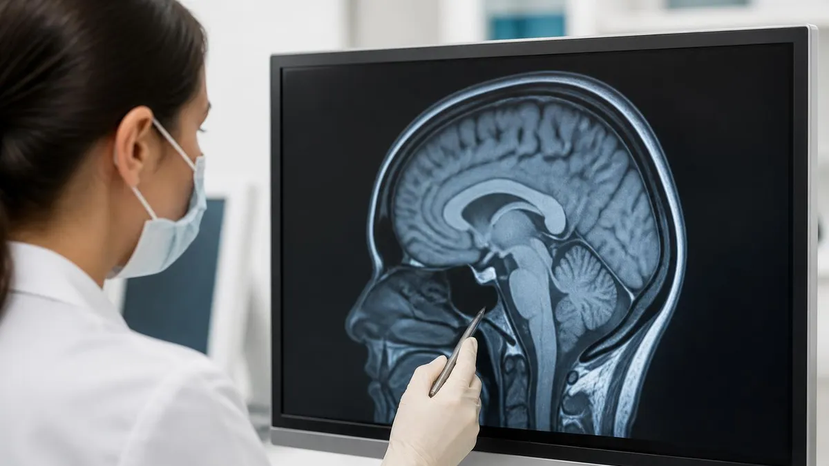
Inside the brain, MRI is the workhorse for diagnosing structural disease. T2-weighted and FLAIR sequences light up areas of demyelination, edema, gliosis, and infarction, which is why neurologists order it for suspected stroke, multiple sclerosis, encephalitis, and brain tumors. Diffusion-weighted imaging detects acute ischemia within minutes of symptom onset, often before a CT scan shows any change. Susceptibility-weighted imaging finds microbleeds that point to cerebral amyloid angiopathy or traumatic brain injury. Magnetic resonance angiography images the circle of Willis and major intracranial arteries without contrast, while magnetic resonance venography catches venous sinus thrombosis that can hide behind a benign-looking headache.
Can an MRI miss a brain tumor? Yes, occasionally. Lesions smaller than three to five millimeters, tumors that share the same signal characteristics as surrounding tissue, and masses hidden in the floor of the third ventricle or the cerebellopontine angle can slip past a routine protocol.
Contrast-enhanced sequences raise the detection rate substantially, which is why a follow-up scan with gadolinium is ordered whenever a patient has persistent neurological symptoms despite a normal non-contrast study. Leptomeningeal disease, where tumor spreads along the surface of the brain rather than forming a solid mass, is another classic blind spot — it requires thin-slice post-contrast imaging and a radiologist who knows to look for the subtle pial enhancement that marks it.
For dementia, MRI plays a supporting role rather than a starring one. Can MRI show Alzheimer's? It can show the consequences — hippocampal atrophy, medial temporal lobe shrinkage, and ventricular enlargement — but those changes appear late and overlap with normal aging. Can MRI diagnose dementia outright? No. The diagnosis is clinical, supported by cognitive testing, blood biomarkers, and increasingly PET imaging of amyloid and tau.
MRI's job is to rule out treatable mimics: normal pressure hydrocephalus, subdural hematoma, vascular dementia, and tumors that present with cognitive decline. A patient who walks into a memory clinic with progressive forgetfulness gets imaged not to confirm Alzheimer's but to make sure something fixable is not being missed before that label gets applied.
What MRI Detects By Body System
Brain tumors, stroke, multiple sclerosis, demyelination, hydrocephalus, traumatic brain injury, spinal cord compression, and disc herniation are all confidently evaluated with MRI.
Cartilage loss, meniscal tears, ligament ruptures, bone marrow edema, sacroiliitis, and early ankylosing spondylitis show up clearly long before plain x-ray changes appear.
Prostate cancer on multiparametric MRI, endometriosis on dedicated pelvic protocols, hepatocellular carcinoma, and rectal cancer staging are core indications.
Functional disorders like ADHD, early Alzheimer's, primary psychiatric illness, and subtle metabolic disease are beyond MRI's structural lens — they need clinical, neuropsychological, or PET workup.
Multiple sclerosis is one of the diseases MRI was practically invented to detect, yet the question "can MS be missed on MRI" still comes up because early disease sometimes presents with a single tiny lesion in the brainstem or upper cervical cord. Standard 1.5T scanners can miss these. Moving to 3T, adding double inversion recovery sequences, and imaging the entire spinal cord lifts sensitivity into the mid-nineties. Even then, about three to five percent of patients with clinically definite MS have an initially normal scan and only develop visible lesions on follow-up.
Brain damage from hypoxia is another area where MRI outperforms CT. Can an MRI show brain damage from lack of oxygen? Yes, and dramatically. Diffusion-weighted imaging picks up cytotoxic edema in the basal ganglia, thalami, and watershed cortical zones within hours of cardiac arrest, near-drowning, or carbon monoxide exposure. The pattern of injury helps prognosticate recovery, which is why hypoxic-ischemic encephalopathy protocols exist in most stroke and neurocritical care centers. Delayed scans done several days after the insult often show even more striking changes as cells finish dying — a window that helps neurologists counsel families about likely outcomes.
Transient ischemic attacks are trickier. Can a TIA be seen on MRI? Sometimes. Roughly one in three patients who present with classic TIA symptoms have a small area of restricted diffusion that confirms a brief ischemic event, which technically reclassifies them as having had a minor stroke.
The other two thirds have negative scans, which does not mean they are out of the woods — TIA remains a clinical diagnosis and an urgent warning of future stroke risk. Same-day MRI within 24 hours of symptom onset catches more events than imaging done a week later, because diffusion changes can fade as tissue either recovers or progresses to frank infarction.
MRI Capabilities And Limits By Region
MRI confidently detects ischemic and hemorrhagic stroke, primary and metastatic brain tumors, multiple sclerosis, encephalitis, abscess, hydrocephalus, congenital malformations, and traumatic brain injury. It can show late-stage Alzheimer's atrophy and vascular dementia changes but cannot make the diagnosis in isolation. Functional psychiatric conditions including ADHD, depression, and schizophrenia leave no reliable structural fingerprint visible on routine brain MRI.
Costochondritis, the inflammation of the cartilage between ribs and sternum, frustrates patients because it produces real chest pain but is mostly a clinical diagnosis. Can you see costochondritis on MRI? Yes, with the right protocol — high-resolution fat-suppressed sequences over the anterior chest wall show edema in the costal cartilages and adjacent bone marrow.
The catch is that most clinicians do not order chest wall MRI for this complaint, partly because the diagnosis is usually made by palpation and partly because the test is not always covered by insurance for non-cardiac chest pain. MRI becomes more useful when the pain is atypical, persistent, or accompanied by red flags suggesting infection or tumor in the chest wall.
Endometriosis sits at the awkward intersection of "MRI helps" and "MRI is not enough." Can you see endometriosis on a MRI? Deep infiltrating disease, ovarian endometriomas, and bowel involvement are usually visible on a dedicated pelvic protocol with antispasmodic medication and a full bladder. Superficial peritoneal implants — the kind that can still cause severe pain — frequently look normal on imaging.
A negative MRI does not rule out endometriosis, and laparoscopy remains the gold standard for definitive diagnosis. Many specialist pelvic radiologists now use a structured reporting template that grades disease in each anatomic compartment, which makes surgical planning far more accurate than a generic narrative report.
Ankylosing spondylitis deserves a similar second look. Patients with chronic inflammatory back pain can spend years bouncing between primary care and physical therapy before someone orders the right scan. A sacroiliac MRI with STIR sequences detects active bone marrow edema long before x-ray shows the late-stage bridging and fusion. Catching it early means starting biologic therapy in the window where joint architecture can still be preserved, which is why rheumatologists increasingly treat persistent inflammatory back pain as an indication for MRI rather than another round of imaging-free trial and error.
If your symptoms persist despite a clean scan, push back. Ask your clinician whether the protocol was right for the question, whether a higher field strength or contrast study is warranted, and whether non-MRI tests — neuropsychological testing for cognitive symptoms, laparoscopy for pelvic pain, lumbar puncture for suspected MS — should be the next step. Imaging is one piece of the puzzle, never the whole picture.
Knowing what your MRI report should contain helps you read it intelligently. Every diagnostic MRI report follows a similar template: indication, technique, comparison, findings by anatomic region, and impression. The impression is the most important section because it summarizes what the radiologist actually thinks. If the impression mentions "recommend correlation with clinical findings" or "consider follow-up imaging in three months," those phrases are not boilerplate — they reflect a level of diagnostic uncertainty that deserves a follow-up appointment.
Patients who treat the report as a one-line yes-or-no answer often miss the qualifying language that radiologists use to flag a finding they could not fully characterize on the available images. Pay particular attention to phrases like "indeterminate," "cannot exclude," and "clinical correlation recommended" — each one is a polite request for more information before a final conclusion can be drawn.
Questions To Ask About Your MRI Report
- ✓Did the protocol match the clinical question (for example, contrast for suspected tumor, mpMRI for prostate)?
- ✓Was the field strength appropriate — 3T for fine neurological detail, 1.5T acceptable for most spine and joint work?
- ✓Are there any incidental findings that need separate follow-up?
- ✓Does the impression directly address the symptom that prompted the scan?
- ✓If the scan is normal but symptoms persist, what non-imaging tests come next?
- ✓Should this scan be repeated at a defined interval to track change?
- ✓Is the comparison to prior imaging mentioned, and if so what changed?
Patients sometimes assume MRI is always the best test. It is not. CT outperforms MRI for acute hemorrhage, pulmonary nodules, kidney stones, and most traumatic injuries because it is faster, cheaper, and more widely available. Ultrasound beats MRI for first-trimester pregnancy, gallbladder disease, and quick bedside evaluation of the thyroid or testes.
PET is the better tool for functional imaging of cancer metabolism, amyloid plaque, and dopamine transporter loss in Parkinson's disease. The right test depends on the question being asked, and the best clinicians work backward from the diagnosis they suspect to the imaging modality that answers it most efficiently.
Cost and access matter too. An MRI in the United States can cost anywhere from $400 at a freestanding outpatient center to several thousand at a hospital outpatient department, and insurance pre-authorization can delay a scan by days or weeks. CT is usually faster to schedule and cheaper, which is part of why emergency rooms default to it.
If your physician orders an MRI but you cannot get one for three weeks, ask whether a CT or ultrasound would answer the immediate question while you wait. Sometimes the answer is yes, sometimes the structural detail of MRI is genuinely irreplaceable, and the conversation is worth having before symptoms get worse.
MRI Compared With CT And Ultrasound
- +Unmatched soft tissue contrast for brain, spine, joints, pelvis
- +No ionizing radiation — safer for repeat scans and younger patients
- +Multi-planar imaging without repositioning the patient
- +Functional sequences (diffusion, perfusion, spectroscopy) add metabolic data
- +Gadolinium contrast highlights vascular and inflammatory lesions
- −Long scan times (30-60 minutes) limit use in unstable patients
- −Claustrophobia and motion artifact reduce image quality
- −Implanted devices and certain metals are absolute or relative contraindications
- −Cost and limited availability compared with CT and ultrasound
- −Cannot diagnose functional disorders like ADHD or early dementia
- −Small lesions and superficial endometriosis can be missed
One subtle point that radiologists wish more patients understood: MRI quality depends on the patient holding still. A motion-degraded scan can mimic disease or hide it entirely. If you struggle with claustrophobia, ask about open-bore scanners, mild sedation, or noise-cancelling headphones. If you have implants, bring documentation — pacemakers, cochlear implants, certain aneurysm clips, and some intraocular foreign bodies are still considered unsafe even in the MRI-conditional era. The technologist will run a safety checklist, but you should know your own hardware history.
Looking ahead, MRI is evolving fast. Artificial intelligence reconstruction is cutting scan times in half without losing detail. Quantitative MRI is moving beyond pretty pictures toward measurable tissue properties that may eventually flag early Alzheimer's or subtle MS plaques that today's eyes miss. Whole-body MRI is becoming a real cancer screening tool for high-risk populations such as Li-Fraumeni and BRCA carriers. The diagnostic ceiling keeps rising, but the basic truth still holds — MRI shows structure exquisitely, and structure is only part of medicine.
Functional MRI, the same technology used in research labs to map brain activity, is a separate question worth touching on. fMRI can correlate blood flow changes with task performance and has been used in surgical planning to map language and motor areas before tumor resection. It is not, however, a diagnostic test for ADHD, autism, depression, or any other primary psychiatric or developmental condition. Clinics that market "functional brain scans" for these conditions are operating outside mainstream evidence-based medicine. Real fMRI lives in research protocols and a narrow set of pre-surgical applications.
The smartest way to use MRI is to treat it as a partner with the clinical exam rather than a substitute for it. When the structural question is sharp — is there a tumor, a torn ligament, a demyelinating lesion, a cancer in the prostate — MRI delivers answers other tests cannot. When the question is functional, behavioral, or biochemical, you need different tools, and a perfectly normal scan should not stop the search for a diagnosis.
Reading your own report with that mindset, and asking the questions in the checklist above, will get you more out of every scan you ever have. The technology is remarkable, but it is still just one chapter in a longer diagnostic story, and the patient who understands what their scan can and cannot show becomes a better partner in their own care.
Bring your symptom timeline, your prior imaging, and your specific questions to every imaging consultation. Ask the ordering physician what they expect to see, what would change management, and what the next step is if the scan comes back clean. That kind of upfront framing is the difference between an MRI that resolves a diagnostic puzzle and an MRI that simply adds another data point to an already confused chart.
MRI Questions and Answers
About the Author
Medical Laboratory Scientist & Clinical Certification Expert
Johns Hopkins UniversityDr. Sandra Kim holds a PhD in Clinical Laboratory Science from Johns Hopkins University and is certified as a Medical Technologist (MT) and Medical Laboratory Scientist (MLS) through ASCP. With 16 years of clinical laboratory experience spanning hematology, microbiology, and molecular diagnostics, she prepares candidates for ASCP board exams, MLT, MLS, and specialist certification tests.
Join the Discussion
Connect with other students preparing for this exam. Share tips, ask questions, and get advice from people who have been there.
View discussion (6 replies)