Abdominal MRI With Contrast: Complete Guide to the Scan, Preparation, and Results
Abdominal MRI with contrast explained: how it works, preparation, what gadolinium shows, costs, safety, and how to read your results. 💡
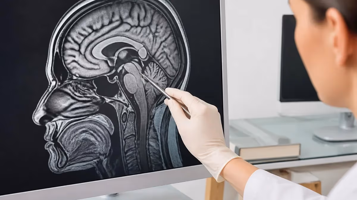
An abdominal MRI with contrast is one of the most detailed imaging studies available for evaluating the liver, pancreas, kidneys, spleen, adrenal glands, and surrounding soft tissues. Using a powerful magnetic field and radiofrequency pulses, the scanner produces cross-sectional images that show organ architecture, vascular flow, and subtle lesions that other modalities frequently miss. When gadolinium-based contrast is added, radiologists can characterize masses, detect inflammation, and map blood supply with remarkable precision, making it a cornerstone tool in modern abdominal diagnostics.
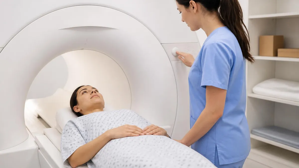
Patients are often referred for this study after ambiguous ultrasound or CT findings, persistent abdominal pain, abnormal liver enzymes, or to monitor known conditions such as cirrhosis, Crohn disease, or pancreatic cysts. Unlike CT, MRI uses no ionizing radiation, which makes it especially attractive for younger adults, pregnant patients in select scenarios, and people who need repeated follow-up imaging. The trade-off is a longer scan time, typically 30 to 60 minutes, and the need to lie still inside a tube-shaped magnet.
The contrast agent itself is a gadolinium chelate injected through a small IV in the arm. As it circulates, the radiologist captures images during arterial, portal venous, and delayed phases. This timing reveals how tissue takes up and releases the dye — a behavior that often distinguishes benign hemangiomas from hepatocellular carcinoma, or simple cysts from cystic neoplasms. For a broader look at how contrast differs from non-enhanced studies, see our guide on mri with and without contrast cpt.
Preparation matters more than most patients expect. You will usually be asked to fast for four to six hours, remove all metal, and complete a thorough safety screening for implants, pacemakers, and prior surgeries. The technologist will also confirm your kidney function through a recent creatinine or eGFR value, because severely impaired kidneys can struggle to clear gadolinium. Patients with claustrophobia may request mild sedation or a wide-bore magnet, both of which are widely available in 2026.
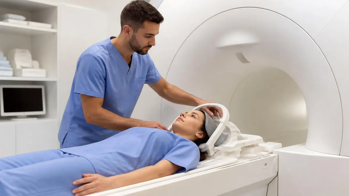
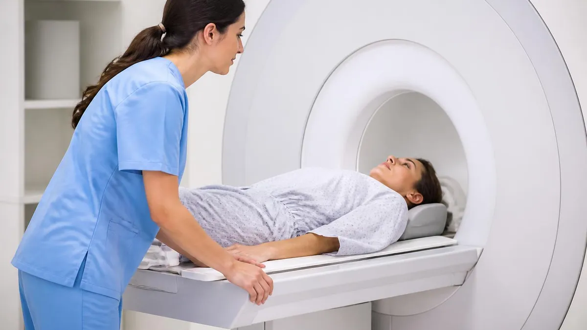
The actual exam is painless. You will lie supine on a padded table that slides into the bore, wear ear protection because the gradient coils are loud, and follow short breath-hold instructions through a microphone. Most protocols include T1- and T2-weighted sequences, in-phase and out-of-phase imaging to detect fat, diffusion-weighted imaging for cellularity, and dynamic post-contrast series. Each sequence answers a different anatomical or pathological question.
Results are typically interpreted within 24 to 48 hours by a subspecialty abdominal radiologist. The final report describes organ size, signal characteristics, enhancement patterns, lymph nodes, and incidental findings such as renal cysts or vertebral hemangiomas. Your ordering physician then correlates those imaging findings with labs, symptoms, and prior studies before recommending the next step, which might be reassurance, biopsy, surveillance, or treatment.
This guide walks through every stage of the process — from why your doctor ordered the study, to how the magnet works, to what each line of your radiology report means — so you arrive informed, prepared, and confident.
Abdominal MRI by the Numbers
Why Doctors Order Abdominal MRI
MRI is the gold standard for distinguishing benign cysts and hemangiomas from focal nodular hyperplasia, adenomas, and hepatocellular carcinoma based on dynamic enhancement patterns.
MRCP, a non-contrast technique often paired with abdominal MRI, maps the bile ducts and pancreatic duct to identify stones, strictures, and cystic neoplasms without endoscopy.
MR enterography uses oral contrast plus IV gadolinium to assess bowel wall thickening, mesenteric inflammation, and fistulas in inflammatory bowel disease without radiation exposure.
Chemical shift imaging detects intracellular fat in adrenal adenomas, while dynamic post-contrast sequences characterize renal cell carcinoma and complex cystic kidney lesions.
Patients with cirrhosis, prior hepatocellular carcinoma, or metastatic disease undergo serial MRI to detect new lesions earlier than ultrasound or CT can reliably identify them.
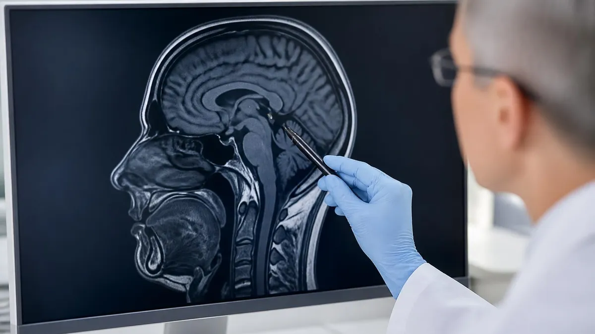
An abdominal MRI works by exploiting the magnetic properties of hydrogen nuclei, which are abundant in the body's water and fat. When you enter the bore, the scanner's main magnet aligns these protons along its axis. Brief radiofrequency pulses then tip them out of alignment, and as they relax back to their original orientation, they emit signals that coils in the table and on your abdomen detect. A computer reconstructs those signals into the grayscale images radiologists analyze.
Different tissues relax at different rates, which is why MRI offers such impressive soft-tissue contrast. T1-weighted sequences make fat appear bright and water dark, useful for anatomy and post-contrast imaging. T2-weighted sequences reverse this, highlighting edema, cysts, and bile. Combining the two — alongside fat-suppression techniques — lets the radiologist isolate inflammation, hemorrhage, or fat deposition inside an organ. The history of these sequences traces back decades, and our overview of the history of MRI shows how each advance opened new diagnostic possibilities.
Modern abdominal protocols also rely on diffusion-weighted imaging (DWI), which measures how freely water molecules move within tissue. Tumors, abscesses, and acutely inflamed tissue restrict diffusion and therefore appear bright on DWI, often before anatomic changes are obvious on conventional sequences. Pairing DWI with apparent diffusion coefficient maps quantifies that restriction and supports cancer detection, especially in the liver and peritoneum.
Breath-holding is essential because the abdomen moves with every inhalation. Most sequences require 15- to 25-second breath-holds, and the technologist coaches you through each one. Free-breathing techniques such as PROPELLER and navigator-triggered acquisitions help patients who cannot hold their breath, though they take longer and may show slightly more motion artifact. The scanner room itself stays around 68°F, and you will be offered a blanket along with the standard hearing protection.
Field strength matters too. A 1.5 Tesla magnet handles most clinical questions reliably, while a 3 Tesla magnet doubles the baseline signal, improving spatial resolution and reducing scan time. The trade-off at 3T is greater susceptibility artifact near bowel gas and metal, which experienced technologists offset with shimming and updated pulse sequences. Many academic centers now route oncology and liver patients to 3T preferentially because of the superior lesion conspicuity.
Finally, image quality depends heavily on patient cooperation. Even small movements blur the picture, so the technologist may use a flexible surface coil strapped gently across your abdomen to keep signal high and reduce the need for repeat sequences. If you feel anxious, communicate before the scan starts — short delays for breathing exercises or a low-dose anxiolytic improve outcomes far more than a rushed, motion-degraded study.
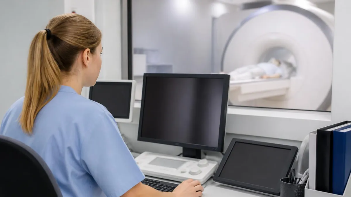
By the end of a typical exam, the scanner has collected hundreds of images across multiple planes and contrasts. The radiologist views them on multi-monitor workstations, often comparing side by side with prior studies to track subtle change over time.
MRI Practice Test Questions
Prepare for the MRI - Magnetic Resonance Imaging exam with our free practice test modules. Each quiz covers key topics to help you pass on your first try.
MRI Knowledge
MRI Exam Questions covering Knowledge. Master MRI Test concepts for certification prep.
MRI Physics
Free MRI Practice Test featuring Physics. Improve your MRI Exam score with mock test prep.
MRI Anatomy and Pathology
MRI Test Prep for MRI Anatomy and Pathology. Practice MRI Quiz questions and boost your score.
MRI Anatomy and Positioning
MRI Questions and Answers on MRI Anatomy and Positioning. Free MRI practice for exam readiness.
MRI Contrast Agents
Free MRI Quiz on MRI Contrast Agents. MRI Exam prep questions with detailed explanations.
MRI Patient Care and Positioning
MRI Practice Questions for MRI Patient Care and Positioning. Build confidence for your MRI certification exam.
Gadolinium Contrast Explained
Gadolinium is a rare-earth metal whose paramagnetic properties shorten T1 relaxation, making tissues that absorb it appear brighter on T1-weighted sequences. In its free form, gadolinium is toxic, so it is bound to a chelating molecule that allows safe injection and rapid renal excretion. Modern agents are either linear or macrocyclic, with macrocyclic chelates holding gadolinium more tightly and now preferred for most abdominal studies in 2026.
Hepatobiliary-specific agents such as gadoxetate disodium add a second imaging window. Hepatocytes take up roughly 50 percent of the dose and secrete it into bile, producing dramatic enhancement of normal liver tissue 20 minutes after injection. Lesions without functioning hepatocytes — including most metastases and hepatocellular carcinomas — appear as dark defects, dramatically improving detection sensitivity.
Abdominal MRI vs CT: Pros and Cons
- +No ionizing radiation, making it safer for repeated follow-up imaging
- +Superior soft-tissue contrast for liver, pancreas, and bowel evaluation
- +Dynamic contrast phases distinguish benign from malignant lesions accurately
- +Diffusion-weighted imaging detects subtle tumors and inflammation
- +MRCP visualizes bile and pancreatic ducts without endoscopy
- +Hepatobiliary contrast agents dramatically improve liver lesion detection
- +Multiplanar imaging without repositioning the patient
- −Longer scan times — typically 30 to 60 minutes versus 5 to 10 for CT
- −Higher cost, often two to four times that of a comparable CT
- −Claustrophobia can prevent completion in roughly 5 percent of patients
- −Incompatible with some pacemakers, cochlear implants, and metal foreign bodies
- −Breath-holding required, which is difficult for very ill patients
- −Limited availability after-hours compared with CT scanners
Abdominal MRI With Contrast: Preparation Checklist
- ✓Confirm your appointment time and arrive 30 minutes early for paperwork
- ✓Fast from solid food for four to six hours before the scan
- ✓Continue prescribed medications with small sips of water unless told otherwise
- ✓Bring a complete list of implants, surgeries, and prior imaging studies
- ✓Provide a recent creatinine or eGFR result if you are over 60 or have kidney disease
- ✓Wear comfortable clothing without metal zippers, snaps, or embroidery
- ✓Remove all jewelry, hairpins, watches, and removable dental work
- ✓Tell the technologist about claustrophobia, pregnancy, or breastfeeding
- ✓Bring a list of allergies, especially any prior reaction to contrast
- ✓Arrange a ride home if you receive sedation for anxiety
Why timing of contrast phases matters
Hepatocellular carcinoma classically shows arterial-phase hyperenhancement followed by washout on portal venous or delayed images — a pattern that is essentially diagnostic in a cirrhotic liver. Missing the arterial phase by even 10 seconds can hide the lesion, which is why precise bolus timing and patient cooperation during breath-holds are critical to a quality study.
Reading an abdominal MRI report can feel overwhelming, but most reports follow a predictable structure. The radiologist starts with a brief clinical history and technique, listing which sequences were performed and the contrast agent and dose used. This opening section helps any future radiologist replicate or compare the study, and it lets your physician confirm that the protocol matched the clinical question being asked.
Next comes the findings section, organized organ by organ. Expect commentary on the liver size, contour, and parenchymal signal; the gallbladder and biliary tree; the pancreas with its duct and any focal lesions; the spleen; the adrenal glands; the kidneys and ureters; the bowel; the peritoneum and retroperitoneum; and finally the vasculature and visualized osseous structures. Even unremarkable organs typically receive a short note confirming they were reviewed and appear normal.
Pay particular attention to language describing enhancement patterns. Phrases like "arterial hyperenhancement with delayed washout" or "progressive centripetal fill-in" carry specific diagnostic weight. The former suggests hepatocellular carcinoma; the latter is classic for a benign hemangioma. Likewise, "restricted diffusion" on DWI typically points to a hypercellular lesion such as a tumor or abscess, though it can also reflect acute infarction or hemorrhage in the right clinical setting.
Size measurements are usually given in three dimensions and compared with prior studies when available. The radiologist may also use standardized reporting systems — LI-RADS for liver lesions in cirrhotic patients, PI-RADS in some pelvic protocols, or O-RADS for ovarian findings extending into the abdomen. These systems map imaging features to probabilities of malignancy, helping your physician decide whether to observe, biopsy, or treat.
Incidental findings are common. Small renal cysts, vertebral hemangiomas, simple liver cysts, and adrenal adenomas frequently appear and rarely require action. The report should describe each one and recommend either no follow-up, a single follow-up scan, or referral to a subspecialist. Society guidelines from the American College of Radiology back most of these recommendations, so ask your physician to walk you through the rationale if anything is unclear.
The impression at the bottom is the most important paragraph. It distills the findings into a numbered list ranked by clinical relevance, beginning with the answer to the referring question and ending with minor incidental findings. If your impression says "no evidence of metastatic disease," that is usually the headline result, even if the body of the report mentions other small abnormalities.
If anything in the report is unclear, request a teleconference with the radiologist. Many institutions now offer direct patient consultation, and a 10-minute conversation can save weeks of uncertainty about what a phrase like "indeterminate T2 hyperintense lesion" actually means for your care plan.
If your eGFR is below 30 mL/min/1.73m², discuss alternatives with your physician before receiving contrast. Macrocyclic agents are considered low-risk even at reduced eGFR, but many facilities still require a documented risk-benefit discussion and signed consent. Acute kidney injury, recent dialysis, or hepatorenal syndrome raises that threshold further.
Cost varies widely across the United States, with cash prices ranging from $1,200 at outpatient imaging centers to more than $4,000 at academic hospitals. Insurance typically covers the study when medically necessary, but high-deductible plans can still leave patients responsible for $500 to $1,500 out of pocket. Calling both your insurer and the imaging facility before the scan to confirm coverage, copay, and any pre-authorization requirements prevents most billing surprises.
Outpatient centers tend to offer lower prices and shorter wait times, though hospital-based scanners may be necessary for sicker patients who need monitoring or sedation. To compare locations, check our guide on how to find an MRI scan near me, which covers accreditation, parking, and how to interpret published cash prices. Accreditation by the American College of Radiology is a strong quality signal regardless of facility type.
Risks beyond contrast reactions are minimal. Hearing damage from gradient noise is prevented by earplugs and headphones. Projectile injuries from ferromagnetic objects are prevented by the rigorous screening process at the scanner door. Burns are extremely rare and almost always involve looped cables or skin-to-skin contact, both of which technologists routinely check and pad before starting sequences.
Pregnancy deserves a special note. Non-contrast MRI is considered safe at any gestational age and is often used to evaluate appendicitis or placental abnormalities when ultrasound is inconclusive. Gadolinium crosses the placenta and is generally avoided unless the diagnostic benefit clearly outweighs theoretical fetal risks. Always disclose pregnancy or possible pregnancy during the screening interview.
Children may also undergo abdominal MRI for tumors, inflammatory bowel disease, or congenital malformations. The lack of radiation makes MRI attractive in pediatrics, though young children sometimes require sedation or general anesthesia to lie still for the full protocol. Pediatric radiologists tailor sequences to minimize scan time and maximize diagnostic yield while keeping the anesthesia exposure short.
Follow-up depends entirely on what the scan shows. A simple cyst may need no follow-up at all. An indeterminate lesion may prompt a repeat MRI in three to six months, while a suspicious mass may lead directly to biopsy or oncology referral. The radiologist's recommendation, combined with your physician's clinical judgment, drives the timeline — never feel rushed into a major decision based on a single line in the report.
Finally, keep a personal copy of every MRI on a disc or cloud account. If you change physicians, move, or seek a second opinion, having the original images — not just the report — speeds up review and avoids the need for an unnecessary repeat scan.
Walking into your appointment prepared makes the entire experience smoother. Eat a light breakfast the day before, drink plenty of water until your fasting window begins, and avoid caffeine if it usually makes you anxious. Pack a small bag with your insurance card, a photo ID, your list of medications and implants, and any prior imaging discs that another facility may have given you. The technologist will appreciate a complete history and you will avoid delays at check-in.
Dress in layers that come off easily. Most facilities provide a gown, but soft drawstring pants without metal can save you from changing entirely. Leave jewelry, smartwatches, hearing aids, magnetic eyelash applicators, and transdermal patches at home or in the locker. If you wear nicotine, nitroglycerin, or hormone patches, the technologist may ask you to remove them temporarily because some contain a thin metal backing that can heat during scanning.
Manage claustrophobia proactively. Practice slow nasal breathing in the days before your appointment, and ask your physician whether a single dose of an anxiolytic such as lorazepam would help. Many facilities offer wide-bore scanners with 70-centimeter openings, prism glasses that let you see out of the bore, and music piped through the headphones. Even a familiar playlist reduces perceived scan time and improves cooperation.
Communicate clearly with the technologist before sliding into the bore. Mention any back pain so they can position pillows. Mention any prior contrast reaction so the team can pre-medicate or switch agents. Confirm the squeeze ball is in your hand and that you understand the breath-hold commands. Once the scan begins, focus on slow breathing between sequences and follow each instruction precisely — a single missed breath-hold can require an entire sequence to be repeated.
Hydrate after the scan to help your kidneys clear gadolinium. Resume your normal diet immediately unless your physician advised otherwise. Mild fatigue or a metallic taste in the mouth lasting a few hours is normal and not a sign of allergy. If you develop a rash, swelling, breathing difficulty, or any new symptom within 24 hours, contact the imaging facility or your physician promptly so they can document the event.
While waiting for results, resist the urge to search every phrase of your future report online. Radiology language is precise but easy to misinterpret without context. Instead, prepare a short list of questions for your physician — what does the impression mean, what is the recommended next step, what is the timeline, and what would change the plan. A 10-minute focused conversation almost always resolves uncertainty better than hours of independent research.
Finally, remember that an abdominal MRI is a tool, not a verdict. Even when findings are abnormal, modern treatments for liver, pancreatic, kidney, and bowel disease have advanced dramatically. Whether the news is reassuring or unexpected, you now have detailed information to guide the next decision — and that is exactly what the scan was designed to provide.
MRI Questions and Answers


About the Author
Medical Laboratory Scientist & Clinical Certification Expert
Johns Hopkins UniversityDr. Sandra Kim holds a PhD in Clinical Laboratory Science from Johns Hopkins University and is certified as a Medical Technologist (MT) and Medical Laboratory Scientist (MLS) through ASCP. With 16 years of clinical laboratory experience spanning hematology, microbiology, and molecular diagnostics, she prepares candidates for ASCP board exams, MLT, MLS, and specialist certification tests.
Join the Discussion
Connect with other students preparing for this exam. Share tips, ask questions, and get advice from people who have been there.
View discussion (6 replies)