
MRI Head Cage: Complete Guide to Head Coils, Function, and Patient Experience
MRI head cage explained: ✏️ what the head coil does, why it's used, what patients experience, and how it produces detailed brain images.
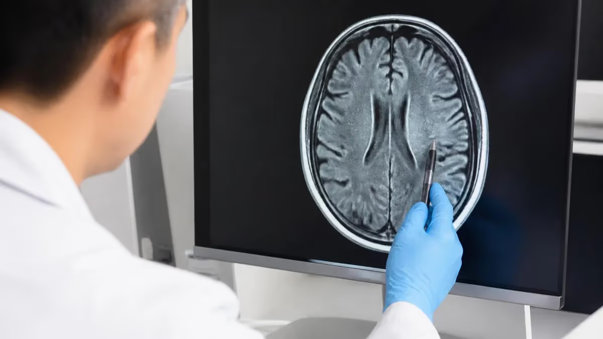
The mri head cage is one of the most recognizable pieces of equipment in any neuropictures of mri machine, yet it is also one of the most misunderstood by patients about to undergo a brain scan. Technically called a head coil or radiofrequency (RF) head coil, this helmet-like device fits over the patient's head once they are positioned on the scanner table. Its purpose is not to restrain the patient but to dramatically improve the quality of the magnetic resonance signal received from the brain, producing sharper, more diagnostically useful images.
Patients often describe the mri head cage as looking like a birdcage, a catcher's mask, or a futuristic helmet with mirrors built in. That visual impression is accurate because the engineering principle behind many head coils is literally called a birdcage resonator. The cage shape allows multiple small antennas, known as channels, to surround the head and capture signals from every angle simultaneously. More channels generally mean faster scans and higher-resolution images of structures like the hippocampus, brainstem, and cortical gray matter.
Understanding the head cage matters for both patients and aspiring MRI technologists. For patients, knowing what the device does helps reduce claustrophobia and anxiety before the exam. For students preparing for registry exams, head coil physics, channel counts, and signal-to-noise ratio (SNR) are core topics that appear repeatedly on certification tests. Reviewing the history of MRI shows how coil technology evolved from single-channel volume coils into modern 64-channel phased arrays.
Modern head coils are designed for both transmit and receive functions, or in many cases for receive-only operation paired with the scanner's body coil for transmission. Receive-only arrays deliver the best signal quality because they sit close to the anatomy of interest. A typical 3 Tesla clinical head coil houses 20 to 64 receive channels arranged in geometric patterns optimized for parallel imaging techniques like SENSE, GRAPPA, and CAIPIRINHA, which reduce scan time substantially.
The head cage also plays an essential role in patient comfort. Built-in mirrors angle upward so the patient can see out of the bore toward the technologist's window, breaking the feeling of enclosure. Padded inserts cushion the temples and cheekbones, immobilizing the head gently to reduce motion artifact without causing discomfort. Some coils include integrated headphones for music or instructions, and channels for functional MRI button-press devices used in research.
This guide walks through everything you need to know about the mri head cage: how it works physically, why it improves image quality, side effects of mri experience during a scan, how technologists position it correctly, safety considerations, and the differences between coil designs across vendors like Siemens, GE, Philips, and Canon. Whether you are a patient preparing for a brain MRI or a student preparing for a board exam, the information below will give you a complete working understanding of this critical piece of equipment.
By the end, you will know the difference between a transmit-receive coil and a receive-only array, why channel count matters, what to do if the cage feels too tight, and how head coils are used for everything from routine brain scans to advanced functional and diffusion imaging studies.
MRI Head Cage by the Numbers
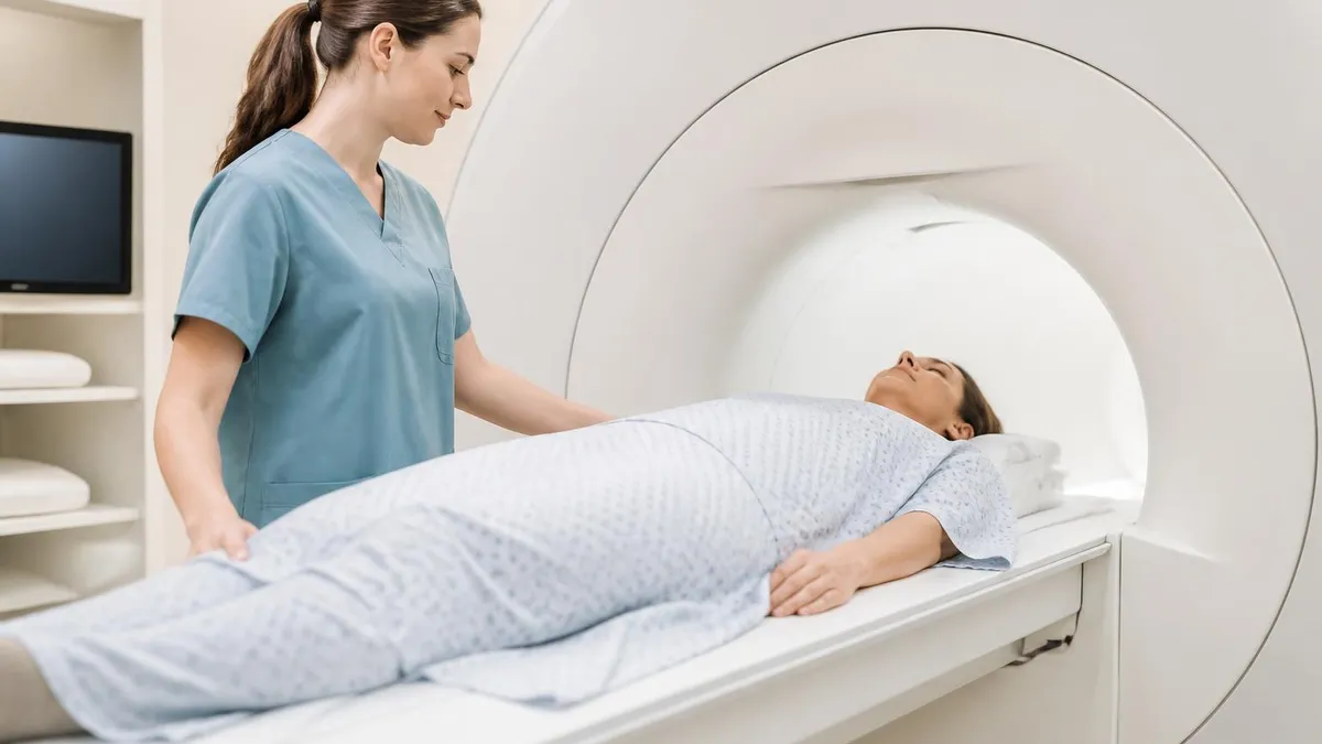
Types of MRI Head Coils
The classic transmit-receive design that surrounds the head with parallel conductors connected by capacitive rings. Produces uniform signal across the entire brain and is still used on lower field systems.
Multiple small receive coils arranged geometrically around the head. Each element captures signal from a different region, then the scanner combines them for higher SNR and faster parallel imaging.
Premium high-density coils used at 3T for advanced applications like functional MRI, diffusion tensor imaging, and high-resolution structural scans. Enables acceleration factors of 4 or more.
Smaller geometry designed for infants and young children. Closer fit to small heads dramatically improves signal-to-noise ratio, allowing shorter exam times that reduce sedation requirements.
Combined coil that images the brain, cervical spine, and neck soft tissues in one setup. Common for stroke protocols, MS workups, and head and neck cancer staging.
To understand how the mri head cage produces images, you need to appreciate the physics happening inside the scanner. The main magnet aligns hydrogen protons in the body along its field direction. A radiofrequency pulse then tips these protons into the transverse plane, causing them to precess and emit a tiny RF signal as they relax back toward equilibrium. The head coil's job is to detect that whisper-quiet signal with maximum efficiency and minimum noise contamination.
Signal-to-noise ratio is the single most important performance metric for any receive coil. Because RF signals from precessing protons are extraordinarily weak, the coil must be placed close to the anatomy and tuned precisely to the Larmor frequency of hydrogen at the scanner's field strength. At 1.5 Tesla, this frequency is approximately 63.87 MHz; at 3 Tesla, it doubles to about 127.74 MHz. Mistuning by even a small amount can cut signal dramatically.
Phased array head coils take advantage of the fact that each small element sees only a portion of the head. Each element has high SNR for its local region but low sensitivity elsewhere. When the scanner combines the signals using sum-of-squares or adaptive reconstruction, the resulting composite image has higher overall SNR than any single large coil could provide. This is why a 32-channel head coil outperforms an 8-channel coil of similar geometry.
Parallel imaging techniques exploit the spatial sensitivity differences between coil elements to skip phase-encoding steps during acquisition. Methods like SENSE, GRAPPA, and CAIPIRINHA reconstruct images from undersampled data, cutting scan time by factors of 2, 4, or even 8. The trade-off is reduced SNR, expressed by the geometry factor or g-factor. Higher channel counts produce more favorable g-factor maps and enable greater acceleration.
The head cage also incorporates passive and active decoupling circuits to prevent the receive coil from interacting with the scanner's body transmit coil during RF excitation. Without proper decoupling, the high-power transmit pulse could damage the receive electronics or create severe artifacts. Diodes, PIN switches, and detuning traps ensure the receive elements only listen during the appropriate acquisition windows.
Patients sometimes ask why the cage cannot simply be larger to feel more open. The answer comes down to physics: the closer the coil sits to the head, the better the SNR. Doubling the distance between the coil and the brain cuts signal sharply because RF sensitivity falls off rapidly with distance. Engineers design head coils to fit snugly while still accommodating roughly the 99th percentile head size. Reading more about MRI medical abbreviation conventions can help decode the technical terminology used in coil specifications and protocols.
Finally, head coil design must coexist with patient safety, ergonomics, and cleanability. The cage is constructed from low-loss dielectric materials, copper conductors, ceramic capacitors, and biocompatible plastic housings that can be wiped down between patients. Cables are routed through the coil plug on the scanner table, with strain-relief features to prevent damage from repeated docking and undocking.
MRI Practice Test Questions
Prepare for the MRI - Magnetic Resonance Imaging exam with our free practice test modules. Each quiz covers key topics to help you pass on your first try.
MRI Knowledge
MRI Exam Questions covering Knowledge. Master MRI Test concepts for certification prep.
MRI Physics
Free MRI Practice Test featuring Physics. Improve your MRI Exam score with mock test prep.
MRI Anatomy and Pathology
MRI Test Prep for MRI Anatomy and Pathology. Practice MRI Quiz questions and boost your score.
MRI Anatomy and Positioning
MRI Questions and Answers on MRI Anatomy and Positioning. Free MRI practice for exam readiness.
MRI Contrast Agents
Free MRI Quiz on MRI Contrast Agents. MRI Exam prep questions with detailed explanations.
MRI Patient Care and Positioning
MRI Practice Questions for MRI Patient Care and Positioning. Build confidence for your MRI certification exam.
Clinical Applications of the MRI Head Cage
The most common use of the head cage is routine brain imaging for headaches, stroke workup, multiple sclerosis surveillance, tumor characterization, and trauma evaluation. Standard protocols include T1-weighted, T2-weighted, FLAIR, diffusion-weighted, and gradient echo sequences. The phased array coil dramatically improves visualization of small lesions in the cortex, brainstem, and deep white matter compared with older volume coils.
For stroke imaging specifically, time matters enormously. Modern head coils paired with parallel imaging can complete a comprehensive stroke protocol in under 10 minutes, including diffusion and perfusion sequences plus MR angiography. This speed has made MRI competitive with CT for acute stroke triage at advanced centers, especially for posterior circulation events where CT sensitivity is lower.
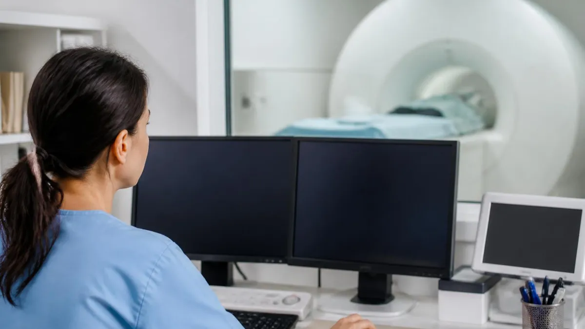
High-Channel Head Coils: Advantages and Trade-offs
- +Significantly higher signal-to-noise ratio than older volume coils
- +Enables aggressive parallel imaging acceleration up to 4x or more
- +Faster scan times reduce patient motion and improve throughput
- +Better visualization of small structures like hippocampus and brainstem
- +Supports advanced applications including fMRI, DTI, and spectroscopy
- +Integrated mirrors and padding improve patient comfort
- +Improved pediatric imaging with smaller dedicated geometries
- −More confining than older single-channel coils, increasing claustrophobia risk
- −Higher cost than basic volume coils, often $80,000 or more
- −Heavier and more cumbersome for technologists to handle
- −Cannot accommodate very large heads or certain hairstyles comfortably
- −Requires more sophisticated reconstruction with potential g-factor artifacts
- −Multiple cables increase risk of damage and downtime
- −Tighter fit may exclude patients with halo fixation devices
Pros and Cons at a Glance
| Pros | Cons |
|---|---|
| Significantly higher signal-to-noise ratio than older volume coils | More confining than older single-channel coils, increasing claustrophobia risk |
| Faster scan times reduce patient motion and improve throughput | Heavier and more cumbersome for technologists to handle |
| Better visualization of small structures like hippocampus and brainstem | Cannot accommodate very large heads or certain hairstyles comfortably |
| Supports advanced applications including fMRI, DTI, and spectroscopy | Requires more sophisticated reconstruction with potential g-factor artifacts |
| Integrated mirrors and padding improve patient comfort | Multiple cables increase risk of damage and downtime |
| Improved pediatric imaging with smaller dedicated geometries | Tighter fit may exclude patients with halo fixation devices |
MRI Head Cage Patient Preparation Checklist
- ✓Remove all metal jewelry, hair clips, earrings, and piercings before entering the scanner room
- ✓Take out any removable dental appliances, hearing aids, and prosthetic eyes if present
- ✓Inform the technologist about any prior head, neck, or eye surgery involving metal implants
- ✓Disclose history of metallic foreign bodies, especially from welding or grinding injuries
- ✓Wash off heavy makeup and eye products containing metallic pigments before the exam
- ✓Remove wigs and hairpieces, which may contain metal clips or magnetic attachments
- ✓Empty pockets of coins, keys, credit cards, and any electronic devices
- ✓Request a sedative in advance if you have a history of significant claustrophobia
- ✓Use the bathroom immediately before scanning since exams can run 30 to 60 minutes
- ✓Ask the technologist about the squeeze ball or call system before the scan starts
Proximity drives image quality
The head coil sits close to your face for a reason — every millimeter of distance reduces signal. A snug fit produces images sharp enough to detect a 2 mm lesion deep in the brainstem. Mirrors inside the cage let you see out of the bore, and the technologist can always hear you through the intercom.
Safety considerations for the mri head cage are largely the same as for any MRI examination, but a few specific issues deserve attention. The first is the absolute requirement that no ferromagnetic objects enter the scanner room. The static magnetic field exerts powerful forces on iron, steel, and other ferromagnetic materials, capable of turning a small object into a dangerous projectile. The head coil itself contains no ferromagnetic components, but anything attached to the patient inside the cage could be hazardous.
Patients with implanted medical devices require careful screening before entering the bore with a head coil in place. Cardiac pacemakers, deep brain stimulators, cochlear implants, programmable shunts, and certain aneurysm clips have specific MR conditional or MR unsafe labels. The technologist must verify model numbers against manufacturer documentation and follow approved protocols, including field strength limits and specific absorption rate (SAR) ceilings.
Specific absorption rate is especially relevant for head imaging because the head cage transmits or receives RF energy that deposits heat in tissues. The FDA limits whole-head SAR to 3.2 watts per kilogram averaged over 10 minutes. Modern scanners monitor SAR continuously and automatically adjust pulse parameters to stay within limits. Patients with metallic dental work, tattoos near the head, or certain cosmetic implants may experience localized warming and should report any heat sensation immediately.
Claustrophobia is the most common patient concern related to the head cage. About 5 to 10 percent of patients experience significant anxiety during a brain MRI, and a small subset cannot complete the exam without sedation. Strategies that help include detailed pre-exam education, allowing a support person in the room when permitted, using prism mirrors to see out, playing music through MR-compatible headphones, and offering oral anxiolytics like lorazepam.
Hearing protection is mandatory for any head MRI because gradient noise can exceed 110 decibels during fast imaging sequences. Standard practice involves foam earplugs combined with circumaural headphones built into the coil or worn separately. Pediatric patients require special attention because their ear canals are smaller and the noise can be more distressing. Some facilities use silent or quiet sequence options that reduce gradient slew rates and noise output.
Patients with claustrophobia who need mri and alzheimers may benefit from learning about mri with and without contrast cpt protocols, since some scans require gadolinium administration that extends the time inside the cage. Open MRI systems are an alternative for severely claustrophobic patients, though image quality is generally lower because of reduced field strength and less optimal coil geometry.
Finally, infection control is a routine but important safety consideration. The head coil contacts the patient's face, hair, and sometimes saliva, so it must be cleaned between every exam with hospital-grade disinfectant wipes. Most facilities use disposable coil liners or paper drapes for additional protection. Patients with active skin infections, lice, or open wounds should inform the technologist so appropriate precautions can be taken.
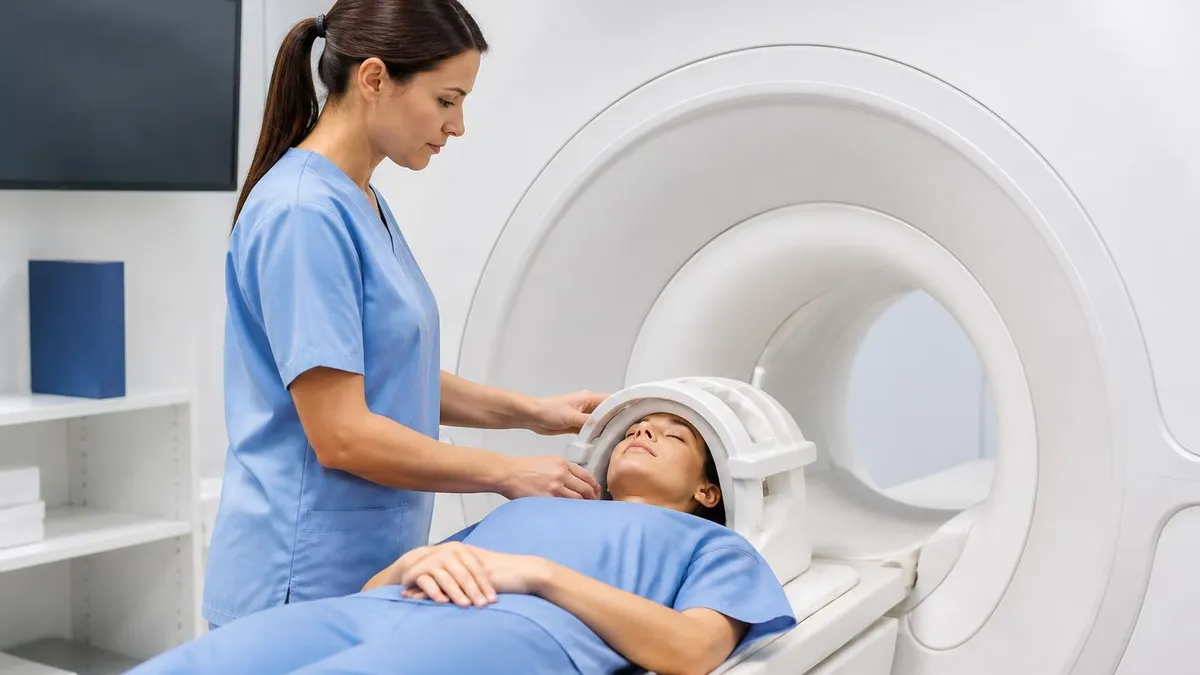
Never enter the MRI scan room with the head coil in place if you have not been screened for metallic implants, foreign bodies, or electronic devices. Even small ferromagnetic objects can become lethal projectiles in the magnetic field. Always disclose every implant, surgery, and embedded object during screening — when in doubt, ask for an X-ray before scanning.
Differences between manufacturers can affect both image quality and patient experience inside the mri head cage. Siemens offers the Head/Neck 20 and Head/Neck 64 coils, the latter providing 64 receive channels split between head and neck regions. GE Healthcare markets head coils with 21 to 48 channels under names like AIR Coil and Head Neck Unit. Philips offers the dStream Head coil line, and Canon provides multi-channel head coils for their Vantage systems. While the underlying physics is identical, geometry, weight, and channel layout differ.
Coil positioning is one of the most important steps performed by the MRI technologist. The patient lies supine on the table with the head centered in the lower half of the coil. The top half is then lowered and locked into place. The nose should be centered, the chin tucked slightly, and the head padded firmly against motion. Misalignment by even a centimeter can degrade SNR and cause asymmetric shading on the final images.
Landmarking refers to setting the table position so that isocenter aligns with the anatomy of interest. For mri for migraines the technologist typically lands at the glabella or nasion. This places the brain at the center of the magnet bore where field homogeneity is best. Off-center landmarking can introduce shading, geometric distortion, and reduced fat saturation efficiency, especially at the periphery of the imaging volume.
For research applications, head coils may be combined with additional accessories like respiratory belts, eye-tracking cameras, button-press response devices, and physiological monitoring equipment. All of these accessories must be MR-compatible and connected through filtered penetrations in the scan room wall. The head coil's design must accommodate these add-ons without compromising patient safety or image quality.
The advent of MRI imaging centers as freestanding outpatient facilities has changed how head coils are used in everyday practice. Outpatient centers often invest in mid-range 1.5T or 3T systems with mid-channel head coils that balance cost, throughput, and image quality. Academic and tertiary hospitals typically maintain a wider range of coils including ultra-high-channel arrays for advanced applications.
Quality control of head coils is performed regularly by medical physicists and service engineers. Standard tests include SNR measurement using a phantom, uniformity assessment, geometric distortion mapping, and channel-by-channel function checks. A failing element in a 32-channel coil might be undetectable to the casual observer but can subtly degrade image quality and parallel imaging performance.
Future developments in head coil technology include even higher channel counts, AI-driven reconstruction methods that compensate for noisy or accelerated data, and motion-tracking systems that adjust scan parameters in real time. Ultra-high-field 7 Tesla systems are pushing head coil engineering to new limits, with custom transmit-receive arrays that overcome challenges like RF field inhomogeneity and increased SAR.
Practical tips for patients about to experience the mri head cage can make the difference between a smooth, successful exam and one that has to be repeated or aborted. The single most useful preparation is mental rehearsal. Visualize lying on the table, hearing the noises, and feeling the cage gently surround your head. Patients who walk in with realistic expectations report less anxiety and are more likely to complete the scan in one attempt without sedation.
Wear comfortable, metal-free clothing to your appointment if you do not want to change into a hospital gown. Soft sweatpants, T-shirts without metal snaps, and cotton socks work well. Avoid clothing with zippers, underwire bras, or metallic decorations on the chest area, which can produce artifacts even on brain images because of stray RF noise pickup.
If you wear glasses, you will need to remove them before the scan starts. If you use them for distance vision and need to see the mirrors clearly, ask the technologist about MR-safe glasses kept on hand at many facilities. Contact lenses are generally fine to leave in place, though some patients find that closing their eyes throughout the exam helps reduce the sensation of confinement.
Communicate with your technologist before and during the exam. Ask how long each sequence will last, when contrast might be given, and how to use the squeeze ball if you need to stop. Technologists genuinely want patients to be comfortable because comfortable patients hold still and produce better images. Speak up immediately about any pain, numbness, unusual warmth, or panic — these are not inconveniences, they are valuable safety signals.
Practice slow, controlled breathing throughout the scan. The cage does not restrict breathing in any meaningful way, but anxiety can create the false sensation that it does. Inhale through the nose for a count of four, hold briefly, and exhale slowly for a count of six. This vagal-nerve breathing pattern lowers heart rate and reduces the urge to move, which is exactly what the scanner needs.
For students and technologists preparing for registry exams, key head coil topics to master include birdcage versus phased array principles, transmit-receive versus receive-only operation, parallel imaging fundamentals, SAR calculations, and the geometry factor. Sample problems often involve calculating Larmor frequency at given field strengths, identifying coil-related artifacts, and choosing the appropriate coil for a specific clinical scenario such as a pediatric stroke or adult tumor staging.
Finally, remember that the head cage is a tool designed to help, not to confine. Modern coils are the product of decades of engineering refinement aimed at making MRI faster, sharper, and more comfortable. The brief discomfort of a snug fit is the price of detecting tiny strokes, characterizing tumors before they become life-threatening, and mapping the function of healthy brain tissue. With the right preparation and the right team, the mri head cage delivers extraordinary value to medicine.
Sample MRI - Magnetic Resonance Imaging Practice Questions
Try these questions from our free MRI - Magnetic Resonance Imaging practice tests. The correct answer and an explanation follow each question.
Require the employment of solenoid coils because of the B0's orientation; these coils are more effective than linear coils and can be electrically combined with other coils to enhance signal homogeneity across an area of interest.
- A. Helmholtz pair
- B. Phased array
- C. Quadrature
- D. Vertical
Answer: D. Vertical
Vertical coils are specifically designed for MRI systems where the main magnetic field (B0) is oriented vertically, often found in open or permanent magnet systems. These coils are more effective than linear coils in this orientation and can be electrically combined with other coils to enhance signal homogeneity across the area of interest, optimizing signal reception for vertical field systems.
What is the function of the shim coils in an MRI system?
- A. Generate the main static magnetic field
- B. Compensate for inhomogeneities in the B0 field to improve field uniformity
- C. Produce the rapidly switching gradients for slice selection
- D. Amplify the RF signal received from the patient
Answer: B. Compensate for inhomogeneities in the B0 field to improve field uniformity
Shim coils generate small corrective magnetic fields that counteract spatial variations in B0, improving homogeneity and image quality.
By adding more loops and circuitry, these coils increase the efficiency with which the MR signal is induced in the coil and raise SNR by about 40% when compared to coils of the same size.
- A. Phased array
- B. Quadrature
- C. Vertical
- D. Helmholtz pair
Answer: B. Quadrature
Quadrature coils (or circularly polarized coils) utilize two receiver coils oriented 90 degrees to each other. By combining the signals from both coils, they can detect the rotating component of the MR signal more efficiently. This design leads to a theoretical 41% increase in signal-to-noise ratio (SNR) compared to a single linear coil of the same size, improving image quality.
When positioning a patient for a lumbar spine MRI, the technologist should place the patient in which position to reduce lumbar lordosis and improve image quality?
- A. Prone with legs flat
- B. Supine with knees flexed and supported by a foam wedge
- C. Lateral decubitus position
- D. Seated upright
Answer: B. Supine with knees flexed and supported by a foam wedge
Flexing the knees with a foam wedge support flattens lumbar lordosis, reducing the curvature and aligning disc spaces parallel to axial slices for optimal visualization.
Take the full MRI - Magnetic Resonance Imaging practice test
MRI Questions and Answers
The History of MRI: From Discovery to Modern Medicine
MRI With and Without Contrast: How It Works, What to Expect
MRI Medical Abbreviation: What MRI Stands For and Why It Matters
Shoulder MRI: What It Shows, Procedure, and Reading the Report
MRI Imaging Centers: Complete Guide to Independent Outpatient MRI Facilities
About the Author

Medical Laboratory Scientist & Clinical Certification Expert
Johns Hopkins UniversityDr. Sandra Kim holds a PhD in Clinical Laboratory Science from Johns Hopkins University and is certified as a Medical Technologist (MT) and Medical Laboratory Scientist (MLS) through ASCP. With 16 years of clinical laboratory experience spanning hematology, microbiology, and molecular diagnostics, she prepares candidates for ASCP board exams, MLT, MLS, and specialist certification tests.
Join the Discussion
Connect with other students preparing for this exam. Share tips, ask questions, and get advice from people who have been there.
View discussion (8 replies)