Side Effects of MRI: What Patients Need to Know About Risks, Reactions, and Recovery
Learn the side effects of MRI scans, including contrast reactions, 🗨️ claustrophobia, noise exposure, and rare complications. Complete patient safety guide.
The side effects of MRI are generally mild and far less concerning than those associated with X-ray or CT imaging because magnetic resonance imaging uses no ionizing radiation. Most patients leave the scanner feeling exactly as they did when they arrived, with no lingering symptoms or recovery period required. However, the procedure is not entirely without risk, and understanding what can happen during and after the scan helps patients make informed decisions, communicate effectively with their radiologist, and recognize the rare situations where medical follow-up becomes necessary.
When physicians order an MRI, they weigh diagnostic benefits against potential discomforts and complications. The strong magnetic field, radiofrequency pulses, gradient coils, and gadolinium-based contrast agents each carry distinct safety profiles. Some patients experience transient sensations like warmth, tingling, or muscle twitching, while others develop more pronounced reactions such as anxiety from confined spaces or nausea from intravenous contrast. Knowing the spectrum of possible responses prepares you mentally and physically for the experience.
This comprehensive guide covers every category of MRI side effect documented in clinical literature, from the trivial to the serious. We examine immediate reactions that occur inside the scanner, delayed effects that emerge hours or days afterward, special concerns for pregnant patients and children, and the specific risks linked to gadolinium contrast. For background on how the technology evolved into today's standard imaging tool, you can read more in our overview of the history of MRI.
Most adverse events are predictable and preventable when patients complete the safety screening questionnaire honestly. Technologists and radiologists rely on accurate disclosure of implants, allergies, kidney function, and previous reactions to tailor the protocol. Even simple details like recent tattoos, transdermal patches, or metallic eye makeup can influence the safety plan. Patients who skip questions or assume information is irrelevant put themselves at higher risk of avoidable complications during the scan.
The body of evidence supporting MRI safety spans more than four decades and millions of scans performed annually worldwide. Regulatory agencies including the FDA, the American College of Radiology, and the International Society for Magnetic Resonance in Medicine continually update guidelines based on new research. Despite this rigorous oversight, individual patient experiences vary widely, and what feels routine to one person may feel overwhelming to another. Personal preparation and clear communication remain the best defense.
Throughout the article, we balance reassurance with realism. MRI remains one of the safest diagnostic procedures in modern medicine, but downplaying potential side effects does patients no favors. By the end of this guide, you should understand exactly what sensations are normal, which symptoms warrant a call to your doctor, how contrast agents affect different populations, and what steps to take if you experience anything unexpected during or after your scan.
MRI Side Effects by the Numbers
Main Categories of MRI Side Effects
Warmth, tingling, muscle twitching, or a metallic taste during the scan. These sensations result from radiofrequency energy and gradient coil activity. They are temporary, harmless, and stop the moment the sequence ends without lingering effects on tissue.
Claustrophobia, panic attacks, and acute anxiety affect a meaningful minority of patients. The narrow bore, loud noise, and 30-60 minute immobility trigger stress responses. Open MRI systems, mild sedation, or breathing techniques typically resolve these issues effectively.
Gadolinium-based contrast can cause nausea, headache, cold sensation at the injection site, or rarely, allergic responses. Patients with reduced kidney function face additional risks including nephrogenic systemic fibrosis, which is why renal screening is mandatory.
Hearing discomfort from gradient noise, vibration sensations, and temporary disorientation when leaving the scanner. Ear protection is mandatory in most facilities. Vibrations can occasionally cause minor discomfort in patients with sensitive musculoskeletal conditions.
Patients with non-MRI-compatible implants face the most serious potential complications including device heating, displacement, or malfunction. Modern screening protocols and conditional-safe device databases have dramatically reduced these incidents over the past decade.
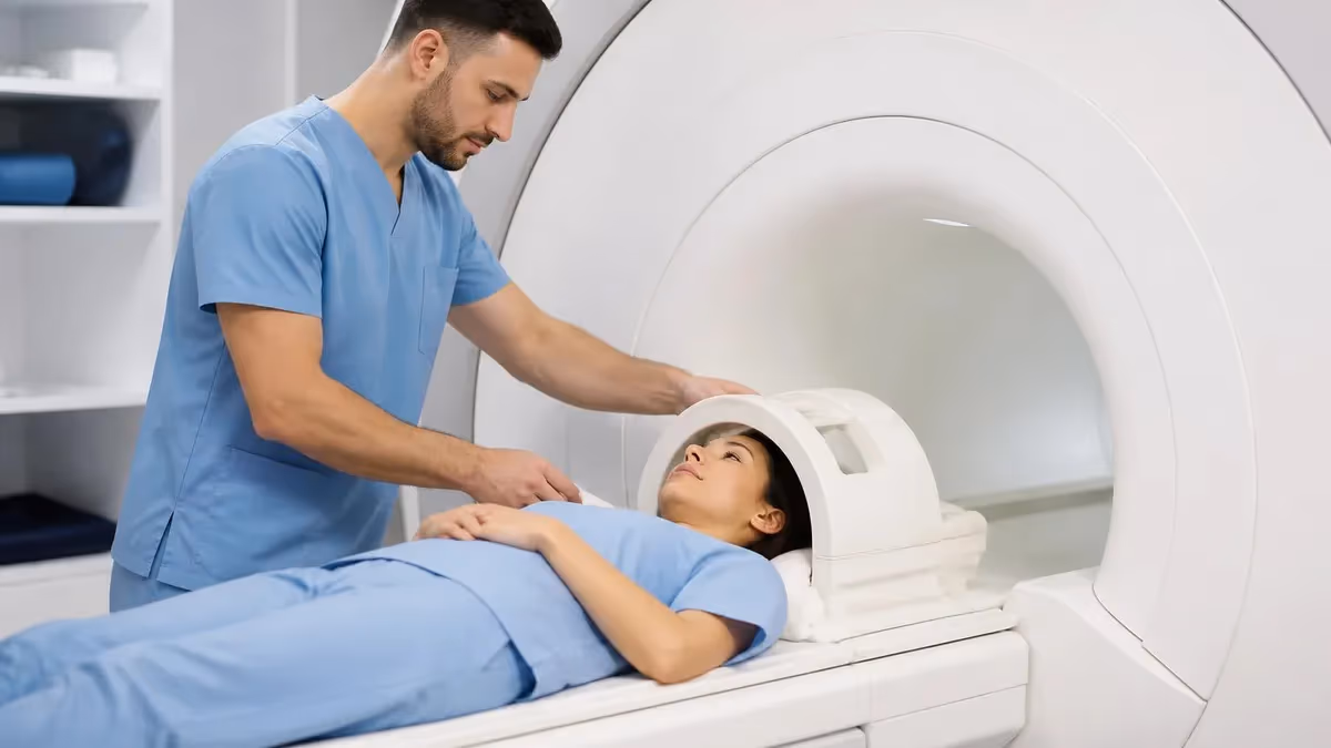
Understanding what happens during an MRI scan helps demystify the sensations patients commonly report. When you enter the scanner, the static magnetic field is already on at full strength, but you cannot feel it directly. Some patients describe a subtle awareness of being inside a large magnet, though this is usually psychological rather than physiological. As the technologist begins each imaging sequence, you will hear rhythmic banging, buzzing, or knocking sounds caused by the rapid switching of gradient coils that encode spatial information into the signal.
The loud noise represents one of the most universal complaints from MRI patients. Scanner acoustic levels routinely reach 110-125 decibels during certain sequences, comparable to a chainsaw or a rock concert. Facilities provide either foam earplugs, padded headphones, or both, and many modern systems offer music streaming through MRI-compatible headsets. Without proper hearing protection, temporary threshold shifts or even permanent hearing damage become real possibilities, particularly with longer cardiac or musculoskeletal protocols.
Warmth and tingling sensations occur when radiofrequency energy deposits into tissue. The specific absorption rate, measured in watts per kilogram, is carefully monitored by the scanner software to stay within FDA limits. Patients with larger body habitus, those undergoing extended scans, or those with metallic tattoos sometimes notice more pronounced warming. Inform the technologist immediately if you feel any burning, focal hot spots, or uncomfortable temperature changes during the examination so adjustments can be made.
Peripheral nerve stimulation produces brief muscle twitching, particularly in the abdomen, back, or extremities, during fast gradient sequences. This phenomenon is well documented and harmless, though it can startle patients who were not warned in advance. The sensation feels like a quick electrical pulse or involuntary muscle contraction and stops the instant the sequence completes. If twitching becomes painful or sustained, the technologist will modify gradient parameters to reduce stimulation. To learn how different protocols compare, see mri with and without contrast cpt.
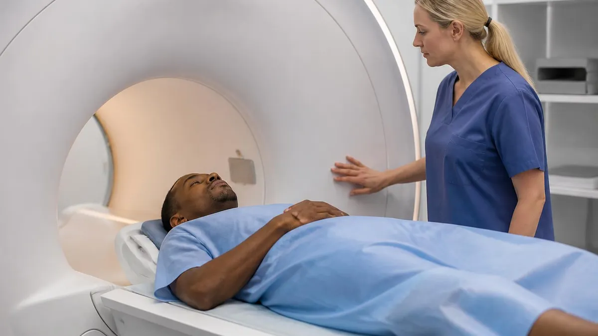
Claustrophobia affects roughly 5-10% of patients to a degree requiring intervention. Symptoms range from mild unease to full panic attacks with rapid heart rate, sweating, and an overwhelming urge to escape. The combination of confined space, immobility, noise, and the inability to see clearly creates a perfect storm for anxious patients. Communication with the technologist via the squeeze bulb or intercom should be tested before the scan begins so you feel in control throughout the procedure.
Disorientation and mild dizziness occasionally occur when patients move quickly through the static magnetic field, particularly when entering or exiting a 3 Tesla scanner. The effect, sometimes called magnetic vertigo, results from interaction between the field and the vestibular system in the inner ear. Symptoms resolve within seconds of moving away from the magnet and pose no lasting harm. Patients prone to motion sensitivity should request slower table movement during positioning and exit.
Finally, some patients experience a brief metallic taste during scanning, particularly when imaging the head or neck region with high field strength systems. This taste sensation results from electrical currents induced in tooth fillings or other oral metalwork. While unsettling at first, the taste disappears within minutes of leaving the scanner and indicates no harm to dental work or surrounding tissue.
MRI Practice Test Questions
Prepare for the MRI - Magnetic Resonance Imaging exam with our free practice test modules. Each quiz covers key topics to help you pass on your first try.
MRI Knowledge
MRI Exam Questions covering Knowledge. Master MRI Test concepts for certification prep.
MRI Physics
Free MRI Practice Test featuring Physics. Improve your MRI Exam score with mock test prep.
MRI Anatomy and Pathology
MRI Test Prep for MRI Anatomy and Pathology. Practice MRI Quiz questions and boost your score.
MRI Anatomy and Positioning
MRI Questions and Answers on MRI Anatomy and Positioning. Free MRI practice for exam readiness.
MRI Contrast Agents
Free MRI Quiz on MRI Contrast Agents. MRI Exam prep questions with detailed explanations.
MRI Patient Care and Positioning
MRI Practice Questions for MRI Patient Care and Positioning. Build confidence for your MRI certification exam.
Gadolinium Contrast: Side Effects of MRI with IV Agents
The most common side effects of MRI contrast injection include a cold sensation traveling up the arm, mild nausea, headache, dizziness, and a metallic taste in the mouth. These symptoms typically begin within minutes of administration and resolve spontaneously within an hour. Approximately 2-4% of patients receiving gadolinium-based contrast agents experience at least one mild reaction, though most are so minor they do not interrupt the scan.
Injection site reactions are also common but usually trivial. Brief stinging, bruising, or a small hematoma at the IV insertion point occurs in some patients but heals within days. Extravasation, when contrast leaks into surrounding tissue, happens rarely but can cause localized swelling and discomfort. Cold compresses and arm elevation typically resolve these issues without medical intervention or lasting effect.
MRI Safety Profile: Benefits vs Side Effect Concerns
- +No ionizing radiation exposure, unlike CT or X-ray imaging
- +Excellent soft tissue contrast for accurate diagnosis
- +Most side effects are mild, transient, and self-limiting
- +Severe adverse reactions are extremely rare with modern protocols
- +Contrast agents have improved safety profiles since 2010
- +Repeat scans possible without cumulative radiation risk
- +Suitable for pediatric and pregnant patients in many cases
- −Loud acoustic noise requires hearing protection throughout
- −Confined space triggers claustrophobia in 5-10% of patients
- −Long scan times of 30-60 minutes demand stillness
- −Gadolinium contrast carries small allergy and kidney risks
- −Implanted devices may be contraindicated or require special protocols
- −Cost is significantly higher than ultrasound or X-ray
- −Cannot be used for patients with non-MRI-safe pacemakers
Patient Checklist to Minimize MRI Side Effects
- ✓Complete the safety screening form honestly, including all implants and surgeries
- ✓Disclose any history of allergic reactions to contrast or medications
- ✓Inform staff if you are pregnant or might be pregnant
- ✓Report kidney disease, dialysis, or recent blood work with creatinine levels
- ✓Remove all metal jewelry, piercings, hearing aids, and dental appliances
- ✓Avoid wearing makeup containing metallic particles or shimmer
- ✓Eat a light meal before scans involving contrast to reduce nausea
- ✓Use the provided earplugs or headphones throughout every sequence
- ✓Practice slow breathing or request music if prone to anxiety
- ✓Tell the technologist immediately about any pain, heat, or unusual sensation
The Squeeze Bulb Is Your Lifeline
Every MRI patient receives a squeeze bulb or call button before the scan begins. Squeezing it pauses the sequence and brings the technologist on the intercom within seconds. Test it before sliding into the bore so you feel confident using it. If anything feels wrong — heat, pain, anxiety, or shortness of breath — squeeze without hesitation. Stopping a scan early is always preferable to powering through dangerous symptoms.
Delayed side effects of MRI typically emerge within 24-72 hours after the scan and are most commonly associated with gadolinium contrast administration rather than the magnetic field itself. Headaches represent the most frequently reported delayed symptom, affecting roughly 1-3% of contrast recipients. These headaches are usually mild, respond to over-the-counter analgesics, and resolve within a day or two. Persistent or severe headaches warrant medical evaluation to rule out other causes.
Skin reactions sometimes appear in the hours following an MRI scan, particularly at the contrast injection site or in patients sensitive to topical ECG electrode adhesives used during cardiac imaging. Localized redness, itching, or mild rash typically resolves with over-the-counter antihistamines or hydrocortisone cream. Generalized urticaria appearing more than an hour after the scan is rare but should prompt a call to your physician, especially if accompanied by any breathing difficulty or facial swelling.
Fatigue is reported by some patients after lengthy MRI examinations, though the cause is not entirely clear. The combination of prolonged immobility, anxiety, fasting for contrast protocols, and the cognitive load of the experience likely contributes. Most patients recover fully with rest, hydration, and a normal meal. Persistent fatigue lasting more than 24 hours is not typical and should be discussed with your healthcare provider as part of broader clinical assessment.
Nephrogenic systemic fibrosis remains the most serious documented long-term side effect of MRI, but it occurs almost exclusively in patients with severe pre-existing kidney impairment. Symptoms develop over weeks to months and include painful skin thickening, joint stiffness, and eventual involvement of internal organs. Since macrocyclic gadolinium agents replaced older linear formulations and eGFR screening became universal, new NSF cases have become extraordinarily rare in the United States and Europe.
Gadolinium retention in brain tissue has emerged as a topic of significant research interest over the past decade. Imaging studies have demonstrated small amounts of gadolinium deposition in the dentate nucleus and globus pallidus of patients receiving multiple contrast-enhanced scans. To date, no specific clinical syndrome has been definitively linked to this retention in patients with normal kidney function, but the FDA recommends limiting contrast use to clinically necessary situations and using macrocyclic agents when possible.
Anxiety and post-traumatic stress symptoms can follow particularly distressing MRI experiences, especially in patients who suffered panic attacks during the scan. These reactions are real and deserve attention even though they do not stem from physiological harm. Cognitive behavioral techniques, exposure therapy, and oral anxiolytics significantly improve subsequent scanning experiences. Patients who struggled with one MRI should advocate for a more supportive protocol before any future imaging is scheduled.
Hearing-related side effects of MRI generally resolve within hours but occasionally persist as temporary threshold shifts. Patients who removed earplugs mid-scan or whose hearing protection was inadequate may notice muffled hearing, ringing, or sensitivity to loud sounds afterward. Most cases resolve without intervention within 24 hours. Persistent tinnitus or hearing loss after MRI is uncommon and warrants audiology evaluation to assess any potential lasting impact on auditory function.
Patients with pacemakers, cochlear implants, neurostimulators, drug infusion pumps, or aneurysm clips must NEVER enter an MRI scanner room without explicit clearance from the radiologist and device manufacturer. Older devices can heat, dislodge, or malfunction in the magnetic field, with potentially fatal consequences. Always carry your device identification card and disclose every implanted item during screening, even decades-old surgical hardware that you may have forgotten about.
Knowing when to contact your doctor after an MRI helps distinguish normal post-scan sensations from genuine medical concerns. Most patients experience no symptoms whatsoever following the procedure and can resume normal activity immediately, including driving home and returning to work. However, certain symptoms warrant prompt medical attention regardless of how mild they may initially appear. Erring on the side of caution is always appropriate when contrast agents or unusual reactions are involved in your case.
Call your physician immediately if you develop hives spreading across your body, facial swelling, difficulty breathing, or wheezing in the hours after a contrast-enhanced MRI. These signs may indicate a delayed allergic reaction requiring antihistamines, steroids, or in severe cases emergency treatment. Most delayed allergic reactions appear within 1-7 days of gadolinium exposure and respond well to standard allergy treatment when addressed promptly by qualified medical personnel.
Persistent headache, nausea, or dizziness lasting more than 24 hours after the scan deserves evaluation. While transient symptoms are normal, sustained discomfort may indicate a contrast reaction, dehydration, or an unrelated condition that coincidentally became symptomatic. Your radiologist or referring physician can determine whether further testing or supportive care is needed. Document the timing, severity, and any accompanying symptoms so the medical team has accurate information to guide their assessment of your situation.
Skin changes at the injection site that progress beyond mild bruising require attention. Significant swelling, increasing pain, warmth, or red streaks tracking up the arm could indicate extravasation with tissue damage or, less commonly, infection. Most extravasation injuries resolve with conservative management, but proper assessment ensures appropriate treatment and documentation. Photograph the area and contact the imaging center where your scan was performed for guidance specific to your situation and any institutional protocols.
Patients with kidney disease who receive gadolinium contrast should monitor for any new skin tightening, joint stiffness, or unusual rashes in the weeks and months following the scan. While modern protocols have made nephrogenic systemic fibrosis extraordinarily rare, early recognition improves outcomes significantly. Discuss any concerning symptoms with both your nephrologist and the radiology department that performed the original imaging study. Honest communication about all post-scan symptoms helps protect future patients too.
If you struggled with severe anxiety, claustrophobia, or panic during the scan, do not simply hope it will be better next time. Talk with your primary care physician about strategies for future imaging, including oral anxiolytics, open MRI options, sedation protocols, or referral to a behavioral therapist specializing in medical procedure anxiety. Many imaging centers now offer dedicated patient comfort programs that significantly reduce anxiety. You can explore local options through our guide to MRI imaging centers.
Finally, keep written records of every MRI you undergo, including dates, scan types, contrast agents used, and any reactions experienced. This information becomes invaluable when scheduling future imaging or evaluating unexplained symptoms years later. Many patients are surprised by how often radiologists and physicians ask for prior scan history. A simple notebook, smartphone note, or patient portal record helps ensure continuity of care across multiple imaging facilities and providers throughout your lifetime.
Practical preparation dramatically reduces the likelihood of experiencing significant MRI side effects. Start by reviewing the appointment confirmation packet sent by the imaging center, which typically includes pre-scan instructions, fasting requirements for contrast studies, and medication guidance. If you take medications for anxiety, ask the prescribing physician whether a single dose 30-60 minutes before your appointment is appropriate. Many patients find this single intervention transforms an unbearable experience into a manageable one.
Hydration is your friend before and after contrast-enhanced MRI. Drinking 16-24 ounces of water in the two hours leading up to the scan helps maintain kidney function and aids in flushing gadolinium from the body afterward. Avoid caffeine on the morning of the scan if anxiety is a concern, as stimulants can amplify the physiological response to confined spaces. Eating a light meal, unless specifically instructed to fast, reduces nausea associated with both anxiety and contrast administration.
Wear comfortable clothing without metal zippers, buttons, snaps, or underwire bras to your appointment. While most facilities provide hospital gowns, arriving prepared saves time and reduces the chance of forgotten metallic items affecting your scan. Leave valuables at home or with a family member, since lockers vary in security across facilities. Bring a list of all implants, surgeries, and medications, and consider asking about your scanning device through resources like the mri safety and compatibility guide.
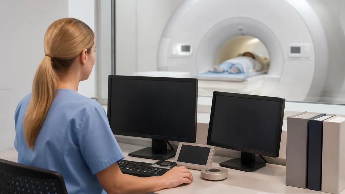
Communication during the scan is critical for patient safety. Before the table moves into the bore, practice using the squeeze bulb so you know exactly how much pressure activates it. Ask the technologist to confirm they can hear you clearly through the intercom. Establish a signal for when you need a break between sequences. Even patients who feel confident before the scan sometimes need to pause partway through, and knowing the system works builds the confidence required to complete the examination.
Breathing techniques significantly reduce claustrophobia and general anxiety inside the scanner. Slow diaphragmatic breathing, with inhalation through the nose for four counts and exhalation through the mouth for six counts, activates the parasympathetic nervous system and calms the body's stress response. Practice this technique in the days before your appointment so it becomes automatic when you need it most. Some patients also find counting backward from 300 keeps the mind engaged and time perception more manageable.
For patients who have struggled with previous MRI experiences, advocate firmly for accommodations that match your needs. Wide-bore scanners with 70 cm openings feel substantially more spacious than older 60 cm systems. Open MRI configurations eliminate the tunnel entirely but produce lower-quality images and longer scan times. Oral anxiolytics, IV sedation, and even general anesthesia are available for severe cases. Discuss these options openly with both your referring physician and the imaging center scheduling team.
After the scan, give yourself permission to rest for the remainder of the day if you experienced significant anxiety or received contrast. While most patients resume normal activity immediately, listening to your body prevents pushing through symptoms that deserve attention. Drink additional fluids, eat a satisfying meal, and avoid alcohol for 24 hours after contrast administration. Schedule any follow-up consultation with your physician promptly so results can be reviewed and next steps planned without unnecessary delay.
MRI Questions and Answers

The History of MRI: From Discovery to Modern Medicine

MRI With and Without Contrast: How It Works, What to Expect

MRI Medical Abbreviation: What MRI Stands For and Why It Matters

MRI Imaging Centers: Complete Guide to Independent Outpatient MRI Facilities

St Jude Pacemaker MRI Compatibility List: A Complete Safety and Scanning Guide
About the Author
Medical Laboratory Scientist & Clinical Certification Expert
Johns Hopkins UniversityDr. Sandra Kim holds a PhD in Clinical Laboratory Science from Johns Hopkins University and is certified as a Medical Technologist (MT) and Medical Laboratory Scientist (MLS) through ASCP. With 16 years of clinical laboratory experience spanning hematology, microbiology, and molecular diagnostics, she prepares candidates for ASCP board exams, MLT, MLS, and specialist certification tests.
Join the Discussion
Connect with other students preparing for this exam. Share tips, ask questions, and get advice from people who have been there.
View discussion (6 replies)