St Jude Pacemaker MRI Compatibility List: A Complete Safety and Scanning Guide
✏️ Complete st jude pacemaker mri compatibility list, MRI conditional device scanning rules, safety screening checklists, and zone management for techs.

The st jude pacemaker mri compatibility list is one of the most frequently referenced safety documents in modern radiology departments, and for good reason. Cardiac implantable electronic devices (CIEDs) from St. Jude Medical, now part of Abbott, include several pacemaker and implantable cardioverter-defibrillator (ICD) platforms that have received MR-Conditional labeling from the FDA. Knowing which generator and lead combinations are scannable, at what field strength, and under which scan parameters is essential before any patient with a St. Jude device crosses into Zone IV of the MRI suite.
MRI safety is not a single checklist item but a layered system involving device screening, implant interrogation, scan parameter restriction, patient monitoring, and post-scan device evaluation. For St. Jude devices, the compatibility list specifies generator models such as the Assurity MRI, Endurity MRI, and certain Accent MRI pacemakers, along with paired Tendril MRI leads. Devices that fall outside this list are typically considered MR-Unsafe or require institutional risk-benefit review under guidance from a cardiac electrophysiologist and an MRI medical director.
Beyond cardiac devices, MRI safety encompasses a much wider landscape: cochlear implants, aneurysm clips, deep brain stimulators, drug-infusion pumps, tissue expanders, retained shrapnel, and dozens of orthopedic implants. Each carries unique conditions for safe scanning, including maximum static field strength (1.5T versus 3T), spatial gradient limits in tesla per meter, and specific absorption rate (SAR) ceilings expressed in watts per kilogram. Skipping any of these checks can cause device malfunction, thermal injury, projectile events, or fatal arrhythmia.
Technologists, radiologists, and MR safety officers should treat the manufacturer compatibility list as a living document. St. Jude and Abbott periodically update labeling, add new models, and revise conditions as post-market data accumulates. The American College of Radiology (ACR) MR Safe Practice Guidelines and the Joint Commission's recommendations both reinforce the need to verify device status against the latest manufacturer documentation, not memory or generic search results.
This guide breaks down the entire MRI safety ecosystem around cardiac devices and implants. You will learn how to interpret MR-Conditional labeling, what specific parameters to enter at the scanner console, how to coordinate with cardiology for device programming, and how to manage screening for both inpatient and outpatient populations. We will also cover screening forms, ferromagnetic detection, zone management, and the unique safety profile of contrast-enhanced cardiac MRI for these patients.
If you are preparing for the ARRT MRI registry or the ARMRIT exam, this material maps directly to the safety domain, which makes up the largest single content area on both certifications. Real-world competence in implant screening is also the single highest-yield safety skill you will use every shift. For a broader look at how the modality evolved into the precision tool it is today, our overview of the history of MRI provides useful context on why these safeguards exist.
By the end of this article, you will understand exactly how to read a St. Jude compatibility table, identify the difference between MR-Conditional and MR-Safe labeling, configure your scanner for a conditional scan, and document the procedure in compliance with ACR Zone IV requirements. We will close with a thorough FAQ section addressing the questions techs and patients most commonly ask before a scan involving an implanted cardiac device.
MRI Implant Safety by the Numbers
St. Jude MR-Conditional Device Categories
A dual-chamber MR-Conditional pacemaker approved for 1.5T full-body scanning when paired with Tendril MRI leads. Requires programming to an MRI-specific mode prior to imaging and reversion afterward by qualified cardiology staff.
Single and dual-chamber platform with broad MR-Conditional labeling at 1.5T. Endurity MRI offers automated lead measurements and was designed with whole-body scanning compatibility, eliminating prior exclusion zones above the chest.
An earlier MR-Conditional generation with conditional 1.5T labeling. Requires confirmation that both the generator and leads are MRI-labeled models; legacy non-MRI leads with an MRI generator do not qualify as a conditional system.
The companion lead family required for any St. Jude conditional pacemaker scan. Mixing non-MRI leads with an MRI generator invalidates conditional status and reclassifies the system as MR-Unsafe under current labeling.
Implantable cardioverter-defibrillator models with 1.5T MR-Conditional labeling under specific conditions. Tachycardia therapies must be suspended during scanning and reactivated immediately after the exam concludes.
Understanding the scan parameter conditions on the st jude pacemaker mri compatibility list requires careful reading of the manufacturer's MRI-Ready System Manual. Every MR-Conditional designation comes attached to a defined set of conditions, and violating any of them voids the conditional label. The four primary conditions are static magnetic field strength, maximum spatial gradient, specific absorption rate, and gradient field slew rate. Each is measured at the scanner and entered as a constraint on the imaging protocol before the patient enters the bore.
For most St. Jude MR-Conditional pacemakers, the static field is limited to 1.5T cylindrical scanners. A small subset of newer Abbott generators carries 3T conditional labeling, but you must verify the model and lead combination against the latest published list. The maximum spatial gradient is typically expressed at 20 T/m, and the whole-body averaged SAR limit is 2.0 W/kg in Normal Operating Mode. Head SAR is usually capped at 3.2 W/kg, and gradient slew rate at 200 T/m/s along any axis.
The MRI technologist is responsible for selecting Normal Operating Mode at the console and confirming that the scanner displays SAR values within the manufacturer's limits before each sequence. If the protocol pushes SAR above the conditional ceiling, the sequence must be modified by reducing flip angle, increasing TR, decreasing the number of slices per TR, or selecting alternative coils. Documentation of the actual SAR values during the exam is a required element of the post-scan record under ACR guidance.
Patient positioning matters as well. Some early MR-Conditional pacemakers carried a chest exclusion zone, meaning the isocenter could not be positioned between specific anatomical landmarks. Newer Assurity and Endurity models eliminated this restriction with full-body labeling, but you should never assume — always reread the conditions for the specific model documented on the patient's device identification card. Patients should bring this card to every MRI appointment, and the card is also accessible through Abbott's online patient portal.
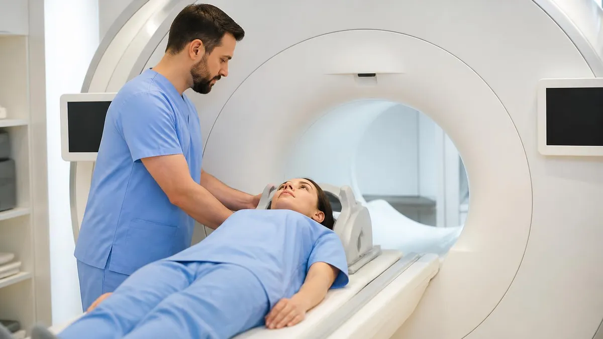
Cardiology coordination is non-negotiable. Before scanning, the device must be interrogated and programmed to an MRI-specific mode, which typically sets pacing to asynchronous mode for pacemaker-dependent patients or disables pacing for non-dependent patients. Tachycardia detection on ICDs is suspended. After the exam, the device is reinterrogated, restored to baseline settings, and checked for any threshold changes. This pre- and post-scan workflow usually adds 45 to 60 minutes of department time per patient.
Contrast administration introduces its own considerations. Gadolinium agents are not contraindicated by the device itself, but renal function and other contrast-specific safety screens still apply. For cardiac-specific imaging that requires gadolinium, refer to our detailed walkthrough of mri with and without contrast cpt for protocol selection, dosing, and adverse reaction management. Many cardiac MRI exams for device patients use delayed enhancement sequences to characterize scar tissue.
Finally, remember that conditional status is a system property, not a generator property. A St. Jude MRI generator implanted with a legacy non-MRI lead is not a conditional system. Abandoned or capped leads in the chest also disqualify many conditional labels because they may act as antennas and heat at the tip. Always confirm the entire system — generator plus every lead, including abandoned hardware — against the manufacturer's current compatibility documentation.
MRI Practice Test Questions
Prepare for the MRI - Magnetic Resonance Imaging exam with our free practice test modules. Each quiz covers key topics to help you pass on your first try.
MRI Knowledge
MRI Exam Questions covering Knowledge. Master MRI Test concepts for certification prep.
MRI Physics
Free MRI Practice Test featuring Physics. Improve your MRI Exam score with mock test prep.
MRI Anatomy and Pathology
MRI Test Prep for MRI Anatomy and Pathology. Practice MRI Quiz questions and boost your score.
MRI Anatomy and Positioning
MRI Questions and Answers on MRI Anatomy and Positioning. Free MRI practice for exam readiness.
MRI Contrast Agents
Free MRI Quiz on MRI Contrast Agents. MRI Exam prep questions with detailed explanations.
MRI Patient Care and Positioning
MRI Practice Questions for MRI Patient Care and Positioning. Build confidence for your MRI certification exam.
MRI Safety and Compatibility in Practice
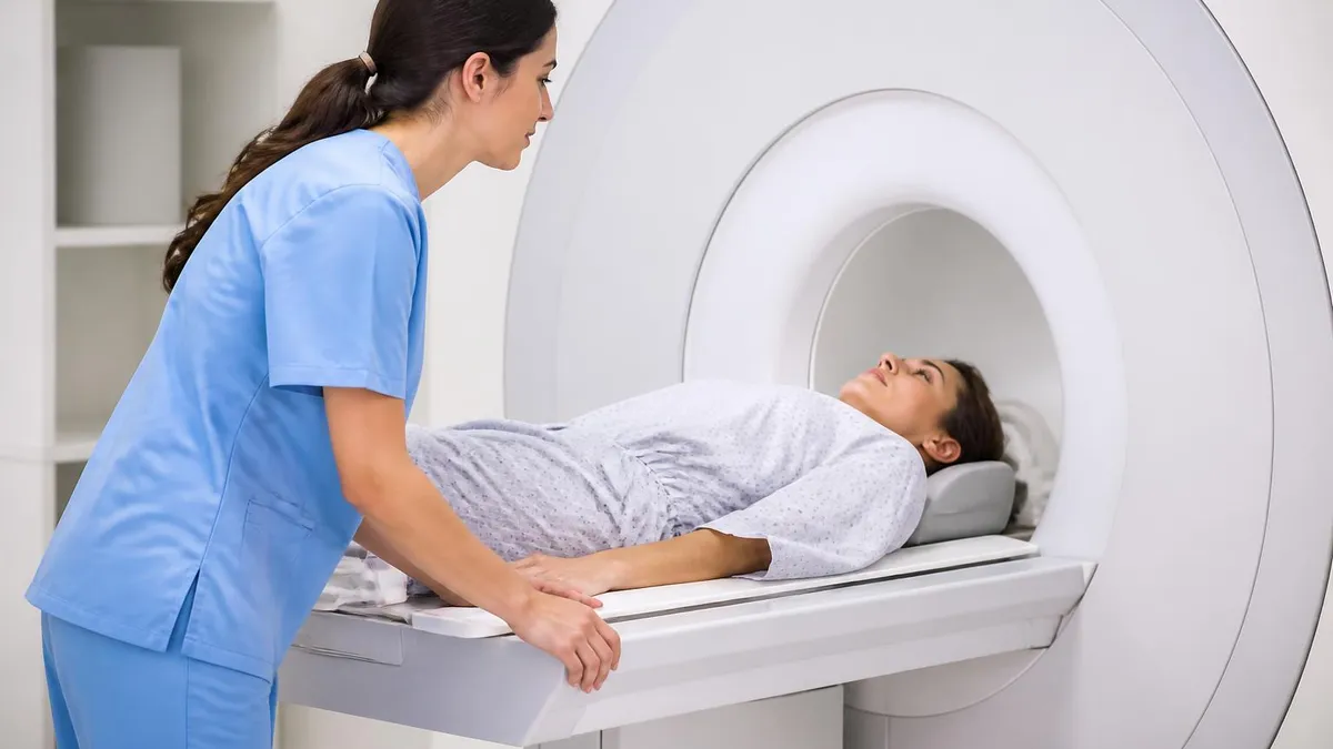
Patient screening begins at appointment scheduling and is repeated at check-in and again immediately before entry into Zone IV. The screening form must explicitly list cardiac devices, neurostimulators, cochlear implants, vascular stents, aneurysm clips, infusion pumps, tissue expanders, and orthopedic hardware. A verbal review with the patient catches items the patient forgot to check off, and a metal detector pass adds a second layer of protection.
For patients with cardiac devices, the screening must capture the model number, implant date, lead models, lead implant dates, and any abandoned leads. The technologist confirms this information against the manufacturer's compatibility documentation. If any element of the system is unverified, the scan does not proceed until the implanting cardiology team or device manufacturer is contacted and the information is documented in the medical record.
Scanning MR-Conditional Cardiac Patients: Benefits vs Burdens
- +Provides access to MRI for cardiac patients who previously had no diagnostic option
- +Allows superior soft-tissue evaluation for stroke, tumor, and musculoskeletal indications
- +Avoids ionizing radiation exposure compared to CT alternatives
- +Enables comprehensive cardiac scar and viability imaging when needed
- +Manufacturer-supplied conditions provide a clear, defensible safety framework
- +Patient outcomes improve when MRI-guided diagnosis replaces invasive testing
- −Adds 45–60 minutes of cardiology coordination per scan
- −Requires dedicated MRI mode reprogramming before and after the exam
- −Limits scanner parameters and may compromise some sequence quality
- −Demands specialized staff training and ongoing competency verification
- −Excludes patients with non-conditional leads or abandoned hardware
- −Increases total cost compared to a standard outpatient MRI
Pre-Scan St. Jude Pacemaker Compatibility Checklist
- ✓Confirm patient identity and MRI order against the requisition
- ✓Obtain the patient's St. Jude device identification card or implant record
- ✓Verify generator model number against the current Abbott MR-Conditional list
- ✓Verify every lead model — including abandoned leads — against the same list
- ✓Confirm time since implantation meets the manufacturer's minimum (often 6 weeks)
- ✓Coordinate with cardiology for MRI-mode programming before the scan
- ✓Set scanner to Normal Operating Mode and verify SAR limits
- ✓Apply MR-compatible ECG electrodes and continuous monitoring
- ✓Brief the patient on the call ball and any sensations to report
- ✓Document conditional parameters, peak SAR, and scan duration in the record
The system must be conditional — not just the generator
An MR-Conditional St. Jude generator paired with any non-MRI or abandoned lead is not an MR-Conditional system. Every component, including capped leads, must appear together in the manufacturer's compatibility table for the scan to qualify as conditional. When in doubt, contact Abbott Technical Services before proceeding.
Even with rigorous screening and protocol control, adverse events can occur during MRI exams involving cardiac devices. The technologist's response in the first thirty seconds determines whether a minor anomaly becomes a sentinel event. The first step in any suspected emergency is to stop the scan immediately, communicate with the patient through the intercom, and assess responsiveness. If the patient is unresponsive or in distress, the standard procedure is to remove them from the magnet bore as quickly as safe transfer allows.
Cardiac symptoms during a conditional scan are uncommon but possible. Patients may report palpitations, lightheadedness, or chest discomfort. These can result from inappropriate device behavior, anxiety, or unrelated cardiac events. The MR safety officer's protocol typically requires immediate termination of the scan, transfer of the patient out of Zone IV, application of standard ACLS monitoring, and emergent device interrogation. Defibrillation, if needed, occurs outside the magnet room because external defibrillator paddles are MR-Unsafe.
Thermal injuries are the most-cited theoretical risk for retained leads and abandoned hardware. RF energy deposited along a conductor can heat the lead tip, particularly when the wavelength of the RF pulse matches the lead length. This is why conditional labels prescribe SAR and B1+rms limits and why abandoned leads frequently disqualify systems from conditional status. Patients should be questioned about any burning sensation along the chest or shoulder during the scan and the exam stopped at the first report.
Projectile events involve ferromagnetic items inadvertently carried into Zone IV. The classic examples are oxygen tanks, IV poles, scissors, and stretcher hardware. While projectile events are not unique to cardiac device patients, anesthetized or sedated patients are at higher risk because they cannot warn staff about objects in their gowns or under blankets. A ferromagnetic detection system at the Zone IV doorway adds a critical layer of defense, and a strict two-person verbal screening immediately before entry remains best practice.
Quench events — the sudden boil-off of liquid helium from the magnet — are rare but high-impact. The boil-off pipe vents helium outside the building, but a failure can release helium into the scan room, displacing oxygen and causing asphyxiation. The technologist must know the location of the manual quench button, the oxygen monitor alarm, and the emergency room evacuation route. Patients with cardiac devices in this scenario are evacuated like any other patient, but post-event device interrogation is mandatory.
Contrast reactions follow the standard gadolinium adverse event algorithm, with attention to renal function, prior reactions, and the specific agent administered. Mild reactions like nausea or urticaria are managed conservatively; severe anaphylactoid reactions require intramuscular epinephrine, oxygen, and rapid transfer out of Zone IV for full resuscitation. Document the agent, dose, lot number, and reaction details. A delayed reaction may not appear until hours later, so patients should receive verbal and written discharge instructions on what to watch for.
Finally, communication after an adverse event is as important as the clinical response. The patient, family, ordering provider, cardiology team, and MR medical director should all be informed. The incident is documented in the safety event reporting system, reviewed by the MR safety committee, and analyzed for protocol gaps. Many institutions schedule a structured debrief within 72 hours so frontline staff can process the event and refine procedures.
A pacemaker-dependent patient scanned without conversion to an asynchronous MRI mode can experience pacing inhibition from RF interference, leading to bradycardia or asystole. Always confirm device programming with cardiology before the patient enters Zone IV, and verify the device has been restored to baseline immediately after the exam.
Comprehensive documentation is the legal and clinical backbone of any conditional MRI scan. The medical record should capture the device model and serial numbers, the lead models and serial numbers, the date of cardiology MRI-mode programming, the names and credentials of MR personnel present, the scan parameters used, peak SAR and B1+rms readings, monitoring data, contrast administered if any, and post-scan device interrogation results. ACR accreditation and Joint Commission surveyors regularly review this documentation during site inspections.
Workflow design separates departments that handle conditional patients smoothly from those that bottleneck. The most successful programs build a dedicated conditional-scan slot into the weekly schedule, often paired with on-site cardiology device technicians. Pre-arrival verification of the compatibility list is handled by a designated MR safety nurse or technologist 24 to 48 hours before the appointment. This proactive workflow avoids day-of cancellations and keeps the schedule from blowing up when paperwork or programming is missing.
Staff training is an ongoing requirement, not a one-time orientation event. The ACR recommends that all MR personnel — Level 1 and Level 2 — complete documented safety training annually, with content covering screening, zone management, ferromagnetic risk, RF and gradient physics, contrast safety, and emergency response. Conditional device scanning is typically covered in advanced training modules and reinforced through case reviews. Many departments maintain a competency log signed off by the MR safety officer.
Quality assurance closes the loop between procedure and outcome. A monthly review of all conditional scans should examine whether documentation was complete, whether parameters stayed within conditional limits, whether monitoring was continuous, and whether post-scan device interrogations showed any threshold or impedance changes. Trends in any of these areas trigger protocol updates. Many programs benchmark their numbers against published academic-center data to identify gaps. For broader context on what the abbreviation MRI actually represents in clinical communication, our explainer on MRI medical abbreviation is a useful staff reference.
Patient education is the often-overlooked element of conditional scanning. Patients with cardiac devices arrive with anxiety about the magnet, the device, and the scan results. A 10-minute pre-scan conversation explaining what the device does, why MRI is safe under the conditional label, what they will feel during the scan, and how the call ball works dramatically reduces patient-reported distress. Pamphlets and short videos in waiting rooms reinforce the verbal briefing.
Interdisciplinary communication is the final pillar. Cardiology, radiology, MR technologists, anesthesia, nursing, and emergency medicine all play roles in the conditional scan workflow. A shared electronic note template — with hard-stop fields for device model, lead model, programming status, and post-scan interrogation — forces complete handoffs and creates an auditable trail. Many institutions add a structured order-set in the EHR that cannot be signed until the required fields are populated.
The future of cardiac device MRI is moving toward fully MR-conditional ecosystems where the generator, leads, and even programmers communicate scan readiness automatically. Abbott, Medtronic, Boston Scientific, and Biotronik continue to expand 3T labeling and full-body access. Until that future arrives, the manufacturer compatibility list remains the single most important document on the MR safety officer's desk, and verifying it for every patient remains the single most important safety habit every MR technologist can practice.
Practical tips for handling St. Jude pacemaker compatibility checks come down to a few habits that experienced MR technologists develop after their first hundred conditional scans. The first habit is to never trust verbal device information without a backup source. Patients often confuse manufacturers, mix up implant dates, or forget about abandoned leads. Always ask for the device card, an EHR-imported implant log, or a cardiology note documenting the exact model and lead combination before clearing the scan.
The second habit is to maintain a quick-reference binder or digital folder of the most current Abbott MR-Conditional list, updated quarterly. Hyperlinks to manufacturer pages go stale, PDFs get superseded, and printed lists from two years ago no longer reflect added models or new labeling. A designated person on the safety team should own the update cycle and notify staff when changes occur. Many programs use a shared cloud folder with version control to keep everyone aligned.
The third habit is to over-communicate with cardiology. A cardiology device team that is surprised by a same-day MRI request will not have the bandwidth to interrogate, reprogram, and recheck the device within the technologist's schedule window. A 48-hour heads-up email with the device model, scan indication, and proposed time slot lets cardiology slot the interrogation into their workflow and reduces day-of friction dramatically. Some institutions formalize this with a shared scheduling channel.
The fourth habit is to memorize your scanner's Normal Operating Mode toggle and SAR display. When patient-specific parameters are tight, you should be able to glance at the console and confirm SAR is under 2.0 W/kg without having to navigate three menus. Practice during routine scans of non-device patients so the workflow is automatic when a conditional patient is on the table. Manufacturers vary in how SAR is displayed, so vendor-specific training matters.
The fifth habit is to use a structured time-out immediately before sliding the patient into the bore. The time-out covers identity, indication, device model, lead model, programming status, monitoring confirmation, and emergency plan. It takes 30 seconds and catches an astonishing number of near-misses. Many programs adopt a written time-out form signed by the technologist and the cardiology device tech before the first sequence begins.
The sixth habit is to perform thoughtful post-scan handoff. The cardiology team needs to reprogram the device promptly, and the patient needs to know when to expect that and what to do if symptoms develop in the interim. A short structured note in the EHR documenting scan completion, peak SAR, any patient symptoms, and the time of post-scan interrogation request creates an unbroken safety chain that protects the patient and the team. Add the cardiology callback number to the discharge instructions.
The seventh habit — and the one that ties all the others together — is intellectual humility. Manufacturers update labeling. Regulatory guidance shifts. New device models enter the market every quarter. The technologist who succeeds long-term treats every conditional scan as a chance to verify, not assume. If anything about the device, leads, or programming status feels uncertain, the right answer is always to pause, escalate to the MR medical director, and document the consultation. A defensible scan is a documented scan.
MRI Questions and Answers


About the Author
Medical Laboratory Scientist & Clinical Certification Expert
Johns Hopkins UniversityDr. Sandra Kim holds a PhD in Clinical Laboratory Science from Johns Hopkins University and is certified as a Medical Technologist (MT) and Medical Laboratory Scientist (MLS) through ASCP. With 16 years of clinical laboratory experience spanning hematology, microbiology, and molecular diagnostics, she prepares candidates for ASCP board exams, MLT, MLS, and specialist certification tests.
Join the Discussion
Connect with other students preparing for this exam. Share tips, ask questions, and get advice from people who have been there.
View discussion (6 replies)