MRI and Alzheimer's: How Brain Imaging Detects, Diagnoses, and Monitors Cognitive Decline
MRI and Alzheimer's disease: 💯 how brain MRI detects hippocampal atrophy, rules out other causes, and tracks progression. Complete guide for 2026 July.
The relationship between MRI and Alzheimer's disease has transformed how clinicians detect, diagnose, and monitor one of the most devastating neurodegenerative conditions of our time. Magnetic resonance imaging provides the only non-invasive window into the structural changes that quietly unfold in the brain years before memory symptoms become severe. For patients, families, and radiologic technologists alike, understanding what MRI can and cannot reveal about Alzheimer's pathology has become essential clinical knowledge in 2026.
Alzheimer's disease affects roughly 6.9 million Americans aged 65 and older, with prevalence projected to nearly double by 2060 according to the Alzheimer's Association. MRI plays a central role in the diagnostic workup recommended by the National Institute on Aging because it can demonstrate medial temporal lobe atrophy, identify vascular contributions to cognitive impairment, and exclude reversible causes such as normal-pressure hydrocephalus, chronic subdural hematoma, or brain tumor mris that mimic dementia symptoms.
Unlike PET imaging, which directly visualizes amyloid plaques or tau tangles, MRI excels at showing the structural consequences of neurodegeneration. The hippocampus, entorhinal cortex, and posterior cingulate gyrus all undergo measurable volume loss as Alzheimer's progresses. Modern 3 Tesla scanners with high-resolution coronal T1 sequences can detect these changes with millimeter precision, allowing radiologists to grade medial temporal atrophy using validated scales like the Scheltens scale that correlate strongly with clinical severity.
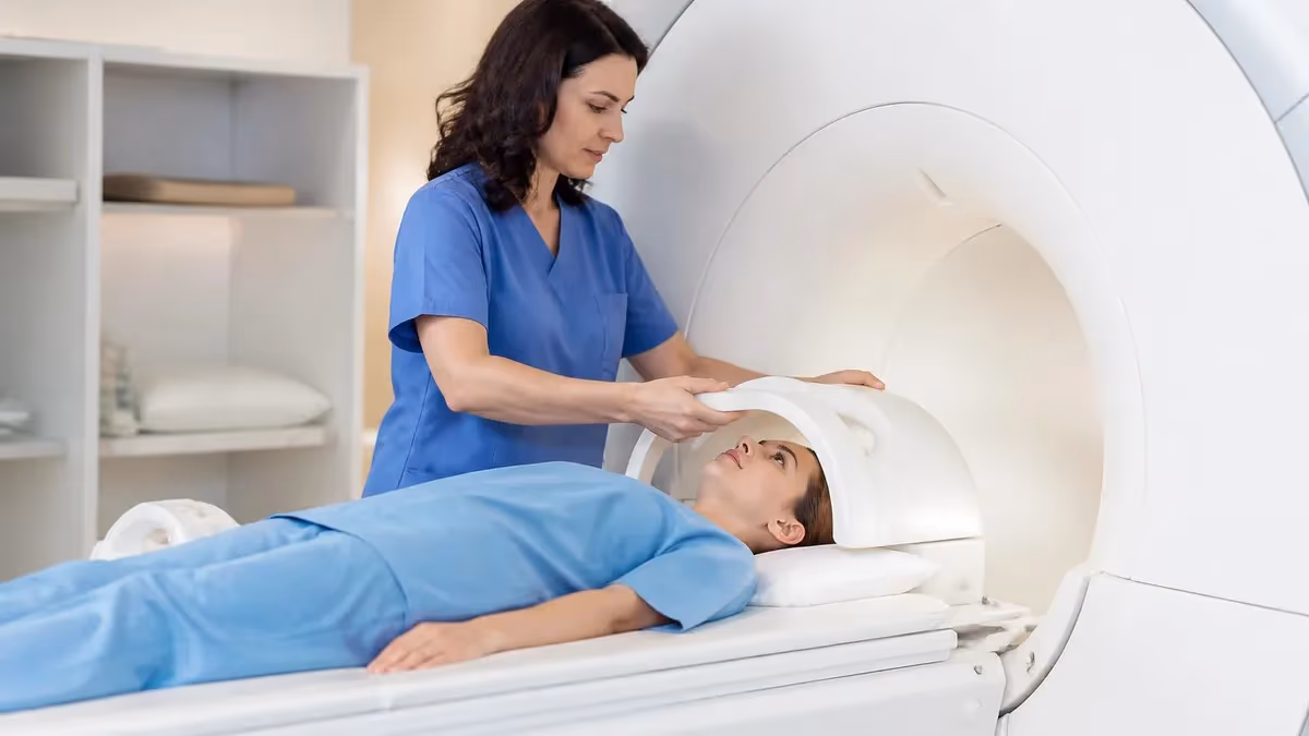
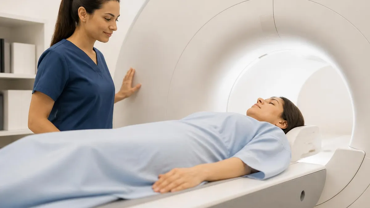
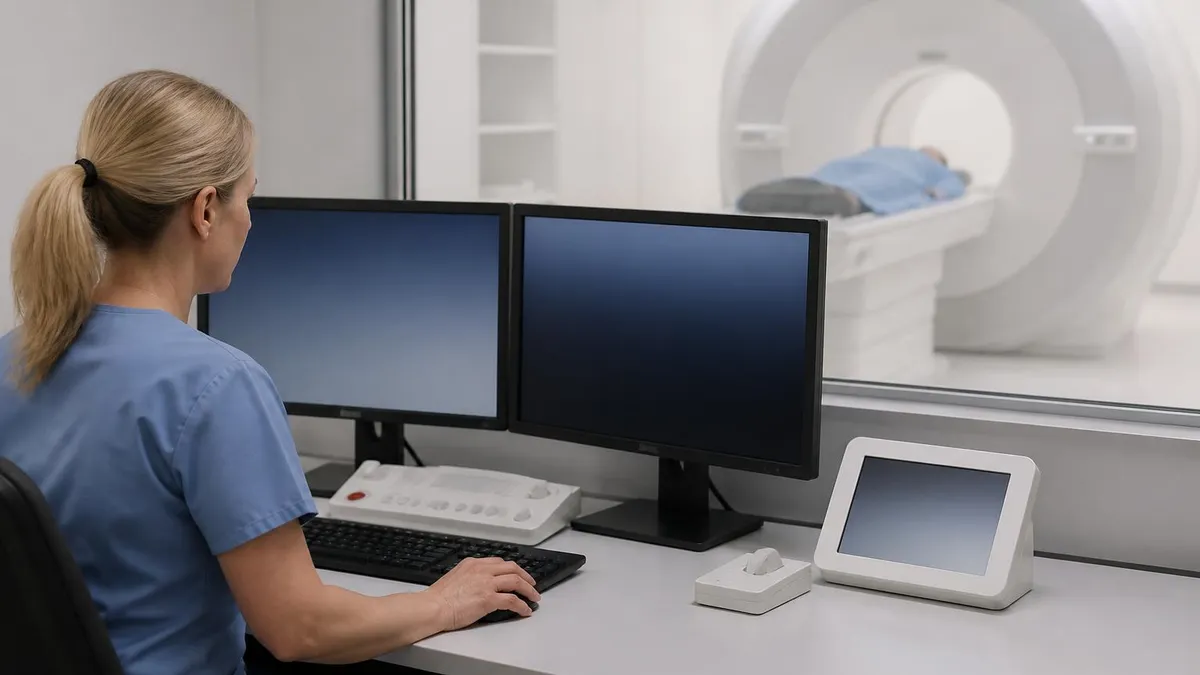
For technologists, scanning a cognitively impaired patient presents unique challenges that go beyond ordinary brain protocols. Patients may be anxious, claustrophobic, unable to lie still for 45 minutes, or unable to follow breath-hold instructions. Choosing the right protocol, optimizing motion-resistant sequences, and knowing when to recommend MRI with or without contrast are skills that directly affect diagnostic yield in this population.
This comprehensive guide walks through every aspect of MRI in Alzheimer's evaluation: which sequences matter most, how radiologists interpret atrophy patterns, what advanced techniques like volumetric analysis and arterial spin labeling add, and how the imaging findings integrate with cerebrospinal fluid biomarkers and the newest blood-based assays. We will also cover practical considerations for technologists working with dementia patients and the registry-relevant physics behind each sequence choice.
Whether you are a radiologic technologist preparing for the ARRT MRI registry, a clinician trying to interpret a neuroradiology report, or a family member trying to understand a loved one's diagnosis, this resource translates current evidence into practical knowledge. The science of imaging Alzheimer's has advanced dramatically since the first volumetric studies in the 1990s, and 2026 brings new clinical decision rules that incorporate MRI findings into staging and treatment eligibility for the newly approved anti-amyloid antibodies.
By the end of this article you will understand which MRI findings carry diagnostic weight, why hippocampal volume loss matters so much, how vascular changes complicate the picture, what ARIA monitoring means for patients on lecanemab or donanemab, and how to recognize when an MRI report points toward Alzheimer's rather than another dementia subtype. The stakes have never been higher: with disease-modifying therapies now available, accurate early imaging diagnosis is no longer academic.
MRI and Alzheimer's by the Numbers
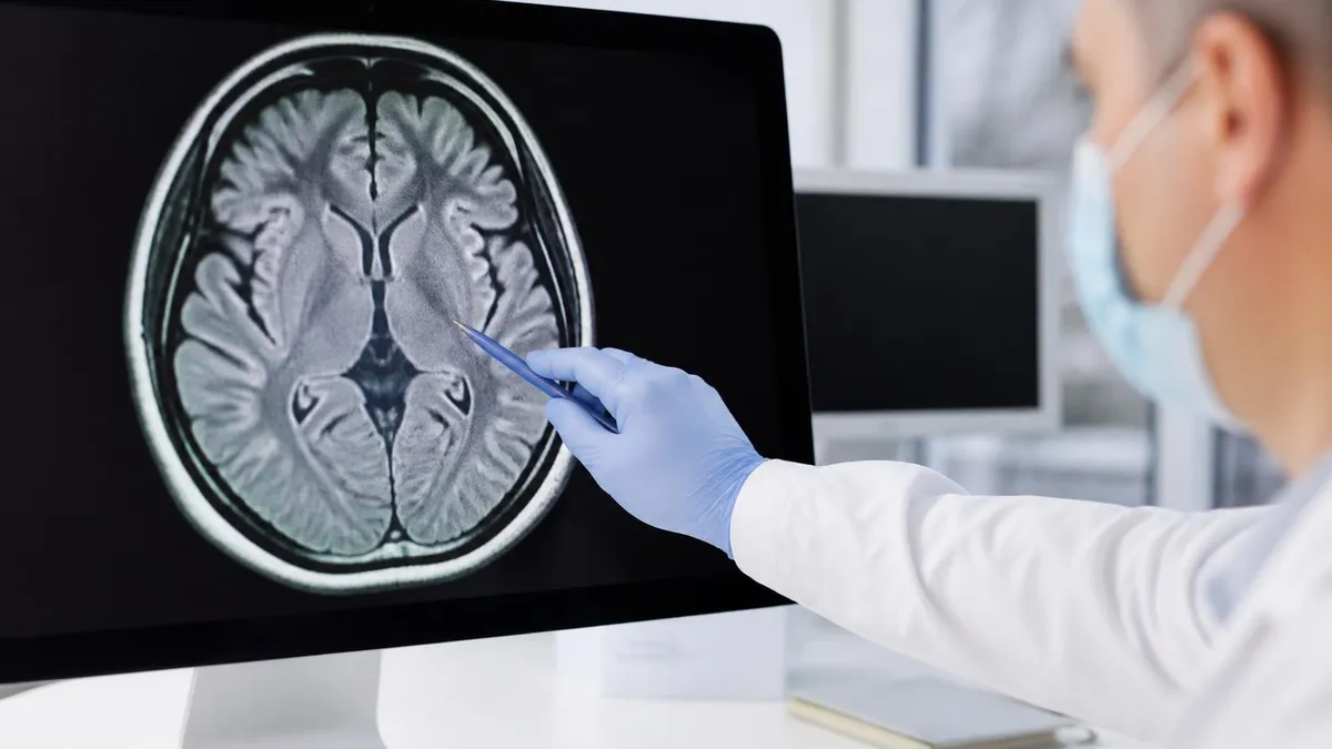
Brain Changes MRI Reveals in Alzheimer's
The hippocampus shrinks earliest and most severely in Alzheimer's. Coronal T1 images show widened temporal horns and reduced hippocampal volume, often asymmetric, with the left side frequently more affected in language-dominant decline.
Posterior cingulate, precuneus, and lateral temporoparietal cortex show measurable thinning. This pattern differs from frontotemporal dementia, which affects frontal and anterior temporal regions more prominently than posterior areas.
As surrounding tissue atrophies, the lateral ventricles expand to fill the space. The temporal horns of the lateral ventricles are particularly sensitive markers when measured against age-matched normative data.
FLAIR sequences reveal periventricular and deep white matter hyperintensities reflecting small vessel ischemic disease. Significant burden suggests mixed vascular and Alzheimer's pathology, common in patients over 75.
SWI or gradient echo sequences detect tiny hemosiderin deposits from cerebral amyloid angiopathy. Lobar microbleeds raise concern for amyloid pathology and influence eligibility for anti-amyloid treatments.
The MRI protocol for evaluating suspected Alzheimer's disease has been refined over three decades into a standardized sequence package that balances diagnostic yield, scan time, and patient tolerance. A typical dementia workup runs 30 to 45 minutes and includes seven to nine sequences chosen specifically to demonstrate atrophy patterns, vascular pathology, and exclude structural mimics. Understanding why each sequence belongs in the protocol helps technologists optimize image quality and helps clinicians interpret reports.
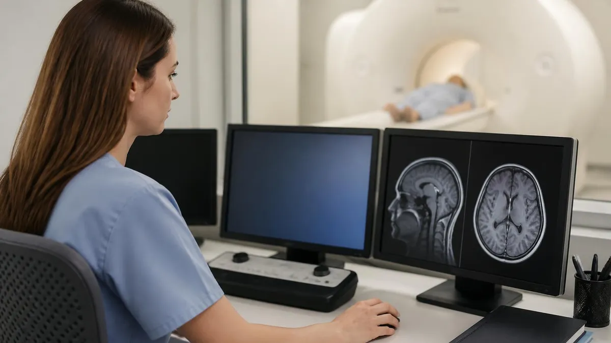
The cornerstone is a high-resolution 3D T1-weighted MPRAGE or BRAVO acquisition with isotropic voxels of 1.0 mm or smaller. This volumetric dataset can be reformatted in any plane, enabling oblique coronal slices perpendicular to the long axis of the hippocampus where atrophy scoring is performed. Many modern systems now run automated volumetric analysis software like NeuroQuant or icobrain dm that generates age and sex-normalized hippocampal volumes within minutes of scan completion.
FLAIR imaging in axial and sagittal planes is mandatory for assessing white matter disease, infarcts, and inflammatory changes. The Fazekas scale grades periventricular and deep white matter hyperintensities from 0 to 3, and a Fazekas 2 or higher burden often points to a vascular contribution that may coexist with Alzheimer's pathology. Reading whats difference between mri and ct scan clarifies why CT cannot replace FLAIR for these subtle white matter findings.
Susceptibility-weighted imaging or T2 star gradient echo sequences are now considered essential rather than optional. These sequences detect cerebral microbleeds with high sensitivity, and the presence, number, and distribution of microbleeds carry significant diagnostic and therapeutic weight. Strictly lobar microbleeds in a patient over 55 with cognitive decline suggest cerebral amyloid angiopathy, which often coexists with Alzheimer's pathology and influences risk stratification for anti-amyloid therapy.
Diffusion-weighted imaging completes the standard dementia protocol by ruling out acute infarction, abscess, or Creutzfeldt-Jakob disease, which can mimic rapidly progressive dementia. Although DWI plays a smaller role in Alzheimer's itself, omitting it risks missing acute strokes in this elderly population. Many sites also add a fast T2-weighted sequence in the axial plane for general anatomic survey and detection of mass lesions.
Contrast administration is generally not required for evaluating Alzheimer's disease. Gadolinium is reserved for cases with suspected infection, tumor, inflammatory disease, or rapidly progressive atypical dementia. The current ACR appropriateness criteria for cognitive impairment list non-contrast MRI as the first-line study, which spares patients the small risks of gadolinium retention and renal complications.
Sequence parameters matter enormously in this population. Patients with dementia often move during long acquisitions, so technologists should favor parallel imaging acceleration, prospective motion correction when available, and split long sequences into shorter blocks. Some facilities use compressed sensing MPRAGE to reduce 3D T1 time from 5 minutes to under 3 minutes while preserving volumetric accuracy, which dramatically improves success rates in agitated or restless patients.
MRI Practice Test Questions
Prepare for the MRI - Magnetic Resonance Imaging exam with our free practice test modules. Each quiz covers key topics to help you pass on your first try.
MRI Knowledge
MRI Exam Questions covering Knowledge. Master MRI Test concepts for certification prep.
MRI Physics
Free MRI Practice Test featuring Physics. Improve your MRI Exam score with mock test prep.
MRI Anatomy and Pathology
MRI Test Prep for MRI Anatomy and Pathology. Practice MRI Quiz questions and boost your score.
MRI Anatomy and Positioning
MRI Questions and Answers on MRI Anatomy and Positioning. Free MRI practice for exam readiness.
MRI Contrast Agents
Free MRI Quiz on MRI Contrast Agents. MRI Exam prep questions with detailed explanations.
MRI Patient Care and Positioning
MRI Practice Questions for MRI Patient Care and Positioning. Build confidence for your MRI certification exam.
MRI and Alzheimer's Atrophy Patterns
The hippocampus is the signature structure in Alzheimer's imaging. On oblique coronal T1 images perpendicular to its long axis, radiologists assess height, width, and the size of the surrounding choroid fissure and temporal horn. The Scheltens medial temporal atrophy scale ranges from 0 (no atrophy) to 4 (severe atrophy with marked CSF expansion), and a score of 2 or higher in patients under 75 has high specificity for Alzheimer's pathology.
Hippocampal atrophy typically begins asymmetrically, with the left side often involved earlier in patients presenting with language and memory complaints. Annual volume loss reaches 4 to 5 percent in clinically symptomatic Alzheimer's compared to 1 percent in healthy aging. Automated software now reports hippocampal volume as a percentile against age-matched controls, with values below the fifth percentile considered abnormal and supportive of an Alzheimer's diagnosis when correlated with clinical findings.
MRI vs Other Imaging in Alzheimer's Workup
- +No ionizing radiation, safe for repeated longitudinal monitoring across years
- +Superior soft tissue contrast reveals subtle hippocampal and cortical atrophy
- +Detects vascular contributions through FLAIR and susceptibility imaging
- +Rules out reversible causes like hydrocephalus, tumors, or chronic subdurals
- +Quantitative volumetry available through automated software like NeuroQuant
- +Required baseline study before starting anti-amyloid antibody therapy
- +Visualizes microbleeds and superficial siderosis affecting treatment eligibility
- −Cannot directly visualize amyloid plaques or tau tangles like PET imaging
- −Long scan times challenging for agitated or claustrophobic dementia patients
- −Contraindicated with non-conditional pacemakers and certain metallic implants
- −Cost typically $800 to $2,000 without insurance, higher than CT
- −Motion artifact common in cognitively impaired patients reduces diagnostic yield
- −Visual atrophy assessment shows significant interreader variability without software
Pre-Scan Checklist for MRI and Alzheimer's Patients
- ✓Confirm patient identity using two identifiers and verify the imaging order matches clinical question
- ✓Complete MRI safety screening with patient and accompanying family member or caregiver present
- ✓Document all implants, prior surgeries, and dental work that could affect image quality
- ✓Verify renal function if contrast is ordered, though most dementia protocols are non-contrast
- ✓Explain the procedure in simple, repeated terms to reduce anxiety and confusion
- ✓Offer earplugs and headphones with calming music to improve tolerance for long acquisitions
- ✓Position carefully with extra cushioning to support hip and shoulder comfort during 45-minute exams
- ✓Use a mirror or prism glasses so patients can see outside the bore and reduce claustrophobia
- ✓Establish hand signal communication and confirm patient understanding before scanning
- ✓Schedule longer time slots than usual to accommodate breaks and patient repositioning
Why Hippocampal Volume Matters in 2026
Hippocampal volume below the fifth percentile for age has a positive likelihood ratio of 4.5 for Alzheimer's pathology when combined with episodic memory impairment. This finding now influences treatment eligibility decisions for lecanemab and donanemab, where baseline MRI atrophy patterns help predict response and risk stratification for ARIA.
ARIA, or amyloid-related imaging abnormalities, has become one of the most important reasons MRI matters in modern Alzheimer's care. With the FDA approvals of lecanemab in 2023 and donanemab in 2024, hundreds of thousands of patients now receive anti-amyloid antibody infusions that require frequent MRI surveillance. Technologists and radiologists must recognize ARIA-E (edema) and ARIA-H (microhemorrhage) findings, because missed abnormalities can lead to symptomatic complications including seizures and cerebral hemorrhage.
ARIA-E appears as patchy FLAIR hyperintensity in the cerebral white matter, typically in the parietal or occipital regions, sometimes extending into the cortex with sulcal effacement. It usually develops within the first three to six months of antibody therapy and typically resolves over weeks if treatment is paused. Mild ARIA-E often allows continuation of therapy, while moderate or severe cases require treatment interruption and close follow-up imaging until resolution.
ARIA-H presents as new microbleeds on susceptibility-weighted imaging or, less commonly, as superficial siderosis along the cortical surface. The official monitoring schedule for lecanemab requires MRI before doses 5, 7, and 14, and additional scans if neurologic symptoms develop. Donanemab follows a similar but slightly different schedule. Familiarity with these protocols matters because patients now move through many imaging departments rather than concentrating at academic centers.
Patients carrying two copies of the apolipoprotein E4 allele face substantially higher ARIA risk, with rates approaching 35 percent compared to 10 percent in non-carriers. Many treatment centers now genotype patients before therapy and require baseline MRI showing fewer than four microbleeds, no prior macrohemorrhage, and no superficial siderosis. Understanding these eligibility criteria helps radiologists write actionable reports that explicitly comment on ARIA risk factors.
The standard ARIA monitoring protocol uses a focused, abbreviated MRI rather than the full diagnostic dementia workup. Typical surveillance scans include axial FLAIR, axial T2 star or SWI, axial T2, and axial DWI, completing in 15 to 20 minutes. This shorter protocol improves throughput and patient tolerance during the year-plus of biweekly infusions. Some centers further accelerate scans using deep learning reconstruction software that maintains image quality at half the acquisition time.
Reporting language for ARIA has become highly standardized. The Barkhof-Sperling ARIA severity scale categorizes ARIA-E by lesion size and number, and ARIA-H by total microbleed count. Radiologists should explicitly state the number of new microbleeds compared to baseline, any new FLAIR hyperintensities suggesting ARIA-E, and any superficial siderosis. The treating neurologist uses this structured information to make immediate decisions about pausing, continuing, or discontinuing therapy.
Beyond ARIA, MRI monitoring during anti-amyloid therapy provides longitudinal data on brain volume and white matter health. Surprisingly, treated patients sometimes show accelerated whole-brain volume loss compared to placebo in the first year, possibly reflecting plaque clearance rather than additional neurodegeneration. Ongoing research is clarifying whether this finding represents a benign treatment effect or a concerning signal, and ongoing MRI tracking will help answer that question over the next several years.
Patients receiving lecanemab or donanemab require MRI scans on a fixed schedule before specific infusions and whenever new neurologic symptoms develop. Missed surveillance imaging has been associated with serious complications including symptomatic edema and intracerebral hemorrhage. Imaging centers must build reliable scheduling and reporting workflows to support these protocols.
Beyond conventional structural imaging, advanced MRI techniques add depth to the Alzheimer's evaluation. Arterial spin labeling (ASL) is a non-contrast perfusion sequence that measures regional cerebral blood flow by magnetically tagging water in arterial blood. In Alzheimer's, ASL typically shows reduced perfusion in the posterior cingulate, precuneus, and lateral temporoparietal cortex, mirroring the pattern seen on FDG-PET. This makes ASL an attractive alternative when PET access is limited.
Diffusion tensor imaging (DTI) measures the directionality of water movement along white matter tracts. In Alzheimer's, DTI reveals microstructural damage in the fornix, cingulum, and uncinate fasciculus before macroscopic atrophy becomes visible. Fractional anisotropy decreases and mean diffusivity increases in these tracts, providing a sensitive early biomarker that has been explored extensively in research settings, though it is not yet a routine clinical sequence in most imaging centers.
Functional MRI using resting-state BOLD signals identifies the default mode network, a constellation of brain regions including the posterior cingulate, medial prefrontal cortex, and angular gyrus. Functional connectivity within this network decreases early in Alzheimer's, and resting-state fMRI is now being studied as a treatment response biomarker for anti-amyloid antibodies. Recognizing the difference between resting-state and task-based fMRI helps technologists understand which protocols their facility supports.
MR spectroscopy measures brain metabolite concentrations including N-acetylaspartate (NAA), myoinositol, creatine, and choline. In Alzheimer's, NAA decreases (reflecting neuronal loss) and myoinositol increases (reflecting glial activity), producing a characteristic NAA/myoinositol ratio reduction in the posterior cingulate. Although MRS is technically demanding and not widely used in routine practice, it provides quantitative metabolic information that complements structural imaging. Learning about advanced mri protocols considerations also helps technologists appreciate how protocols are tailored to specific clinical questions.
Quantitative susceptibility mapping (QSM) post-processes phase data from SWI sequences to quantify tissue iron content. Alzheimer's brains show increased iron in the hippocampus, putamen, and caudate, and this iron deposition may contribute to neurodegeneration through oxidative stress. QSM remains primarily a research tool, but it is being incorporated into longitudinal cohort studies tracking disease progression.
Ultra-high-field 7T MRI provides unprecedented spatial resolution that can resolve individual hippocampal subfields including CA1, CA3, and the dentate gyrus. Research at 7T demonstrates that the CA1 subfield atrophies first in Alzheimer's, before other subfields and before total hippocampal volume drops measurably at 3T. Clinical 7T availability is still limited to specialized academic centers, but the technology is expanding rapidly and will likely reshape early diagnostic imaging within the decade.
Machine learning and artificial intelligence are accelerating the clinical utility of all these techniques. Algorithms trained on thousands of scans can now flag suspicious hippocampal atrophy patterns, detect microbleeds automatically, and generate predicted brain age that compares to chronological age as a global aging biomarker. Several FDA-cleared software packages provide these analyses within minutes of scan completion, putting quantitative neuroimaging into routine workflow for the first time.
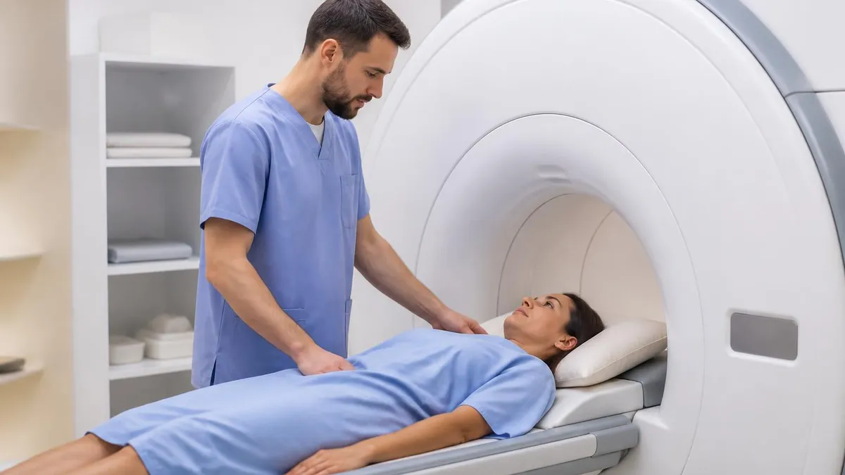
Practical preparation for an MRI study in a patient with cognitive impairment begins long before the patient enters the scanner. A successful exam requires coordination among the ordering clinician, scheduling staff, technologists, and family caregivers. Skipping any of these steps risks an incomplete or non-diagnostic study, repeat exams, and unnecessary stress for an already vulnerable population. Building a dementia-friendly imaging workflow pays dividends in image quality and patient satisfaction.
Scheduling matters more than most clinicians realize. Patients with Alzheimer's typically function best in the morning, when fatigue and sundowning are minimal. Booking dementia patients in the first two hours of the day reduces no-shows and improves cooperation. Allowing 60 minutes rather than the standard 30 to 40 minutes provides buffer time for positioning, repositioning, and breaks without falling behind schedule. These minor scheduling adjustments dramatically improve completion rates.
Communication strategies should be adapted to cognitive level. Use short, simple sentences. Avoid jargon. Repeat instructions multiple times in slightly different words. Demonstrate before requiring action, for example by showing how to hold the squeeze ball and pressing it yourself first. Maintain eye contact and speak slowly. Family members can often translate or rephrase instructions more effectively, so welcome a trusted caregiver into the control room or even into the scan room when safety screening permits.
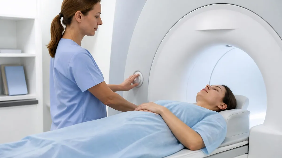
Anxiety management is essential. Approximately 20 percent of patients without dementia experience significant claustrophobia, and rates climb in patients with cognitive impairment. Options include verbal reassurance during the scan over the intercom, prism glasses to see outside the bore, a warm blanket to provide tactile comfort, soft padding around the head coil, and oral anxiolytic premedication when prescribed by the ordering provider. Open or wide-bore scanners help when available.
Image quality optimization starts with motion-resistant pulse sequences. Use the shortest possible 3D T1 acquisition that maintains diagnostic resolution. Apply parallel imaging acceleration aggressively. Consider PROPELLER or BLADE motion-correcting T2 and FLAIR sequences if your platform supports them. Some modern systems offer real-time motion tracking with prospective correction, dramatically improving scan success rates in restless patients. Discussing these options with your applications specialist can transform your dementia imaging workflow.
Reading and acting on the report requires teamwork between radiology and the ordering clinician. A well-structured report should comment on hippocampal volume (ideally with quantitative software), medial temporal atrophy score, posterior atrophy grade, global cortical atrophy grade, Fazekas white matter score, microbleed count and distribution, and any incidental findings. Including these standardized elements ensures the clinician receives all the information needed to integrate imaging with clinical and biomarker data.
Finally, longitudinal comparison transforms a single MRI from a snapshot into a trajectory. Annual or biennial follow-up imaging in patients with mild cognitive impairment reveals whether atrophy is progressing at a pathologic rate. Side-by-side comparison with prior studies, ideally using the same protocol and scanner, dramatically increases diagnostic confidence. Encouraging patients to bring prior outside imaging on disc enables this comparison even when they change healthcare systems, an underappreciated but transformative aspect of clinical care.
MRI Questions and Answers
MRI MARS Protocol: Complete Guide to Metal Artifact Reduction Sequences for Orthopedic Imaging
MRI With or Without Contrast: What to Expect

The History of MRI: From Discovery to Modern Medicine
What's the Difference Between MRI and CT Scan? A Complete Comparison Guide

MRI With and Without Contrast: How It Works, What to Expect
About the Author
Medical Laboratory Scientist & Clinical Certification Expert
Johns Hopkins UniversityDr. Sandra Kim holds a PhD in Clinical Laboratory Science from Johns Hopkins University and is certified as a Medical Technologist (MT) and Medical Laboratory Scientist (MLS) through ASCP. With 16 years of clinical laboratory experience spanning hematology, microbiology, and molecular diagnostics, she prepares candidates for ASCP board exams, MLT, MLS, and specialist certification tests.
Join the Discussion
Connect with other students preparing for this exam. Share tips, ask questions, and get advice from people who have been there.
View discussion (6 replies)