MRI Technology Explained: How Magnetic Resonance Imaging Works in 2026 July
Explore mri technology — how magnetic resonance imaging works, key hardware, pulse sequences, safety, advances, and what techs need to know in 2026 July. ❓
MRI technology has transformed diagnostic medicine over the last four decades, giving clinicians a non-ionizing window into soft tissue, vasculature, and metabolism that no other modality can match. Unlike CT or X-ray, magnetic resonance imaging uses powerful magnets, radiofrequency pulses, and gradient coils to manipulate hydrogen nuclei inside the body, then reconstructs the returning signals into cross-sectional images of extraordinary detail. For technologists, radiologists, and patients alike, understanding the underlying physics is essential to interpreting both the strengths and the limits of what we see on the console.
The core principle is deceptively elegant. Place a patient inside a strong static magnetic field, typically 1.5 or 3 Tesla, and a small majority of the body's protons align with that field. A precisely tuned radiofrequency pulse tips those protons out of alignment, and as they relax back, they emit signals that gradient coils spatially encode. The scanner's computer transforms those signals through a Fourier transform into the gray-scale images radiologists read on PACS.
Modern MRI suites are far more than magnets and coils. They include cryogen-cooled superconducting wire, multi-channel phased-array receivers, parallel imaging algorithms, and increasingly deep-learning reconstruction pipelines that cut scan times in half without sacrificing resolution. A scanner installed in 2026 routinely captures sequences in five minutes that took twenty minutes on a 2010 system, and image quality has improved at every field strength.
This guide walks through the components, sequences, safety considerations, clinical applications, and emerging trends shaping MRI today. Whether you are a registry candidate, a working technologist refreshing fundamentals, or a curious patient who wants to know what is happening inside that tube, you will leave with a clearer mental model of what makes magnetic resonance imaging tick. We will cover hardware, pulse design, contrast mechanisms, and the practical workflow inside the control room.
One of the things that makes MRI uniquely useful is its tissue contrast flexibility. By manipulating timing parameters such as repetition time (TR) and echo time (TE), a technologist can weight an image toward T1, T2, proton density, diffusion, or perfusion. A single anatomical region can therefore yield a dozen different views, each highlighting a different biological feature — edema, hemorrhage, fat, cartilage, blood flow, or restricted diffusion that suggests acute stroke.
For a deeper look at what these sequences actually reveal on clinical images, review our companion guide on Common MRI Findings: Brain, Spine and Joints Guide. That resource pairs well with this article because it bridges the gap between physics and interpretation.
Throughout the sections ahead, we will return to the same theme: MRI is a system. The magnet, the gradients, the coils, the sequence, the reconstruction, and the human operator all contribute to the final image. Weak performance in any one of those domains shows up as artifact, low signal-to-noise, or missed pathology, so technologists who understand the whole chain consistently produce better studies than those who only know how to press the start button.
MRI Technology by the Numbers
Core Components of an MRI System
Generates the static B0 field, typically 1.5T or 3T. Uses niobium-titanium wire cooled by liquid helium near absolute zero to maintain zero electrical resistance and a stable, homogeneous field.
Three orthogonal coils (X, Y, Z) that superimpose linear field variations onto B0, enabling slice selection and spatial encoding. Rapid switching produces the loud knocking sound patients hear during scans.
Body and surface coils deliver radiofrequency pulses at the Larmor frequency and detect the faint return signal. Multi-channel phased arrays dramatically improve signal-to-noise for parallel imaging.
Passive iron and active electromagnetic shims correct field inhomogeneity caused by patient anatomy and room conditions. Good shimming is critical for fat suppression and spectroscopy quality.
High-performance workstations apply Fourier transforms, parallel imaging algorithms, and increasingly deep-learning reconstruction to convert raw k-space data into the DICOM images sent to PACS.
To understand mri technology, start with hydrogen. The human body is mostly water and fat, both rich in hydrogen atoms, and each hydrogen nucleus is a single proton that behaves like a tiny spinning bar magnet. Outside the scanner, these protons point in random directions and cancel each other out. Inside a 1.5T or 3T magnet, a slight majority align with the static field, producing a net magnetization vector — the raw material every MRI image is built from.
A radiofrequency pulse tuned to the Larmor frequency, which depends on field strength (~63.87 MHz at 1.5T and ~127.74 MHz at 3T for hydrogen), tips that net magnetization away from alignment. When the pulse stops, protons relax back toward equilibrium along two independent pathways: T1 (longitudinal recovery) and T2 (transverse decay). Different tissues have different T1 and T2 values, and that is the entire basis of MRI contrast.
Gradient coils encode spatial information by making the magnetic field strength vary linearly across the patient. Because the Larmor frequency depends on field strength, protons in different locations precess at slightly different frequencies. The scanner samples this spatially encoded signal into k-space, a frequency-domain matrix, and then applies a 2D or 3D Fourier transform to reconstruct the familiar anatomical image.
Slice selection happens when the gradient is applied during the RF pulse — only protons in a thin slab match the pulse frequency and get excited. Phase encoding and frequency encoding then localize signal within that slice. The interplay of these three gradients, repeated hundreds of times per scan, is what fills k-space and ultimately produces a sharp anatomical map.
Pulse sequences are recipes that determine how RF pulses and gradients are timed. A spin-echo sequence uses a 90-degree pulse followed by a 180-degree refocusing pulse, recovering signal lost to local field inhomogeneity. A gradient-echo sequence skips the refocusing pulse for speed but keeps that inhomogeneity sensitivity, making it useful for detecting hemorrhage and calcification through susceptibility effects.
The single most important practical insight is this: nothing in MRI is automatic. Every parameter — TR, TE, flip angle, matrix, field of view, bandwidth, slice thickness — represents a trade-off between contrast, resolution, signal-to-noise, scan time, and artifact. Understanding the physics lets a technologist make those trade-offs intentionally rather than relying on a vendor preset that may not match the clinical question.
For working technologists curious about how this physics knowledge translates into a career, our overview on How to Become an MRI Technician: Schools, Certification, Salary walks through credentialing and education pathways in detail. Strong physics fundamentals separate competent technologists from exceptional ones in every clinical setting.
MRI Practice Test Questions
Prepare for the MRI - Magnetic Resonance Imaging exam with our free practice test modules. Each quiz covers key topics to help you pass on your first try.
MRI Knowledge
MRI Exam Questions covering Knowledge. Master MRI Test concepts for certification prep.
MRI Physics
Free MRI Practice Test featuring Physics. Improve your MRI Exam score with mock test prep.
MRI Anatomy and Pathology
MRI Test Prep for MRI Anatomy and Pathology. Practice MRI Quiz questions and boost your score.
MRI Anatomy and Positioning
MRI Questions and Answers on MRI Anatomy and Positioning. Free MRI practice for exam readiness.
MRI Contrast Agents
Free MRI Quiz on MRI Contrast Agents. MRI Exam prep questions with detailed explanations.
MRI Patient Care and Positioning
MRI Practice Questions for MRI Patient Care and Positioning. Build confidence for your MRI certification exam.
Pulse Sequences and Image Contrast
T1-weighted sequences use short TR (typically 400–600 ms) and short TE (10–20 ms) to emphasize differences in longitudinal relaxation. Tissues with short T1, like fat, appear bright, while tissues with long T1, like cerebrospinal fluid, appear dark. T1 weighting is the workhorse for anatomical detail because boundaries between gray matter, white matter, fat, and fluid are crisp and well defined on every modern scanner.
Gadolinium-based contrast agents shorten T1 dramatically in tissues with intact blood supply or disrupted blood-brain barrier. Post-contrast T1 imaging therefore highlights tumors, infections, active demyelination, and vascular structures. Radiologists routinely compare pre- and post-contrast T1 sequences side by side to localize enhancement, characterize lesions, and monitor treatment response in oncology and neurology workflows.
MRI vs Other Imaging Modalities: Pros and Cons
- +No ionizing radiation, safe for repeat imaging and pediatric patients
- +Superior soft tissue contrast unmatched by CT, ultrasound, or X-ray
- +Multiplanar imaging in any orientation without repositioning the patient
- +Multiple contrast mechanisms in one exam — T1, T2, DWI, perfusion
- +Excellent for neurological, musculoskeletal, and oncologic imaging
- +Functional and metabolic information through fMRI and spectroscopy
- −Long scan times compared to CT, often 20–45 minutes per study
- −Strict safety screening required for implants and ferromagnetic objects
- −Claustrophobia affects roughly 5–10% of patients in closed-bore systems
- −Significantly higher equipment and operating costs than CT or ultrasound
- −Limited use in critically ill patients on ventilators or with metallic life-support
- −Acoustic noise during gradient switching can reach 110 dB without hearing protection
MRI Safety Screening Checklist
- ✓Verify patient identity and confirm the ordered exam matches the clinical question
- ✓Screen for cardiac pacemakers, ICDs, neurostimulators, and cochlear implants
- ✓Document all prior surgeries with implanted hardware, clips, stents, or coils
- ✓Check for embedded metallic foreign bodies, especially metal workers requiring orbital radiographs
- ✓Confirm pregnancy status and document trimester for any gadolinium discussion
- ✓Review renal function (eGFR) before administering gadolinium-based contrast agents
- ✓Remove all external metal: jewelry, watches, hearing aids, dentures, hairpins, and piercings
- ✓Provide hearing protection rated to attenuate at least 30 dB of acoustic noise
- ✓Establish two-way communication and place the call button in the patient's hand
- ✓Verify Zone IV access is restricted to screened, MRI-safe personnel only
Higher Tesla means more signal — but also more artifact.
Signal-to-noise scales roughly linearly with field strength, so a 3T scanner delivers about twice the SNR of a 1.5T system, enabling thinner slices and faster scans. However, 3T also doubles susceptibility artifacts near metal, increases specific absorption rate (SAR), and amplifies chemical shift. Choosing the right field strength is a clinical decision, not just a marketing one.
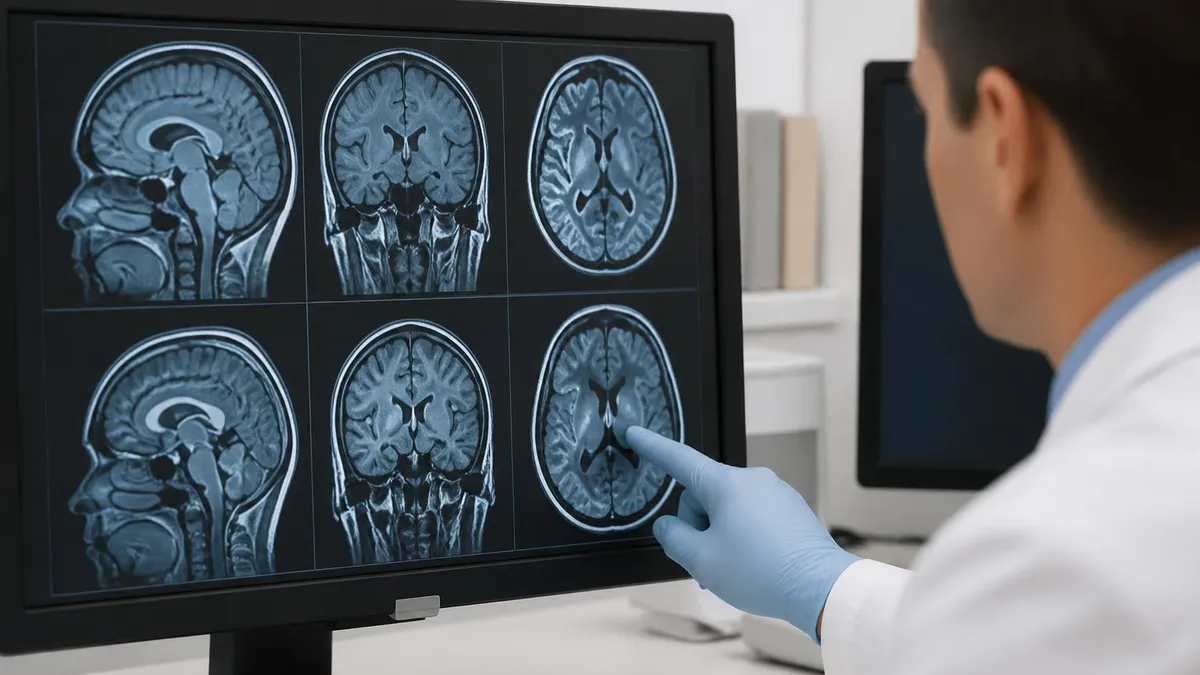
Clinical MRI touches nearly every organ system, but neurological imaging remains the largest application. Brain MRI is the gold standard for evaluating stroke, multiple sclerosis, brain tumors, epilepsy, traumatic brain injury, and dementia. Diffusion-weighted imaging identifies acute ischemia within minutes, while perfusion imaging maps tissue at risk. Spine MRI characterizes disc herniations, spinal stenosis, cord compression, and demyelinating lesions with detail no other modality can match.
Musculoskeletal MRI is the second-largest clinical use case. Knee, shoulder, hip, ankle, and wrist exams reveal meniscal tears, ligament injuries, cartilage damage, labral pathology, tendinopathy, and stress fractures. Sports medicine clinics now rely on MRI for nearly every soft tissue injury, and many high-volume orthopedic practices have dedicated extremity scanners that image only a single joint at a time, increasing throughput and patient comfort.
Body imaging encompasses abdominal, pelvic, and thoracic MRI. Liver MRI with hepatobiliary contrast agents like gadoxetate disodium characterizes focal lesions, screens for hepatocellular carcinoma in cirrhosis, and evaluates biliary anatomy. Pelvic MRI is the standard for prostate cancer staging using multiparametric protocols and for assessing endometriosis, fibroids, and gynecologic malignancies. Cardiac MRI quantifies ejection fraction, myocardial viability, and infiltrative cardiomyopathies.
Breast MRI plays a focused role in high-risk screening, problem solving after ambiguous mammography, and pre-surgical staging of newly diagnosed cancers. It is highly sensitive but moderately specific, so it is paired with mammography and ultrasound rather than replacing them. Abbreviated breast MRI protocols completed in under ten minutes are expanding access for screening populations who previously could not tolerate or afford full diagnostic exams.
Vascular MRI uses both contrast-enhanced and non-contrast techniques. Time-of-flight angiography images carotid arteries and the circle of Willis without injection. Contrast-enhanced MRA evaluates the aorta, renal arteries, and peripheral runoff vessels. Phase-contrast techniques quantify flow velocity, useful for congenital heart disease and CSF flow studies in normal pressure hydrocephalus and Chiari malformations evaluation by neurosurgical referral teams.
Functional and metabolic imaging extends MRI beyond anatomy. fMRI maps language and motor cortex before tumor resection. MR spectroscopy quantifies N-acetylaspartate, choline, creatine, and lactate to characterize brain tumors and metabolic disorders. Diffusion tensor imaging traces white matter tracts. These advanced applications are concentrated in academic centers but are spreading into community practice as software simplifies acquisition and reconstruction.
For a comprehensive look at what conditions modern MRI can identify across all these applications, see our companion article on What MRI Can Detect: Conditions & Diagnostic Capabilities. It catalogs the specific pathologies each protocol is designed to find and explains why MRI outperforms competing modalities for each indication.
Patients with severely reduced kidney function (eGFR below 30 mL/min/1.73m²) face an elevated risk of nephrogenic systemic fibrosis from certain gadolinium-based contrast agents. Always verify recent renal labs, document informed consent, and use the lowest necessary dose. Group II macrocyclic agents carry the lowest documented risk and are now preferred for at-risk populations in U.S. practice.
The pace of MRI innovation has accelerated dramatically in the 2020s. Deep-learning image reconstruction is now standard on every major vendor's platform, taking undersampled k-space data and producing diagnostic-quality images in a fraction of the time. A brain MRI that took 25 minutes in 2018 routinely runs in 8–10 minutes in 2026, with equivalent or better image quality, expanding throughput and reducing motion artifact in pediatric and uncooperative patients.
Compressed sensing, parallel imaging, and simultaneous multi-slice acquisition all contribute to these speed gains. Simultaneous multi-slice excites and reads several slices at once, dramatically cutting diffusion and functional MRI acquisition times. Wave-CAIPI techniques accelerate 3D sequences. The combined effect is that screening protocols once considered impractical — like whole-body MRI for cancer surveillance — are becoming clinically feasible at reasonable cost in many U.S. centers.
Ultra-high-field 7T MRI received FDA clinical approval in 2017 and continues to expand. At 7T, the higher SNR enables sub-millimeter resolution of hippocampal subfields, cortical layers, and small vessel anatomy that 3T cannot resolve. Indications remain focused on neurological and musculoskeletal applications because B1 inhomogeneity, increased SAR, and susceptibility artifacts limit body imaging. Research is steadily addressing these challenges through parallel transmit and dielectric pads.
At the other end of the spectrum, low-field MRI is having a renaissance. Portable 0.064T point-of-care scanners now operate at the bedside in intensive care units, emergency departments, and even ambulances. These systems use AI-heavy reconstruction to overcome low intrinsic SNR and produce diagnostically useful brain images for stroke triage, hemorrhage detection, and ICU monitoring. They cost a fraction of conventional scanners and require no shielded suite.
Helium-free magnet designs are reducing dependence on increasingly scarce liquid helium. Sealed cryogenic systems use a few liters of helium rather than the 1,500-liter open dewars of legacy magnets, dramatically lowering installation cost and refill burden. Some manufacturers now market essentially zero-boil-off magnets that may never need a top-off during the scanner's life, an important development for facilities in remote areas.
Artificial intelligence is also reshaping the radiologist workflow. Auto-segmentation tools measure tumor volumes, brain structures, and cartilage thickness automatically. Triage algorithms flag suspected intracranial hemorrhage, pulmonary embolism on MR angiography, and acute stroke for prioritized reading. These tools augment rather than replace radiologists, but they shorten turnaround time and reduce missed findings in high-volume environments.
If you are curious about how all these acquisition advances translate to the actual patient experience and what comes off the scanner, our guide to Full Body MRI: What It Scans, How Long It Takes, Cost, and What to Expect walks through one of the most rapidly growing applications enabled by these speed and reconstruction innovations across modern radiology practice.
For technologists who want to deepen their grasp of mri technology in practice, a few habits separate strong operators from average ones. First, learn to read the protocol rather than just running it. Open the parameter card, look at TR, TE, flip angle, bandwidth, matrix, and slice thickness, and ask why each value was chosen. When you understand the trade-offs, you can adapt intelligently when a patient cannot hold still, has an implant, or has unusual anatomy.
Second, build a mental library of artifacts. Wraparound, chemical shift, motion ghosting, Gibbs ringing, susceptibility blooming, and zipper artifacts each have characteristic appearances and specific fixes. Recognizing them in real time on the console — rather than after the patient is dressed and gone — is a hallmark of an experienced technologist and prevents costly repeat exams. Keep a personal reference of before-and-after examples from your own scanner.
Third, develop fluency with coil selection and positioning. The wrong coil or a poorly positioned one can cost you 50% of available signal-to-noise, no matter how powerful the magnet. Spend time centering anatomy in the coil isocenter, immobilizing the area of interest, and using cushioning that respects the patient's comfort while reducing motion. Good positioning often matters more than expensive hardware for image quality and diagnostic confidence.
Fourth, cultivate strong patient communication. Explain the duration of each sequence before it starts, warn patients about loud knocking, and check in between sequences. A calm patient who knows what is coming holds still better, which directly improves image quality. For claustrophobic patients, prone positioning, mirrors, music, and headphones with familiar voices can be the difference between a completed exam and a cancelled appointment that wastes valuable scanner time.
Fifth, study the safety screening process until it is second nature. The vast majority of serious MRI incidents involve missed implants, missed metallic foreign bodies, or unscreened personnel entering Zone IV. Never assume a previous screen is complete; verify it yourself for every patient, every visit. Treat the screening form as a clinical document, not paperwork, and escalate any uncertainty about an implant to the radiologist or MR safety officer before proceeding.
Sixth, stay current with continuing education. MRI is one of the fastest-evolving modalities in radiology, and a technologist trained even five years ago may be unaware of compressed sensing, deep-learning reconstruction, abbreviated protocols, or new contrast agents. ARRT requires structured CE for a reason — use it to actually learn rather than to check a box. Vendor webinars, ASRT modules, and physics refresher courses pay back the time investment many times over.
Finally, take care of yourself. MRI suites are physically demanding workplaces with heavy patients, long shifts, and the cognitive load of constant safety vigilance. The technologists who sustain decade-long careers are the ones who manage ergonomics, sleep, and stress as carefully as they manage their scanners. The job is rewarding and intellectually rich, but only if you build the habits that let you keep doing it well, year after year, exam after exam.
MRI Questions and Answers
About the Author
Medical Laboratory Scientist & Clinical Certification Expert
Johns Hopkins UniversityDr. Sandra Kim holds a PhD in Clinical Laboratory Science from Johns Hopkins University and is certified as a Medical Technologist (MT) and Medical Laboratory Scientist (MLS) through ASCP. With 16 years of clinical laboratory experience spanning hematology, microbiology, and molecular diagnostics, she prepares candidates for ASCP board exams, MLT, MLS, and specialist certification tests.
Join the Discussion
Connect with other students preparing for this exam. Share tips, ask questions, and get advice from people who have been there.
View discussion (6 replies)