MRI With vs Without Contrast: Complete Guide to How Each Scan Works, What It Shows, and When You Need Gadolinium
🟢 What does an MRI without contrast show vs with contrast? Compare uses, safety, gadolinium risks, costs, and when each MRI scan is ordered.
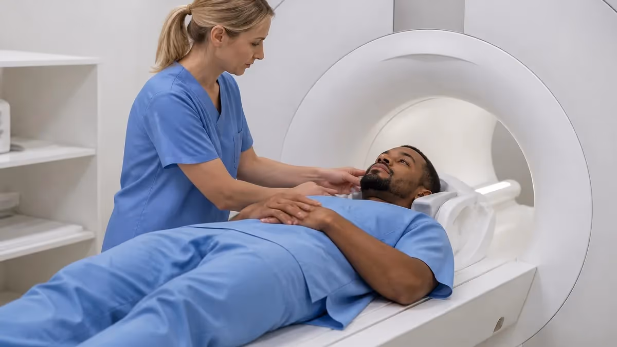
If your doctor just handed you a referral for an MRI, you may be wondering what does an MRI without contrast show compared to one performed with gadolinium contrast agent. The short answer is that a non-contrast MRI reveals exceptional detail of soft tissue, bone marrow, cartilage, ligaments, tendons, brain matter, the spinal cord, and fluid collections, all without injecting any dye. That makes it the default exam for most musculoskeletal complaints, routine brain screening, and follow-up imaging where structural anatomy is the primary question.
Contrast-enhanced MRI, by contrast, uses a gadolinium-based contrast agent injected through an IV to make blood vessels, inflammation, infection, and certain tumors light up brilliantly against surrounding tissue. The contrast crosses leaky or abnormal capillaries that healthy tissue does not have, so areas of disease appear bright on T1-weighted images. Radiologists rely on this enhancement pattern to detect small metastases, characterize masses, evaluate multiple sclerosis activity, and assess post-surgical scar versus recurrent disease.
Choosing between the two studies is not arbitrary. The ordering physician weighs the suspected diagnosis, the patient's kidney function, prior allergic reactions, pregnancy status, and whether earlier imaging already answered the structural question. In many cases the protocol calls for both: sequences are first acquired without contrast, then repeated after gadolinium is administered, giving the radiologist a before-and-after comparison that maximizes diagnostic confidence on a single visit.
For patients, the practical differences matter too. A non-contrast scan typically takes 20 to 40 minutes, requires no IV stick, no fasting in most centers, and carries essentially zero pharmacologic risk. A contrast study adds 10 to 20 minutes, an IV line, a screening lab for kidney function in higher-risk patients, and a small but non-zero possibility of gadolinium retention or allergic reaction that the technologist will review with you beforehand.
This guide walks through both protocols in depth: the physics that make each one work, the conditions that benefit most from contrast, the safety considerations around gadolinium, the cost differences in the United States healthcare system, and the questions you should ask your ordering provider before scheduling. Whether you are an imaging student preparing for the registry or a patient trying to understand a recent order, you will leave with a clear framework for interpreting why a particular study was chosen.
We also address common patient anxieties: claustrophobia, scanner noise, the loud knocking that frightens first-timers, and what happens if the radiologist calls back asking for additional contrast sequences. By the end you will recognize that with-contrast and without-contrast MRI are complementary tools, not competing ones, and that the right exam is the one matched precisely to your clinical question.
Throughout the article we will refer back to real protocols used in US imaging centers, registry-style review points helpful for technologists in training, and patient-facing tips drawn from American College of Radiology appropriateness criteria. Treat this as both a primer and a reference you can return to before your appointment or before your next clinical rotation.
MRI With vs Without Contrast by the Numbers
How Each MRI Protocol Works
T1, T2, PD, STIR, and FLAIR sequences are acquired in multiple planes. Each weighting highlights different tissue properties — fluid bright on T2, fat bright on T1 — letting the radiologist map anatomy and detect edema, hemorrhage, or structural damage without any injection.
A technologist inserts a small IV in the antecubital vein and injects roughly 0.1 mmol/kg of a gadolinium-based agent. The contrast circulates within seconds, shortening T1 relaxation times so abnormal tissue appears bright on post-contrast T1-weighted images.
After injection, T1-weighted sequences are repeated in the same planes, often with fat suppression. Comparing pre- and post-contrast images reveals enhancement patterns that point to tumors, infections, demyelinating lesions, and vascular abnormalities invisible on standard sequences.
Dynamic contrast-enhanced MRI, MR angiography, and perfusion studies require precise timing of contrast boluses. These advanced protocols evaluate blood flow, vessel patency, and tissue perfusion for stroke, cancer staging, and cardiac imaging applications.
A non-contrast MRI shows remarkable structural detail without any injected dye, which is why it remains the workhorse of musculoskeletal, spine, and routine neurologic imaging in the United States. On a knee study, for example, the radiologist can confidently diagnose meniscal tears, anterior cruciate ligament rupture, cartilage defects, bone bruises, joint effusions, and Baker cysts simply by interpreting T1, T2, and proton-density weighted sequences in three orthogonal planes.
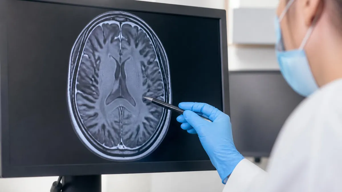
For the brain, a non-contrast protocol detects acute stroke on diffusion-weighted imaging within minutes of symptom onset, identifies chronic ischemic changes on FLAIR, picks up hemorrhage on susceptibility-weighted sequences, and visualizes hydrocephalus, mass effect, or congenital malformations with high reliability. Many emergency departments order non-contrast brain MRI first because it answers most acute questions without the delay of IV placement and contrast injection.
Spine imaging is another area where non-contrast studies dominate. Disc herniation, spinal stenosis, spondylolisthesis, vertebral compression fractures, marrow edema, and degenerative facet arthropathy all appear vividly on standard sequences. Adding contrast is generally reserved for suspected infection, tumor, or post-operative recurrence — clinical scenarios where enhancement patterns dramatically change management.
Abdominal and pelvic non-contrast MRI is increasingly used for liver lesion characterization with hepatobiliary protocols, for renal cyst evaluation, and for MR enterography in patients who cannot receive contrast. Diffusion-weighted imaging adds functional information that often substitutes for what gadolinium would otherwise reveal, particularly in patients with chronic kidney disease who cannot safely receive standard contrast agents.
Cardiac MRI without contrast can quantify ventricular volumes, ejection fraction, valvular function, and myocardial thickness with extraordinary accuracy using cine steady-state free precession sequences. Even without late gadolinium enhancement, T1 and T2 mapping techniques now allow detection of myocardial edema, iron overload, and diffuse fibrosis, expanding what a non-contrast cardiac study can answer. To appreciate how the technology evolved to this level, the history of MRI traces decades of incremental physics and engineering breakthroughs.
Pediatric imaging also leans heavily on non-contrast protocols because children are more sensitive to any long-term gadolinium exposure and because many pediatric questions — congenital anomalies, hydrocephalus, joint pain — can be answered without enhancement. Fast sequences and feed-and-swaddle techniques have made non-contrast pediatric MRI a frontline tool in many children's hospitals across the country.
Finally, screening MRI in high-risk populations frequently relies on non-contrast protocols. Abbreviated breast MRI sometimes uses post-contrast images, but screening brain MRI for headaches, hearing loss, or family history of aneurysm is often non-contrast, as is MR cholangiopancreatography for biliary anatomy. The takeaway: non-contrast MRI is not a lesser study, it is a precision tool with a clearly defined and very wide diagnostic scope.
MRI Practice Test Questions
Prepare for the MRI - Magnetic Resonance Imaging exam with our free practice test modules. Each quiz covers key topics to help you pass on your first try.
MRI Knowledge
MRI Exam Questions covering Knowledge. Master MRI Test concepts for certification prep.
MRI Physics
Free MRI Practice Test featuring Physics. Improve your MRI Exam score with mock test prep.
MRI Anatomy and Pathology
MRI Test Prep for MRI Anatomy and Pathology. Practice MRI Quiz questions and boost your score.
MRI Anatomy and Positioning
MRI Questions and Answers on MRI Anatomy and Positioning. Free MRI practice for exam readiness.
MRI Contrast Agents
Free MRI Quiz on MRI Contrast Agents. MRI Exam prep questions with detailed explanations.
MRI Patient Care and Positioning
MRI Practice Questions for MRI Patient Care and Positioning. Build confidence for your MRI certification exam.
When Contrast Is Required for MRI
Gadolinium contrast is essential when evaluating suspected primary brain tumors, metastatic disease, or characterizing indeterminate masses anywhere in the body. Most tumors have abnormal, leaky vasculature that allows contrast to pool inside the lesion, producing the bright enhancement that distinguishes active tumor from surrounding edema, necrosis, or post-treatment changes on T1-weighted sequences.
For known cancer patients, contrast MRI is the standard of care for surveillance after surgery or radiation. It separates expected post-treatment scar from recurrent disease, identifies small leptomeningeal deposits invisible on non-contrast sequences, and guides biopsy targeting. Oncologists also rely on dynamic contrast-enhanced perfusion studies to assess treatment response before tumor size changes become apparent.
Should You Choose MRI With or Without Contrast?
- +Contrast dramatically improves detection of small tumors and metastases
- +Enhancement patterns help characterize indeterminate lesions
- +Active versus chronic inflammation can be distinguished
- +Post-surgical scar versus recurrent disease becomes clear
- +Vascular structures and perfusion can be evaluated
- +Multiple sclerosis disease activity is accurately staged
- −Adds 10-20 minutes to total exam time
- −Requires IV placement and kidney function screening
- −Small risk of allergic reaction or extravasation
- −Gadolinium retention concerns in some patient groups
- −Contraindicated in severe kidney disease without precautions
- −Higher cost and additional insurance authorization steps
Pre-Scan Preparation Checklist for MRI With or Without Contrast
- ✓Confirm with the ordering provider whether contrast is needed for your specific question
- ✓Bring a current medication list including any prior contrast reactions
- ✓Disclose any kidney disease, dialysis history, or recent creatinine results
- ✓Notify staff if you are pregnant or breastfeeding
- ✓Remove all metal jewelry, hairpins, watches, and dental appliances if requested
- ✓Wear loose clothing without metal zippers, snaps, or underwire
- ✓Eat lightly beforehand unless your facility requires fasting
- ✓Arrive 30 minutes early to complete the MRI safety screening form
- ✓Discuss claustrophobia openly so sedation or open MRI options can be considered
- ✓Plan for a driver if you receive sedatives or anti-anxiety medication
Always confirm the contrast decision in writing.
Insurance pre-authorization, scheduling, and even the IV setup all hinge on whether the order specifies contrast. A simple phone call to confirm "MRI brain with and without contrast" versus "MRI brain without contrast" can prevent rescheduling, denied claims, or a repeat visit. Always verify the exact CPT code on your order matches what the imaging center has booked.
Gadolinium-based contrast agents have been used safely in tens of millions of MRI scans worldwide since the late 1980s, but no medication is risk-free. The most important safety screen is renal function, because patients with severely reduced glomerular filtration rate were historically at elevated risk for nephrogenic systemic fibrosis, a rare but disabling condition affecting the skin, joints, and internal organs. Modern macrocyclic agents and pre-scan creatinine screening have made this complication extremely uncommon today.
The next consideration is allergic-type reactions. True gadolinium allergy is rare — about 1 in 10,000 patients experience moderate reactions and severe anaphylaxis is even less common, occurring in roughly 1 in 100,000 administrations. Mild reactions like nausea, hives, or transient warmth are more frequent but generally self-limited. Patients with prior contrast reactions, asthma, or multiple allergies should disclose this history so the radiologist can decide whether premedication or an alternative agent is warranted.
Gadolinium retention has become a major topic of patient discussion over the past decade. Studies have shown trace deposits in brain tissue, particularly the dentate nucleus and globus pallidus, even in patients with normal kidney function. The clinical significance of these deposits remains uncertain, and major regulatory agencies including the FDA have not restricted gadolinium use for medically indicated exams, but they do recommend using the lowest necessary dose and preferring macrocyclic agents.
Pregnancy is another area of caution. Gadolinium crosses the placenta and is excreted into amniotic fluid, where the fetus may swallow it repeatedly. Although no proven fetal harm has been documented, the American College of Radiology recommends avoiding gadolinium during pregnancy unless absolutely necessary, with the decision made jointly by the obstetrician, radiologist, and patient based on the clinical urgency.
Breastfeeding mothers can almost always continue nursing after gadolinium administration. Less than 0.04% of the maternal dose is excreted into breast milk, and an even smaller fraction is absorbed by the infant gastrointestinal tract. Most professional societies no longer recommend pumping and dumping after routine doses, though anxious mothers may choose to do so for 24 hours for personal peace of mind.
Extravasation, where contrast leaks into the soft tissue around the IV instead of the vein, occurs in roughly 0.05% of injections. Most events cause only temporary swelling and discomfort, but large extravasations can rarely cause compartment syndrome requiring surgical attention. Technologists monitor the injection site carefully and stop immediately if the patient reports unusual pain or burning at the IV.
Finally, patients with implants or devices need a separate safety screening regardless of contrast use. Pacemakers, cochlear implants, certain aneurysm clips, and some neurostimulators may be contraindicated or require specific scanning protocols. The MRI technologist will review every device against the manufacturer's MR conditional labeling before the scan begins, ensuring patient safety remains the highest priority throughout the imaging visit.

Severe kidney disease, prior contrast reaction, pregnancy, implanted devices, and recent surgeries can all change whether contrast is safe or even possible. Disclose them before your appointment — ideally when scheduling — so the radiologist can choose the safest protocol. Last-minute disclosures may force the technologist to cancel the scan and reschedule, delaying your diagnosis by days or weeks.
The cost of an MRI in the United States varies enormously depending on geography, facility type, insurance coverage, and whether contrast is administered. National averages for non-contrast MRI range from about $400 at independent imaging centers to over $3,500 at hospital-based outpatient departments. Adding gadolinium typically increases the bill by $100 to $300, reflecting the agent cost, IV supplies, and additional technologist and radiologist time.
Insurance pre-authorization is often required for both contrast and non-contrast MRI, but contrast studies sometimes face stricter scrutiny because of the higher overall cost. If your insurer denies authorization, the ordering provider can appeal with clinical documentation explaining why contrast is necessary. Patients with high-deductible plans often save significantly by choosing freestanding imaging centers over hospital outpatient departments for the same study quality.
Scheduling timelines also differ. Non-contrast MRI typically books faster, sometimes same-week, because the scan slot is shorter and no contrast preparation is needed. Contrast studies require a clear pathway for renal function review, IV placement, and post-injection monitoring, which can extend the appointment by 20 to 30 minutes and limit available slots. Plan accordingly if your diagnosis is time-sensitive.
For patients without insurance, many imaging centers offer cash-pay discounts, sometimes reducing the bill by 40 to 60 percent. Asking for the cash price and comparing across local facilities can produce dramatic savings. Some centers also offer payment plans, charity care, or sliding-scale fees for qualifying patients. Always ask before assuming the full sticker price is your only option.
If you are anxious about the experience itself, consider what to expect on the day of the scan. The scanner makes a series of loud knocking, banging, and humming sounds caused by rapid gradient coil switching, which can be unsettling for first-time patients. Reading about the mri machine noise in advance and using the provided earplugs or headphones makes a major difference in patient comfort during longer protocols.
Open MRI scanners and wide-bore systems offer alternatives for patients with claustrophobia or larger body habitus, though image quality may be slightly reduced on lower field strength magnets. Many centers also offer mild oral sedation with prescribed lorazepam, music distraction, prism glasses to see outside the bore, and the ability to bring a support person into the scanner room when safety screening allows.
Finally, the report turnaround matters. Most outpatient MRI reports are completed within 24 to 48 hours and posted to your patient portal. Urgent or stat exams can be read within an hour. If contrast revealed an unexpected finding, the radiologist may call your ordering provider directly the same day, and you should not be alarmed by a quick callback — it usually means the team is acting promptly on the new information to keep your care moving forward.
Putting all of this together, the most useful mindset for patients and clinicians is to treat with-contrast and without-contrast MRI as different tools answering different questions. If the clinical concern is purely structural — a torn ligament, a herniated disc, a suspected fracture, baseline anatomy — a non-contrast study usually suffices. If the question involves active inflammation, tumor characterization, infection, vascular detail, or post-surgical recurrence, contrast adds critical information that non-contrast sequences cannot replicate.
For technologists preparing for the ARRT MRI registry or ARMRIT certification, expect questions on indications for contrast, gadolinium dosing in mmol/kg, contraindications based on estimated GFR, recognition of nephrogenic systemic fibrosis risk factors, and the differences between macrocyclic and linear chelates. Practice cases often combine imaging findings with patient history to test whether you would suggest adding or omitting contrast for that scenario.
Patients can advocate effectively by asking three questions before any MRI: What specific diagnosis are we trying to confirm or rule out? Will contrast change the management based on the result? Have you ordered the exact protocol that answers that question? Most physicians welcome these conversations because they confirm that the patient is engaged and reduce the chance of having to repeat an inadequate study later.
Aftercare for a non-contrast MRI is essentially nothing — you can resume all activities immediately, including driving, eating, and returning to work. After a contrast study, hydration is encouraged to help your kidneys clear the gadolinium more quickly, and the IV site may be tender for a few hours. Most patients experience no noticeable effects beyond mild fatigue from lying still for the duration of the scan.
Follow-up MRI is common for chronic conditions like multiple sclerosis, low-grade gliomas, breast cancer screening in high-risk patients, and post-operative monitoring. Consistency of protocol matters enormously: comparing apples to apples — same field strength, same sequences, same contrast or none — gives the radiologist the most reliable read on whether disease is progressing, stable, or improving over time.
If a follow-up reveals new findings, do not panic before the full report is reviewed. Many enhancing areas turn out to be inflammation, infection, or treatment-related changes rather than recurrent disease. Discuss the report with the ordering provider, ask whether a specialist second opinion is warranted, and remember that contrast MRI is a screening and characterization tool, not a definitive diagnosis — biopsy or further imaging often follows ambiguous findings.
Whether you are a student preparing for boards, a patient walking into your first scan, or a clinician deciding what to order, the central principle holds: match the protocol to the question. A well-chosen MRI — whether with or without contrast — answers the clinical question accurately, safely, and with minimal burden on the patient, which is exactly what modern radiology is designed to deliver. For broader context on imaging choices, consider when MRI alternatives like CT, ultrasound, or PET may better serve your specific question.
MRI Questions and Answers

MRI Medical Abbreviation: What MRI Stands For and Why It Matters

The History of MRI: From Discovery to Modern Medicine

Knee MRI Images: A Complete Guide to Reading, Understanding, and Interpreting Knee Scans

Noise of MRI Machine: Why MRI Scanners Are So Loud and What to Expect
MRI Alternatives: When CT, Ultrasound, X-Ray, and PET Make More Sense Than an MRI
About the Author
Medical Laboratory Scientist & Clinical Certification Expert
Johns Hopkins UniversityDr. Sandra Kim holds a PhD in Clinical Laboratory Science from Johns Hopkins University and is certified as a Medical Technologist (MT) and Medical Laboratory Scientist (MLS) through ASCP. With 16 years of clinical laboratory experience spanning hematology, microbiology, and molecular diagnostics, she prepares candidates for ASCP board exams, MLT, MLS, and specialist certification tests.
Join the Discussion
Connect with other students preparing for this exam. Share tips, ask questions, and get advice from people who have been there.
View discussion (6 replies)