CPR ROSC: Return of Spontaneous Circulation Explained for Rescuers and Healthcare Providers in 2026 July
🟢 CPR ROSC explained: ACLS algorithm steps, post-resuscitation care, infant CPR considerations, AED use, and how to recognize return of spontaneous
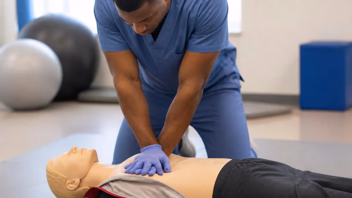
CPR ROSC stands for the moment in cardiopulmonary resuscitation when the patient achieves Return of Spontaneous Circulation, meaning the heart resumes generating a sustained, palpable pulse and measurable blood pressure without ongoing chest compressions. For rescuers following the acls algorithm, ROSC is the pivotal transition point between active resuscitation and post-cardiac arrest care. Recognizing ROSC quickly and correctly determines whether the team continues compressions, escalates medication dosing, or pivots to airway management, hemodynamic stabilization, and targeted temperature management protocols.
The term ROSC was formalized by resuscitation councils to standardize how providers describe the physiological recovery of circulation after cardiac arrest. Before ROSC terminology became widespread, providers used vague phrases like "got a pulse back" or "converted," which were inconsistent across hospitals and EMS systems. Today, ROSC is documented with specific criteria: a palpable central pulse lasting longer than one minute, return of measurable blood pressure, or a sudden rise in end-tidal CO2 above 35-40 mmHg during continuous capnography monitoring.
Understanding ROSC matters whether you are a layperson learning hands-only CPR, a BLS-certified responder, or an advanced provider working through the acls algorithm in a code. For laypeople, ROSC means the moment you can stop compressions and place the victim in the recovery position while waiting for EMS. For professionals, ROSC signals the start of an entirely new clinical phase focused on preventing rearrest, protecting the brain, and identifying the cardiac arrest cause.
This guide walks through every aspect of CPR ROSC including how to recognize it, what to do in the first ten minutes after achieving it, why infant CPR ROSC criteria differ from adult, how AED use influences ROSC rates, and which post-arrest interventions improve neurologically intact survival. We will also address common myths, such as the false belief that any movement during CPR indicates ROSC, and explain why pulse checks alone are unreliable without confirmatory data points.
The American Heart Association, European Resuscitation Council, and the national cpr foundation all emphasize ROSC as the single most important short-term outcome measure during cardiac arrest care. Studies published in Circulation and Resuscitation journals show that hospitals tracking ROSC rates and time-to-ROSC consistently achieve better survival-to-discharge numbers, particularly when paired with strong basic life support, early defibrillation, and structured post-arrest debriefing programs.
Finally, this article focuses on practical, evidence-based information that healthcare providers and trained rescuers can apply in real codes. Whether you are preparing for pals certification, refreshing your BLS card, or simply trying to understand what happens in those critical seconds when a heart restarts, the following sections break down ROSC into clear, actionable knowledge. We will use real-world examples, current 2025-2026 guidelines, and case-based reasoning to help you internalize the concepts.
By the end of this guide you will be able to define ROSC precisely, list the three primary indicators used to confirm it, describe the immediate post-ROSC checklist, explain how capnography changes signal circulatory return, and understand why ROSC is the beginning of recovery rather than the end of resuscitation. Let us begin with the numbers that frame why ROSC matters so much in modern cardiac arrest care.
CPR ROSC by the Numbers

From Cardiac Arrest to ROSC: The Critical Timeline
Recognition and Activation
High-Quality CPR Begins
Defibrillation for Shockable Rhythms
Advanced Interventions
ROSC Confirmation
Post-Arrest Care
Return of spontaneous circulation is not a single instant but a physiological cascade in which the heart's electrical activity reorganizes, mechanical contraction resumes, and systemic perfusion becomes self-sustaining. During chest compressions, rescuers are mechanically squeezing the heart and thoracic cavity to push blood through the body. At ROSC, the myocardium itself resumes coordinated contraction, generating cardiac output without external pressure. The transition is sometimes obvious and sometimes subtle, which is why objective monitoring matters far more than gut feeling.
The most reliable real-time signal of ROSC in a monitored patient is a sudden, sustained rise in end-tidal CO2 measured by continuous waveform capnography. When circulation returns, blood flow to the lungs increases dramatically, carrying accumulated CO2 from tissues back to alveoli where it is exhaled. A jump from baseline values around 10-20 mmHg during CPR up to 35-40 mmHg or higher within one or two breaths is highly specific for ROSC and often precedes detectable pulse return by 15-30 seconds.
Pulse checks during CPR have long been controversial because they are unreliable. Studies show even experienced providers misidentify pulses about 20 percent of the time when palpating quickly during a pulse check. That is why the acls algorithm recommends limiting pulse checks to no more than 10 seconds and pairing them with capnography, arterial line tracings, or point-of-care ultrasound whenever possible. A respiratory rate that returns spontaneously and gasping or coughing also raise suspicion for ROSC.
Blood pressure return is the third pillar of ROSC confirmation. In a patient with an arterial line, the appearance of a clear pulsatile waveform with systolic pressures above 60 mmHg is diagnostic. Without an arterial line, manual cuff measurements during the brief pulse check window or automated noninvasive cycles immediately after stopping compressions confirm circulation. A systolic pressure below 90 after ROSC signals post-arrest shock requiring immediate intervention.
It is essential to understand that ROSC can be transient. Many patients achieve ROSC for seconds or minutes only to rearrest. This is why teams continue intensive monitoring, maintain pads and IV access, and prepare to resume compressions instantly. Roughly one in five patients who initially achieve ROSC will rearrest within the first hour, and the cardiac arrest cause is often still present, whether that is acute coronary occlusion, electrolyte derangement, or massive pulmonary embolism awaiting definitive treatment.
Patient movement during CPR is sometimes misinterpreted as ROSC. CPR-induced consciousness, where the patient grimaces, reaches up, or even opens their eyes during effective compressions, is increasingly recognized. This phenomenon proves your compressions are perfusing the brain but it does NOT mean ROSC has occurred. Stopping compressions in this scenario can be catastrophic. Always confirm with capnography or pulse before declaring ROSC, even when the patient appears to wake up.
Finally, ROSC physiology in pediatric and infant patients differs slightly. Children rarely arrest from primary cardiac causes; most pediatric arrests are respiratory in origin, so reversing hypoxia often produces dramatic ROSC. Capnography thresholds are similar but compression-induced ETCO2 baselines tend to be higher in small children due to differences in dead space ventilation. Providers preparing for pals certification should commit these nuances to memory.

CPR Practice Test Questions
Prepare for the CPR Cardiopulmonary Resuscitation Practice exam with our free practice test modules. Each quiz covers key topics to help you pass on your first try.
CPR Basic CPR
CPR Exam Questions covering Basic CPR. Master CPR Test concepts for certification prep.
CPR and First Aid
Free CPR Practice Test featuring and First Aid. Improve your CPR Exam score with mock test prep.
CPR (Cardiopulmonary Resuscitation) Adult ...
CPR Mock Exam on (Cardiopulmonary Resuscitation) Adult CPR and AED Usage. CPR Study Guide questions to pass on your first try.
CPR (Cardiopulmonary Resuscitation) Airway...
CPR Test Prep for (Cardiopulmonary Resuscitation) Airway Obstruction and Choking. Practice CPR Quiz questions and boost your score.
CPR (Cardiopulmonary Resuscitation) Cardio...
CPR Questions and Answers on (Cardiopulmonary Resuscitation) Cardiopulmonary Emergency Recognition. Free CPR practice for exam readiness.
CPR (Cardiopulmonary Resuscitation) Child ...
CPR Mock Test covering (Cardiopulmonary Resuscitation) Child and Infant CPR. Online CPR Test practice with instant feedback.
CPR (Cardiopulmonary Resuscitation) High-P...
Free CPR Quiz on (Cardiopulmonary Resuscitation) High-Performance Team Dynamics. CPR Exam prep questions with detailed explanations.
CPR (Cardiopulmonary Resuscitation) Legal ...
CPR Practice Questions for (Cardiopulmonary Resuscitation) Legal and Ethical Considerations. Build confidence for your CPR certification exam.
CPR for Specific Populations
CPR Test Online for for Specific Populations. Free practice with instant results and feedback.
CPR Airway & Breathing Management
CPR Exam Questions covering Airway & Breathing Management. Master CPR Test concepts for certification prep.
CPR Assessment & Recognition of Cardiac Ar...
Free CPR Practice Test featuring Assessment & Recognition of Cardiac Arrest. Improve your CPR Exam score with mock test prep.
CPR Chest Compressions & Defibrillation
CPR Mock Exam on Chest Compressions & Defibrillation. CPR Study Guide questions to pass on your first try.
CPR Post-Resuscitation Care & Recovery
CPR Test Prep for Post-Resuscitation Care & Recovery. Practice CPR Quiz questions and boost your score.
The ACLS Algorithm Pathway to ROSC
When the rhythm is ventricular fibrillation or pulseless ventricular tachycardia, the acls algorithm prioritizes immediate defibrillation. After each shock, providers resume compressions for two minutes before reassessing rhythm. Epinephrine 1 mg is administered after the second shock and repeated every 3-5 minutes. Amiodarone 300 mg or lidocaine 1-1.5 mg/kg is considered for refractory shockable arrests, typically after the third shock. Reversible causes are evaluated in parallel rather than waiting for completion.
ROSC in shockable arrests often occurs after defibrillation converts the chaotic rhythm into an organized one capable of generating output. The capnography trace usually shows a dramatic spike at the moment of conversion. Providers must continue compressions for the full two-minute cycle after each shock, even if they suspect ROSC, unless ETCO2 rise and other indicators are clearly present. Premature pulse checks reduce overall compression fraction significantly.
Bystander CPR Before EMS Arrival: Impact on ROSC
- +Doubles or triples likelihood of survival when initiated within the first few minutes
- +Maintains coronary perfusion pressure, dramatically improving ROSC chances after defibrillation
- +Buys critical time for AED retrieval and EMS arrival
- +Reduces neurological injury by sustaining cerebral blood flow
- +Hands-only CPR is easy enough for any adult to perform without prior training
- +Dispatcher-assisted CPR over the phone increases bystander participation significantly
- −Bystander hesitancy or fear of liability still delays initiation in many cases
- −Compression quality without training is often suboptimal, with shallow depth and incorrect rate
- −Fatigue sets in within 1-2 minutes, reducing compression depth and effectiveness
- −Many bystanders incorrectly perform mouth-to-mouth when hands-only would be sufficient
- −Public misconceptions about ribs breaking deter aggressive compression depth
- −Without an AED nearby, even excellent CPR rarely produces ROSC in shockable rhythms

Post-ROSC Immediate Action Checklist for Healthcare Providers
- ✓Confirm ROSC with at least two indicators: ETCO2 rise, palpable pulse, measurable blood pressure
- ✓Continue advanced airway management and verify endotracheal tube placement with waveform capnography
- ✓Set ventilator to achieve SpO2 between 92-98 percent and PaCO2 between 35-45 mmHg
- ✓Obtain 12-lead ECG within 10 minutes to identify STEMI or other actionable ischemic changes
- ✓Establish two large-bore IVs or central access for hemodynamic support and infusion therapy
- ✓Initiate vasopressors if mean arterial pressure remains below 65 mmHg after fluid resuscitation
- ✓Start targeted temperature management between 32-36 degrees Celsius in comatose patients
- ✓Draw arterial blood gas, lactate, electrolytes, troponin, glucose, and complete metabolic panel
- ✓Identify and treat the underlying cardiac arrest cause using the Hs and Ts framework
- ✓Communicate with family using clear, compassionate language and avoid premature prognostication
Survival to Discharge Depends Heavily on Post-Arrest Care
Achieving ROSC is a tremendous milestone, but only about half of patients who reach ROSC in the hospital survive to discharge, and fewer leave neurologically intact. Aggressive, protocolized post-cardiac arrest care including TTM, coronary angiography when indicated, and delayed neuroprognostication is the single biggest determinant of meaningful recovery. Hospitals with cardiac arrest centers consistently outperform those without them on these outcomes.
Preventing rearrest in the minutes and hours after CPR ROSC is one of the most challenging aspects of resuscitation medicine. The conditions that caused the original cardiac arrest often persist, the myocardium is stunned, the brain has been hypoxic, and inflammatory cascades resembling sepsis are unleashed. This combination, known as the post-cardiac arrest syndrome, makes patients fragile and prone to recurrent arrest, dysrhythmias, and circulatory collapse. Vigilance and proactive management during the first six to twelve hours are essential.
Hemodynamic instability is the most common driver of rearrest. Patients frequently develop vasodilatory shock similar to sepsis, requiring norepinephrine or epinephrine infusions to maintain perfusion. Mean arterial pressure targets of at least 65 mmHg, and ideally 80-100 mmHg in suspected anoxic brain injury, help preserve cerebral perfusion. Fluid resuscitation must be balanced carefully because excessive volume worsens pulmonary edema and right ventricular dysfunction in a stunned heart.
Ventilation strategy directly affects rearrest risk. Hyperventilation lowers PaCO2, causing cerebral vasoconstriction and reducing brain perfusion at exactly the wrong moment. Hypoventilation allows hypercapnia and acidosis, both of which lower the threshold for malignant arrhythmias. The acls algorithm recommends a respiratory rate of 10 breaths per minute initially with adjustments guided by capnography and serial blood gases. Avoiding both extremes is harder than it sounds in a chaotic resuscitation room.
Coronary reperfusion is critical when STEMI or suspected acute coronary occlusion is the cardiac arrest cause. Studies consistently show that immediate cardiac catheterization improves survival in these patients, even when they remain comatose. Many cardiac arrest centers now have protocols to take post-ROSC STEMI patients directly to the cath lab from the emergency department. Delays here translate directly into increased rearrest, expanded infarct size, and worse neurologic outcomes.
Electrolyte and metabolic optimization reduces arrhythmia recurrence. Potassium is often shifted intracellularly after epinephrine administration and during reperfusion, leaving patients hypokalemic and at risk for further VF. Targets of potassium 4.0-4.5 mEq/L and magnesium above 2.0 mg/dL are common. Glucose is kept between 140-180 mg/dL because both hypoglycemia and severe hyperglycemia worsen neurologic outcomes. Lactate clearance is monitored as a marker of adequate perfusion.
Targeted temperature management between 32 and 36 degrees Celsius for 24 hours has been a cornerstone of post-ROSC neuroprotection since landmark trials in 2002. More recent data including the TTM2 trial suggest 36 degrees may be equivalent to 33 degrees for most patients, though some subgroups may benefit from deeper cooling. The most important rule is strict avoidance of fever, which clearly worsens brain injury after cardiac arrest in every study performed.
Finally, do not forget the basics: pad placement should be confirmed, defibrillator left armed and ready, IV access secured, and the resuscitation team should remain in the room or immediately available. Rearrest can happen suddenly, sometimes with little warning beyond a falling ETCO2 or new ectopy on the monitor. Teams that debrief briefly after ROSC, clarify roles, and prepare for recurrence consistently respond faster and achieve better outcomes when rearrest occurs.
Approximately 20 percent of patients who achieve ROSC will rearrest, with the majority of recurrences happening within the first hour. Do not relax monitoring, leave the bedside without coverage, or remove defibrillation pads during this critical window. Maintain continuous ECG, capnography, and arterial blood pressure monitoring at all times until the patient is hemodynamically stable in an ICU setting.
Special populations require modified approaches to ROSC recognition and post-arrest care. Infant CPR, for example, follows different physiological rules than adult CPR. Most infant cardiac arrests are secondary to respiratory failure rather than primary cardiac causes, meaning effective ventilation often produces dramatic ROSC. The compression-to-ventilation ratio is 30:2 for a single rescuer but shifts to 15:2 for two rescuers, and compression depth is approximately 1.5 inches or one-third the anteroposterior chest diameter.
Pediatric ROSC criteria mirror the adult criteria but with attention to age-appropriate vital signs. A newborn with a heart rate above 100 is generally considered to have adequate circulation, while infants and small children should have age-adjusted blood pressure targets. Capnography remains valuable but baseline ETCO2 during compressions tends to be slightly different due to smaller dead space. Providers preparing for pals certification should drill these distinctions until they become automatic.
Pregnant patients in cardiac arrest receive standard high-quality CPR with one critical modification: manual left uterine displacement during compressions if the uterus is at or above the umbilicus. This relieves aortocaval compression and improves venous return. If ROSC is not achieved within four minutes of arrest in a viable pregnancy, perimortem cesarean delivery is considered to improve maternal and fetal outcomes. Post-ROSC care addresses both maternal and fetal physiology.
Drowning victims and hypothermic patients deserve prolonged resuscitation efforts. The classic adage "not dead until warm and dead" applies because severe hypothermia can be neuroprotective and patients have recovered after extended downtimes with appropriate rewarming. ROSC in these scenarios may take 60 minutes or more of CPR combined with extracorporeal rewarming. Avoid premature termination of resuscitation in cold-water drowning, especially in children.
Opioid overdose arrests have surged with the fentanyl crisis. Naloxone reverses respiratory depression and can restore circulation in opioid-induced arrests, although profound hypoxia may have already caused secondary cardiac arrest by the time naloxone is given. High-quality CPR and ventilation remain primary. For those wondering what does aed stand for in this context, automated external defibrillators still play a role because mixed-drug overdoses sometimes induce shockable rhythms via QT prolongation or stimulant toxicity.
Patients with implanted devices such as pacemakers, ICDs, or LVADs complicate ROSC recognition. ICDs may discharge during arrest, which is helpful, but their presence does not change CPR technique except to avoid placing pads directly over the device. LVAD patients may not have a palpable pulse even with circulation, so ROSC determination relies on Doppler, end-organ perfusion, and device-specific parameters rather than traditional pulse checks. These patients require specialty input quickly.
Finally, elderly patients and those with significant comorbidities have lower ROSC rates and worse outcomes after ROSC. This does not mean resuscitation is futile, but goals-of-care conversations before arrest, when possible, ensure that life support interventions align with patient values. After ROSC, ongoing discussions with family about prognosis, neurological recovery potential, and quality of life become central to ethical post-arrest care.
Practical preparation for CPR ROSC scenarios begins long before the code happens. Healthcare teams should participate in regular mock cardiac arrest drills with realistic monitors, capnography simulation, and structured debriefs afterward. The act of recognizing ROSC under pressure is a perishable skill, and teams that drill monthly consistently outperform those that drill annually. Make sure your simulation includes the moment of ROSC transition, not just compressions and shocks.
For laypeople and BLS-certified rescuers, the goal is not to memorize the entire acls algorithm but to perform high-quality compressions, deploy an AED rapidly, and recognize obvious signs of recovery such as the victim breathing normally or moving purposefully. If those signs appear, stop compressions, place the victim in the recovery position on their side, and continue to monitor breathing. Be ready to resume CPR immediately if breathing stops again before EMS arrives.
Equipment readiness is non-negotiable. AEDs should be checked monthly for battery and pad expiration. Crash carts should be inventoried daily in clinical settings. Capnography supplies including sampling lines and sensors must be stocked. The most beautiful resuscitation algorithm fails if the defibrillator battery is dead or the capnography sensor is missing. Standardize your equipment checks and audit them regularly to prevent these basic failures.
Documentation during and after a code matters more than many providers realize. Time of arrest, time of first compression, number of shocks, total epinephrine dose, time of ROSC, initial ETCO2, and rhythm at ROSC are all data points that feed quality improvement programs and clinical research. Use a code-recording template or designate a recorder during every resuscitation. Hospitals submitting data to registries like Get With The Guidelines consistently improve their outcomes over time.
Team dynamics during resuscitation deserve explicit attention. Clear role assignment, closed-loop communication, and empowering any team member to speak up about concerns all improve performance. The team leader should not be performing compressions or pushing drugs but should be standing back, watching the monitor, capnography, and team interactions. Crew resource management principles from aviation translate directly to resuscitation medicine and dramatically reduce errors.
Continuing education is essential. CPR guidelines update every five years through ILCOR consensus statements, and individual organizations like the American Heart Association and the national cpr foundation publish updated training materials regularly. Whether you maintain BLS, ACLS, or PALS certification, plan to refresh skills annually rather than only at the two-year renewal mark. Hands-on practice with feedback devices significantly improves real-world compression quality.
Finally, remember that resuscitation is emotionally taxing whether ROSC is achieved or not. Both successful and unsuccessful codes leave marks on responders. Build a culture of post-code debriefing that addresses not only clinical performance but also emotional impact. Brief hot debriefs immediately after the event, followed by formal cold debriefs days later, support team resilience. Healthy responders provide better care over the long arc of a career, so this investment pays returns far beyond any single code.
CPR Questions and Answers

CPR Card Lookup: How to Verify, Replace, and Access Your CPR Certification in 2026

Adult CPR: Complete Step-by-Step Guide to Hands-Only and Standard CPR in 2026

CPR - Cardiopulmonary Resuscitation: Complete Study Guide 2026

Inappropriate CPR Songs: What Not to Play, Why It Matters, and Better Beat-Per-Minute Alternatives

AHA CPR Recertification: How to Recertify CPR Online with the American Heart Association in 2026
About the Author
Registered Nurse & Healthcare Educator
Johns Hopkins University School of NursingDr. Sarah Mitchell is a board-certified registered nurse with over 15 years of clinical and academic experience. She completed her PhD in Nursing Science at Johns Hopkins University and has taught NCLEX preparation and clinical skills courses for nursing students across the United States. Her research focuses on evidence-based exam preparation strategies for healthcare certification candidates.
Join the Discussion
Connect with other students preparing for this exam. Share tips, ask questions, and get advice from people who have been there.
View discussion (5 replies)