What Is Magnetic Resonance Imaging: MRI Explained for Beginners 2026 July
🔎 What is magnetic resonance imaging guide: how MRI works, what it shows, types of MRI, safety, and when MRI is preferred over other imaging methods.
Magnetic resonance imaging (MRI) is medical imaging technology using powerful magnets and radio waves to create detailed images of body's internal structures, particularly soft tissues like brain, spinal cord, joints, and organs. Unlike X-rays or CT scans, MRI uses no ionizing radiation, making it particularly safe for repeated imaging and certain patient populations. Whether you're patient preparing for first MRI scan, healthcare student learning imaging modalities, or simply curious about medical imaging technology, understanding MRI fundamentals helps appreciate this critical medical diagnostic tool.
For magnetic resonance imaging specifically, several patterns matter. Powerful magnetic field (typically 1.5T or 3T strength). Radio waves manipulate hydrogen atoms in body. Computer constructs images from emitted signals. No ionizing radiation. Specific superior soft tissue imaging. Each MRI element supports detailed diagnostic imaging. Quality MRI capability substantially valuable for diagnosing many medical cos where soft tissue detail matters substantially over alternative imaging modalities.
For MRI versus other imaging specifically, MRI distinct from other imaging modalities. CT (Computed Tomography): uses X-rays, faster, better for bone and acute trauma. Ultrasound: uses sound waves, real-time, common for pregnancy and abdomen. X-ray: simple radiation imaging, fast, primarily for bone. PET: radioactive tracer for metabolic imaging. Each modality serves specific clinical purposes. Quality understanding helps appreciate when specific modality preferred for specific clinical questions.
This guide covers magnetic resonance imaging comprehensively: how MRI mri mrcp cpt code MRI shows, types of MRI, safety considerations, and when MRI preferred. Whether you're starting MRI knowledge or extending existing understanding, you'll find practical context here for understanding this fundamental medical imaging technology.
Stands for: Magnetic Resonance Imaging
Technology: Powerful magnetic field + radio waves
Best for: Soft tissue imaging (brain, spine, joints, organs)
Radiation: None (uses no ionizing radiation)
Procedure time: 20-90 minutes typical depending on body part
For specific MRI technology specifically, MRI uses quantum physics of atomic nuclei. Powerful magnetic field aligns hydrogen atoms in body water and fat. Radio frequency pulses tip atom alignment. Atoms emit radio signals returning to alignment. Computer processes signals into images. Specific multiple imaging sequences highlight different tissue characteristics. Each technology element produces detailed images. Quality technology produces superior soft tissue detail compared to other imaging modalities. The how does an MRI work guide covers technology in depth.
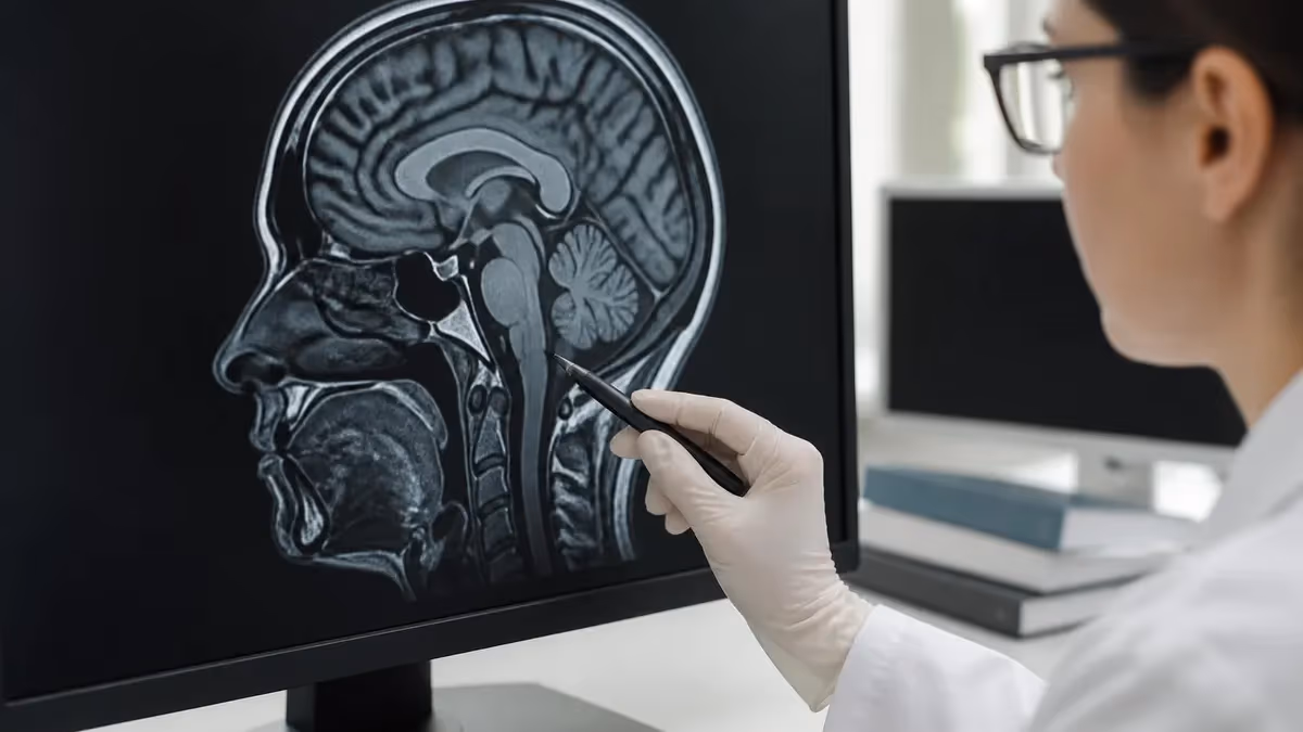
For specific what MRI shows specifically, MRI excellent for several tissue types. Brain (tumors, stroke, multiple sclerosis, dementia). Spine (disc herniations, spinal cord, tumors). Joints (ligaments, cartilage, tendons). Soft tissue tumors. Heart (specialized cardiac MRI). Specific other applications. Each MRI application leverages superior soft tissue detail. Quality MRI capability for these tissues substantially better than CT alternative for soft tissue evaluation.
For specific MRI types specifically, several MRI types serve specific needs. Standard MRI for general imaging. Functional MRI (fMRI) for brain function. MR angiography (MRA) for blood vessels. MR spectroscopy (MRS) for chemical analysis. Specific specialty MRI applications. Each MRI type extends imaging capability. Quality specialized MRI types provide diagnostic information not available from standard imaging for specific clinical questions warranting specialized approach. The MRI meaning guide covers MRI fundamentals.
For specific MRI safety specifically, MRI safety considerations substantial given powerful magnetic field. Metal objects can become projectiles. Metallic implants may not be MRI-compatible. Patients with pacemakers historically excluded (modern MR-conditional pacemakers may be acceptable). Specific contraindications must be assessed before scanning. Each safety consideration prevents potentially serious incidents. Quality safety screening through detailed patient history essential. The MRI safety guide covers safety details.
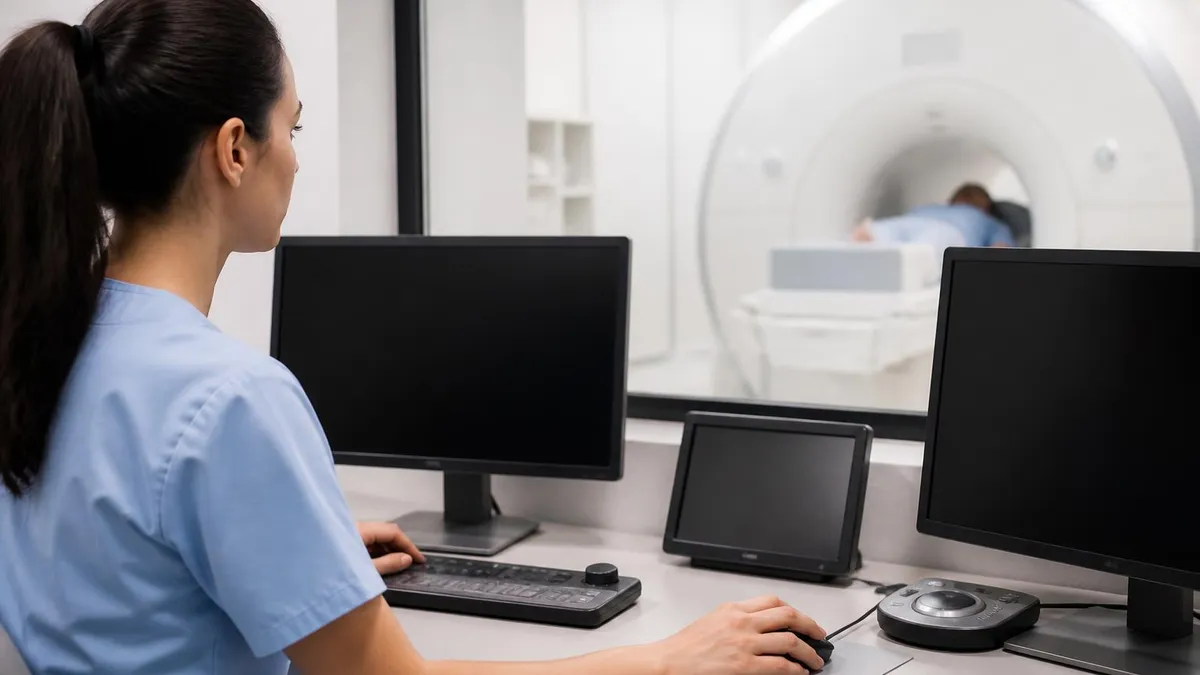
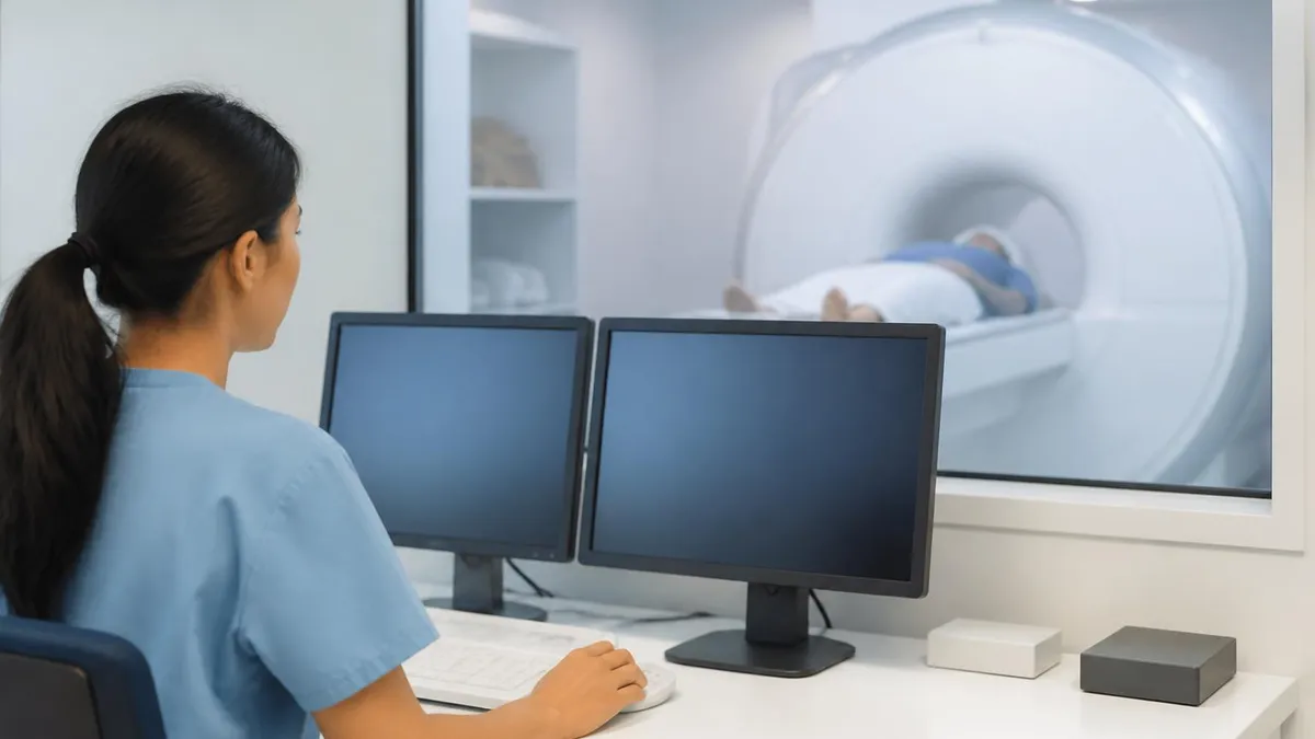
For specific patient experience specifically, MRI patient experience involves several elements. Lying still on table during scan. Wearing hearing protection due to loud noise. Confined space in closed scanner. Communication device available for patient comfort. Specific procedure length 20-90 minutes. Each experience element affects patient comfort. Quality understanding helps patients prepare mentally for procedure reducing anxiety substantially through knowledge of what to expect during MRI scan.
MRI Common Applications
Tumors, stroke evaluation, multiple sclerosis, dementia assessment, subtle brain conditions. MRI substantially superior to CT for brain soft tissue detail in most non-emergency contexts.
Disc herniations, spinal stenosis, spinal cord conditions, tumors. Substantial use in evaluating back pain causes. Excellent visualization of spinal anatomy and pathology.
Knee, shoulder, hip, ankle, wrist evaluation. Ligament tears, cartilage damage, soft tissue conditions around joints. Common orthopedic imaging modality.
Abdominal organs, pelvis, breast, cardiac applications. Various specialized body MRI applications. Less common than CT for body imaging but specific advantages for certain conditions.
For specific MRI advantages specifically, MRI offers several imaging advantages. Excellent soft tissue contrast. No ionizing radiation. Multiplanar imaging (any plane). Functional imaging capability. Specific advantages over other modalities for many conditions. Each advantage serves specific clinical needs. Quality MRI advantages make it preferred imaging for many specific clinical situations involving soft tissue evaluation requiring detailed tissue characterization.
For specific MRI disadvantages specifically, MRI also has disadvantages. Long scan times (typically 20-90 minutes). Loud noise during scanning. Claustrophobic environment in closed scanner. Substantial cost ($1,000-$3,000 typical). Specific contraindications limit some patients. Each disadvantage affects MRI use. Quality balance between advantages and disadvantages determines appropriate MRI use vs alternative imaging methods for specific clinical situations.
For specific contrast use specifically, some MRI scans use contrast agents. Gadolinium-based contrast most common for MRI. Injected intravenously before or during scan. Enhances visualization of certain conditions. Specific contrast considerations for kidney function. Each contrast use serves specific diagnostic purpose. Quality contrast use significantly improves diagnostic accuracy for many conditions but introduces additional considerations. The MRI with contrast guide covers contrast details.
For specific scanner types specifically, several MRI scanner types. Closed scanners traditional and most common. Open scanners accommodate claustrophobic and larger patients. Stand-up scanners for weight-bearing imaging. Wide-bore scanners accommodate larger patients. Specific scanner types serve different needs. Each scanner type has tradeoffs. Quality scanner selection matches patient needs and clinical requirements. The open MRI guide covers open scanner option.
For specific MRI cost specifically, MRI cost varies substantially. Typical scan $1,000-$3,000 in U.S. Insurance coverage common but with copays/deductibles. Specific facility costs (hospital vs imaging center). Geographic cost variations affect patient out-of-pocket expense. Each cost factor matters for affordability. Quality cost awareness helps patients understand financial implications. The MRI cost guide covers detailed pricing.
MRI Considerations by Body Part
Brain MRI specifics:
- Time: 30-60 minutes typical
- Common uses: Stroke, tumors, MS, dementia evaluation
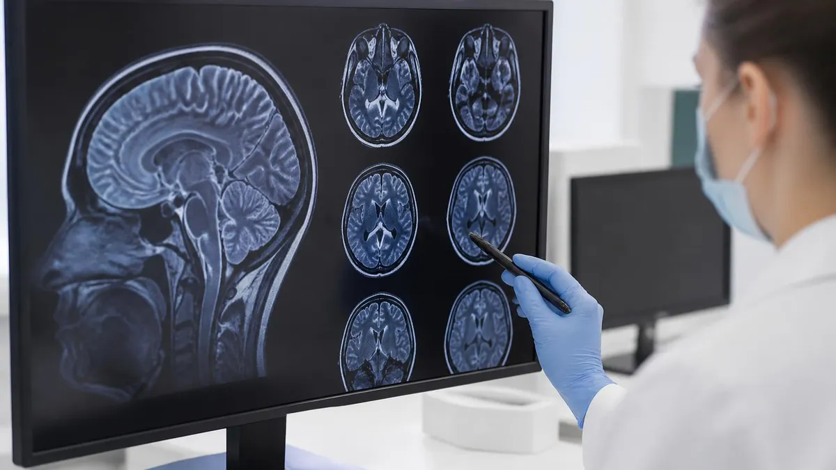
- Sequences: T1, T2, FLAIR, DWI typical
- Contrast: Often used for tumor evaluation
- Resolution: Sub-millimeter possible with 3T scanner
For specific MRI noise specifically, MRI scanners produce loud knocking and buzzing sounds. Sounds caused by gradient coils rapidly switching during imaging. Sound levels can exceed 100 decibels requiring hearing protection. Each scan involves substantial noise. Quality hearing protection (headphones, earplugs) standard during scans. Some scanners include music systems for patient comfort during long scans.
For specific claustrophobia specifically, claustrophobic patients face MRI challenges. Closed scanner enclosed space difficult for some. Several mitigation strategies available. Sedation sometimes used. Open scanners alternative when available. Specific anxiety management techniques. Each mitigation supports patient ability to complete scan. Quality claustrophobia management substantially extends MRI accessibility to patients otherwise unable to complete scans.
For specific implant safety specifically, metallic implants substantial MRI safety consideration. Modern MR-conditional implants designed for MRI safety. Some older implants absolutely contraindicate MRI. Pacemaker situations particularly important. Specific implant verification before MRI essential. Each implant has specific MRI compatibility status. Quality implant disclosure substantial — disclose all implants and devices before MRI even if seeming irrelevant.
For specific pregnancy specifically, MRI generally avoided in first trimester. Precautionary rather than evidence-based. No ionizing radiation makes MRI preferred over X-ray/CT during pregnancy when imaging needed. Specific pregnancy considerations vary. Each pregnancy MRI decision requires consideration. Quality pregnancy disclosure essential before MRI — staff makes appropriate decisions based on full information about pregnancy status.
For specific MRI tech career specifically, MRI technologists operate scanners. Substantial training required including imaging certifications. Patient interaction skills important. Specific technical skills for scanner operation. Image quality optimization responsibilities. Each career element shapes MRI tech work. Quality career path through formal training and certification produces qualified MRI techs essential for proper scanner operation. The MRI technologist guide covers career details.
Critical to disclose all metallic implants, pacemakers, surgical clips, and medical history before MRI. Strong MRI magnetic field can damage some implants, cause heating, or attract ferromagnetic objects with potentially fatal consequences. Modern MR-conditional implants designed for MRI safety but require specific protocols. Some older implants absolutely contraindicate MRI. Pregnancy disclosure important. Tattoos with metallic ink may heat during scanning. Quality complete medical history disclosure prevents serious incidents during scanning. Don't withhold information thinking it's irrelevant — let MRI staff determine relevance based on complete disclosure.
For specific scan day preparation specifically, several preparation elements support good MRI experience. Wear comfortable clothing without metal. Avoid jewelry on scan day. Bring list of medications and implants. Allow extra time for paperwork and changing. Specific facility instructions vary. Each preparation element prevents day-of issues. Quality preparation reduces stress and ensures smooth procedure.
For specific MRI history specifically, MRI development represents major scientific achievement. NMR (nuclear magnetic resonance) physics discovered 1940s. Application to medical imaging developed 1970s. First clinical MRI scanners 1980s. Substantial improvement over decades. Specific Nobel Prizes awarded to key MRI developers. Each historical milestone advanced MRI capability. Quality understanding of MRI development helps appreciate this remarkable technology emerged through decades of physics, engineering, and medical research collaboration.
For specific MRI advances specifically, MRI technology continues advancing. Higher field strengths providing better resolution. Faster imaging sequences reducing scan times. AI-enhanced image reconstruction. Specific specialized applications expanding clinical use. Each advancement extends MRI capability. Quality MRI advancement continues making this technology increasingly valuable for diverse medical applications.
For specific MRI accessibility specifically, MRI accessibility expanding beyond major hospitals. Outpatient imaging centers increasingly common. Specific rural areas sometimes still limited. Each accessibility expansion supports patient access. Quality accessibility improvements substantially better for patients than past when MRI primarily available only at major hospitals requiring substantial travel for many patients.
For specific MRI interpretation specifically, MRI images interpreted by radiologists. Specific subspecialty radiologists for complex cases. Reports communicated to ordering physician. Patient receives results from physician typically. Each interpretation step requires expertise. Quality radiologist interpretation essential — specific expertise particularly important for complex cases benefiting from subspecialty radiologist expertise.
MRI Preparation Checklist
- ✓Disclose all implants, devices, surgeries, and medical history
- ✓Remove all metal objects before entering MRI suite
- ✓Inform technologist of pregnancy status
- ✓Discuss anxiety/claustrophobia concerns for accommodations
- ✓Follow facility-specific instructions about eating, medications
For specific procedure timing specifically, MRI procedure timing varies. Brain MRI: 30-60 minutes typical. Spine MRI: 30-60 minutes per spine region. Knee MRI: 20-45 minutes. Cardiac MRI: 60-90 minutes. Specific times affected by sequences performed and patient cooperation. Each scan has typical time range. Quality time expectations help patients plan scan day appropriately.
For specific result timing specifically, MRI results typically available within days of scan. Radiologist reads images and creates report. Report sent to ordering physician. Physician communicates results to patient. Specific urgency affects result timing. Each result step takes time. Quality understanding helps patients set realistic expectations for receiving results — typically not immediate same-day delivery.
For specific MRI alternatives specifically, several MRI alternatives exist when MRI not appropriate. CT for many conditions. Ultrasound for some applications. X-ray for simple bone conditions. Each alternative serves specific situations. Quality alternative selection depends on specific clinical question. The MRI vs CT scan guide covers comparison.
For specific specialized MRI specifically, several specialized MRI applications. Cardiac MRI for heart imaging. Breast MRI for breast cancer evaluation. MR Angiography for blood vessel imaging. MR Spectroscopy for chemical analysis. Each specialized application serves specific needs. Quality specialized MRI provides diagnostic information not available from standard MRI for specific clinical questions warranting specialized approach. The cardiac MRI guide covers cardiac imaging.
For specific image quality factors specifically, several factors affect MRI image quality. Patient cooperation (lying still). Magnet strength (3T substantially better than 1.5T for some applications). Specific imaging sequences. Specific protocols for body part. Each quality factor matters. Quality MRI through patient cooperation plus appropriate scanner and protocol substantially better than rushed scans with poor patient cooperation producing diagnostically limited images requiring repeat scanning.
For specific MRI room safety specifically, MRI room substantial safety zone designation. Zones I-IV with increasing access restrictions. Zone IV magnet room substantial restrictions. Specific safety screening before entering MRI room. Each safety zone protects against incidents. Quality safety zone respect through screening compliance prevents serious incidents — substantial injuries documented from ferromagnetic objects becoming projectiles when magnet attracts unscreened metal items into magnet bore.
For specific MRI quench specifically, quench events possible safety consideration. Superconducting magnet warming above critical temperature causes rapid magnetic field loss. Massive helium release fills room. Emergency response needed. Specific protocols for handling quench events. Each safety consideration affects facility design. Quality understanding helps facilities prepare for rare but serious events requiring rapid evacuation and emergency ventilation system activation.
For specific MRI signal generation specifically, hydrogen atoms emit radio signals at specific frequency. Signal strength varies by tissue type. Specific tissue characteristics affect signal patterns. Computer measures and processes these signals. Each signal element provides diagnostic information. Quality signal analysis enables differentiation between healthy and diseased tissue and various tissue types appearing visually distinct on resulting images.
For specific T1 vs T2 weighting specifically, fundamental MRI imaging concept. T1-weighted: fat appears bright, water appears dark. Best for anatomical detail. T2-weighted: water appears bright, fat appears dark depending on technique. Best for pathology detection. Specific other sequences (FLAIR, DWI, others) highlight specific tissue characteristics. Each weighting emphasizes different aspects. Quality understanding of T1 vs T2 helps appreciate why MRI uses multiple sequences during single examination.
For specific imaging sequences specifically, MRI uses multiple sequences during single scan. T1-weighted, T2-weighted, FLAIR, DWI, others. Specific sequences highlight specific tissue characteristics. Each sequence provides specific diagnostic information. Quality multiple sequences during single exam provide comprehensive tissue characterization not possible with single sequence alone — MRI substantially powerful through multi-sequence approach unique among medical imaging modalities.
For specific MR angiography specifically, MRA images blood vessels. Various techniques visualize arteries and veins without invasive procedures. Particularly valuable for brain, neck, abdominal vessel evaluation. Specific MRA techniques (TOF, contrast-enhanced) suit different applications. Each MRA element extends MRI capability. Quality MR angiography particularly valuable when CT angiography contraindicated or specific MRI advantages relevant for clinical question requiring vascular imaging.
For specific functional MRI specifically, fMRI measures brain activity through blood flow changes. Active brain regions show increased blood flow detected through specific imaging sequences. Used for research and clinical brain mapping. Specific tasks during scan reveal brain function.
Each fMRI element extends MRI capability. Quality fMRI applications include brain function research, neurosurgery planning, various neuroscience applications using same physical scanner with specialized imaging protocols. Substantially extends MRI capability beyond anatomy visualization to functional brain understanding through brain activity measurement during specific tasks throughout neuroscience and clinical neurology contexts using specialized fMRI capabilities for various brain function studies and clinical applications throughout modern neuroscience research and clinical care utilizing fMRI capability extensively across many medical centers.
MRI Quick Facts
MRI Imaging Strengths
Excellent visualization of brain, spinal cord, joints, soft tissue organs. Substantial advantage over CT for soft tissue evaluation. Best modality for many soft tissue conditions.
Uses magnetic field and radio waves rather than ionizing radiation. Safer for repeated imaging. Particularly valuable for younger patients and those needing multiple scans over time.
Images any plane (axial, sagittal, coronal, oblique) without repositioning patient. Substantial advantage over X-ray and many other modalities requiring patient repositioning for different views.
fMRI measures brain activity through blood flow changes. MR spectroscopy measures chemical compounds. Specific functional capabilities beyond just anatomy visualization.
MRI Imaging Considerations
- +Excellent soft tissue contrast superior to CT for many tissues
- +No ionizing radiation (safer for repeated imaging)
- +Multi-planar imaging capabilities
- +Functional imaging available (fMRI for brain function)
- +Specialized applications for cardiac, vascular, breast imaging
- −Long scan times (20-90 minutes typical)
- −Loud noise requiring hearing protection
- −Claustrophobic environment in closed scanners
- −Substantial cost ($1,000-$3,000 typical)
- −Contraindications for some implants and devices
MRI Questions and Answers
About the Author
Medical Laboratory Scientist & Clinical Certification Expert
Johns Hopkins UniversityDr. Sandra Kim holds a PhD in Clinical Laboratory Science from Johns Hopkins University and is certified as a Medical Technologist (MT) and Medical Laboratory Scientist (MLS) through ASCP. With 16 years of clinical laboratory experience spanning hematology, microbiology, and molecular diagnostics, she prepares candidates for ASCP board exams, MLT, MLS, and specialist certification tests.
Join the Discussion
Connect with other students preparing for this exam. Share tips, ask questions, and get advice from people who have been there.
View discussion (6 replies)