Functional Magnetic Resonance Imaging: How fMRI Maps Brain Activity 2026 July
Functional magnetic resonance imaging (fMRI) maps brain activity using BOLD signal. Learn how it works, what it shows, and how to read fMRI scans. 🆕
Functional magnetic resonance imaging, commonly abbreviated as fMRI, is a non-invasive neuroimaging technique that measures brain activity by detecting tiny changes in blood oxygenation and flow. Unlike a standard structural MRI that captures anatomy, fMRI captures function in near real time, producing a dynamic map of which regions of the brain are working hardest during a specific task, thought, or emotion. It has transformed neuroscience, cognitive psychology, and clinical neurology since the early 1990s.
The core principle behind fMRI is the blood-oxygen-level-dependent signal, almost always called BOLD. When a cluster of neurons fires, the local demand for oxygen rises, and the brain responds by flooding that region with oxygen-rich blood. Because oxygenated and deoxygenated hemoglobin have different magnetic properties, the MRI scanner can detect this subtle shift and translate it into a colored activity map overlaid on a high-resolution anatomical scan of the same brain.
fMRI is used in a remarkable range of settings. Neurosurgeons use it for presurgical mapping before tumor or epilepsy operations, marking the locations of speech, motor, and memory regions to avoid during resection. Researchers use it to study attention, decision making, language acquisition, addiction, depression, autism, and Alzheimer's disease. Sports scientists, marketers, and even legal teams have explored it, although clinical and research applications remain by far the most validated uses today.
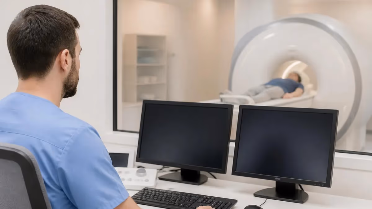
For radiology professionals, understanding fMRI matters because the modality is increasingly ordered alongside diffusion tensor imaging, perfusion sequences, and magnetic resonance spectroscopy in advanced neuroimaging protocols. Knowing how the BOLD signal is acquired, what paradigms patients perform inside the bore, and how technologists prepare patients for these long, demanding studies separates a confident MR technologist from one who simply runs the protocol. A solid grasp of the history of MRI helps frame why BOLD imaging was such a leap.
This guide walks through the physics of the BOLD effect, the hardware and pulse sequences that make fMRI possible, the difference between task-based and resting-state designs, common clinical and research applications, and the realistic limitations every reader should keep in mind. Whether you are a registry candidate, a graduate student, or a clinician who reads fMRI reports, this article aims to give you a working mental model and the vocabulary to use it confidently.
We will keep the math light but the concepts honest. fMRI is genuinely complicated, and oversimplifying it has led to a lot of bad press and reproducibility problems in the field. By the end, you should understand why a 1.5 percent change in MR signal can be statistically meaningful, why head motion of two millimeters can ruin a study, and why an fMRI map is a probability statement, not a photograph of thought.
If you are preparing for the ARRT MRI registry or the ARMRIT exam, fMRI shows up in advanced procedure sections and in the rationale behind echo planar imaging questions. Treat this article as a foundation, then drill the details with practice questions that test sequence parameters, safety, and patient management.
Functional MRI by the Numbers
The BOLD Effect and the Physics Behind fMRI
Deoxygenated hemoglobin distorts the local magnetic field, shortening T2* and reducing MR signal in nearby tissue. When neurons fire, oxygen consumption rises briefly, then blood flow overshoots.
Active neurons trigger local arterioles to dilate within seconds, flooding the region with oxygenated blood. The net result is a paradoxical drop in deoxyhemoglobin and an increase in MR signal where work is happening.
fMRI uses gradient-echo echo planar imaging (GRE-EPI) because T2* is exquisitely sensitive to the small magnetic susceptibility differences caused by changing blood oxygenation. EPI captures one whole slice in tens of milliseconds.
The BOLD response is sluggish compared to neural firing. After stimulation, signal rises over 4-6 seconds, peaks, then dips below baseline for 10-20 seconds. This shape is modeled by the canonical HRF.
Because the raw signal change is only 1-3 percent, fMRI relies on dozens of repeated trials and a general linear model to separate real activation from physiological noise, scanner drift, and head motion.
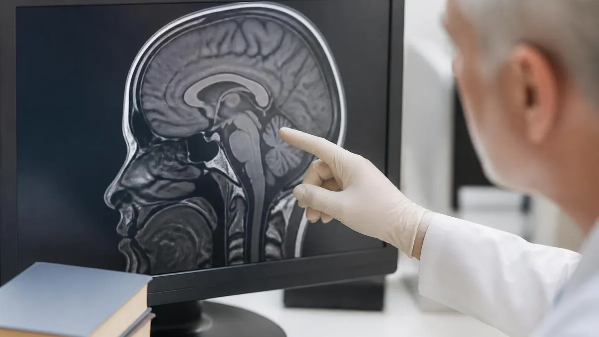
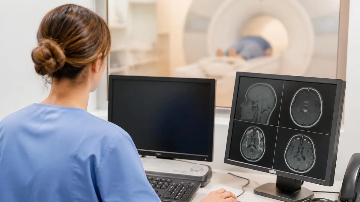
Inside the scanner, a functional MRI examination begins with a high-resolution anatomical scan, typically a 3D T1-weighted MPRAGE or BRAVO, lasting five to eight minutes. This structural reference is essential because the functional maps generated later are low resolution and low contrast, and they must be coregistered to detailed anatomy to be interpretable. Without that anatomical underlay, a colored blob in the middle of the brain tells you almost nothing about which gyrus or sulcus is involved.
The functional scans themselves are acquired with single-shot gradient-echo echo planar imaging. EPI is the engine of fMRI because it can collect an entire slice of k-space after a single radiofrequency excitation, which means a whole brain volume of 30 to 50 slices can be sampled every 1.5 to 2.5 seconds. Modern multiband or simultaneous multislice techniques push this further, acquiring two, four, or even eight slices in parallel and dropping the TR to under one second on advanced 3T platforms.
During functional runs, the patient performs a paradigm. A paradigm is simply a structured sequence of task and rest blocks, or randomly timed events, that the patient experiences while in the bore. Tasks might involve tapping the fingers, watching flashing checkerboards, listening to spoken sentences, naming pictures shown on a projection mirror, or silently generating verbs. Each paradigm is designed to selectively activate the brain region of clinical or research interest, such as the motor cortex, visual cortex, or Broca's area.
Stimulus delivery requires MRI-safe equipment. Most centers use a back-projection system with a mirror mounted on the head coil so the patient can see a screen at the foot of the bore. Audio is delivered through pneumatic or shielded electronic headphones that double as hearing protection. Responses are recorded with fiber-optic button boxes that contain no ferromagnetic components. The technologist runs the paradigm software on a stimulus computer outside the scan room, synchronized to the scanner's trigger pulse so every TR is timestamped.
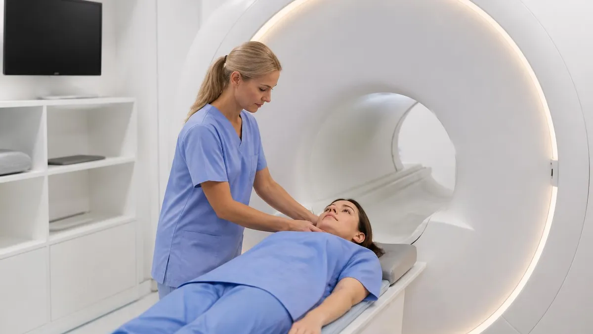
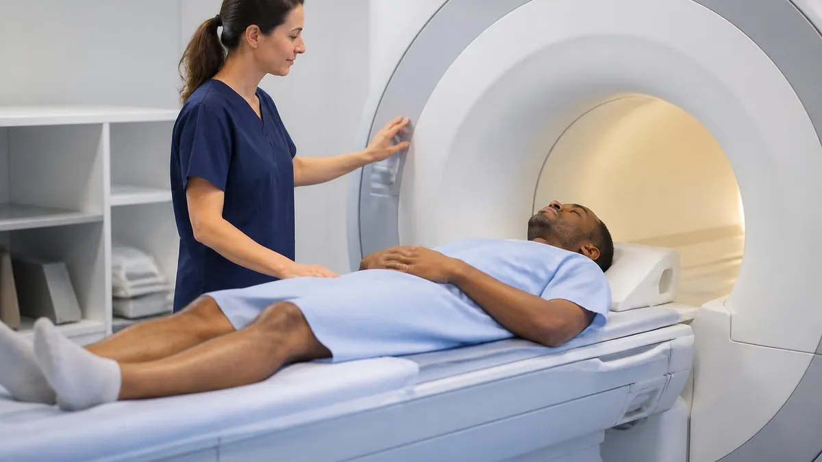
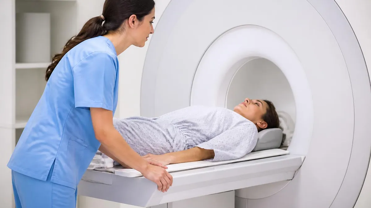
Patient preparation is more involved than for a routine brain MRI. The technologist must explain each task in plain language, practice it outside the scanner if possible, and confirm the patient understands what to do and when. Even small misunderstandings, like tapping during rest blocks, can ruin a run. The screening process is identical to any MRI, mirroring what you would do for an mri with and without contrast cpt, with extra attention to glasses, dental work, and anything that could distract a long study.
Motion is the enemy of fMRI. Because the technique looks for one to three percent signal changes, head movement of even one or two millimeters can create false activations or wipe out real ones. Foam pads, vacuum cushions, surgical tape across the forehead, and clear coaching all help. Some centers use real-time motion monitoring software that alerts the technologist when displacement exceeds a threshold, prompting a quick reminder before the next run.
Total scan time for a presurgical fMRI typically runs 30 to 60 minutes, with three to six functional runs of four to eight minutes each, plus the structural sequences. For research, sessions can stretch to 90 minutes, although fatigue and motion increase sharply after the first hour. Good technologists treat fMRI as a collaboration with the patient, not a passive scan.
MRI Practice Test Questions
Prepare for the MRI - Magnetic Resonance Imaging exam with our free practice test modules. Each quiz covers key topics to help you pass on your first try.
MRI Knowledge
MRI Exam Questions covering Knowledge. Master MRI Test concepts for certification prep.
MRI Physics
Free MRI Practice Test featuring Physics. Improve your MRI Exam score with mock test prep.
MRI Anatomy and Pathology
MRI Test Prep for MRI Anatomy and Pathology. Practice MRI Quiz questions and boost your score.
MRI Anatomy and Positioning
MRI Questions and Answers on MRI Anatomy and Positioning. Free MRI practice for exam readiness.
MRI Contrast Agents
Free MRI Quiz on MRI Contrast Agents. MRI Exam prep questions with detailed explanations.
MRI Patient Care and Positioning
MRI Practice Questions for MRI Patient Care and Positioning. Build confidence for your MRI certification exam.
Task-Based, Resting-State, and Real-Time fMRI
Task-based fMRI is the classic paradigm. The patient alternates between active task blocks and rest blocks, or performs discrete events at known times. A general linear model fits an expected BOLD response to each task and tests where in the brain the data correlate significantly with that predicted signal, producing colored statistical maps overlaid on anatomy.
Common clinical task paradigms include finger tapping for motor mapping, silent verb generation or sentence completion for language lateralization, and word listening for auditory processing. Each paradigm is validated against gold standards like intraoperative cortical stimulation or the Wada test, although fMRI is not yet a complete replacement for those procedures.
Strengths and Weaknesses of fMRI Compared to Other Brain Mapping Methods
- +Non-invasive with no ionizing radiation, safe for repeated use
- +Excellent spatial resolution of 2 to 4 millimeters in routine clinical use
- +Whole-brain coverage in every volume, not limited to a small region
- +Can be combined with structural MRI in the same session for coregistration
- +Validated for presurgical motor and language mapping at most major centers
- +Resting-state designs work in patients who cannot perform tasks
- +Widely available on standard 1.5T and 3T clinical scanners
- −Indirect measure of neural activity through blood flow, not firing
- −Poor temporal resolution of seconds compared to milliseconds for EEG
- −Susceptibility artifacts near sinuses, ear canals, and metal implants
- −Extremely sensitive to head motion, even one millimeter degrades data
- −BOLD signal change is small, requiring long scans and statistical models
- −Cannot reliably localize deep brainstem or small subcortical nuclei
- −Expensive scanner time and specialized analysis software required
Functional Magnetic Resonance Imaging Patient Prep Checklist
- ✓Complete a thorough MRI safety screening including implants, tattoos, and prior surgery
- ✓Confirm the patient can lie still for at least 45 to 60 minutes without distress
- ✓Explain each task paradigm in plain language and rehearse it outside the scanner
- ✓Verify vision and hearing are adequate to perceive the stimuli reliably
- ✓Provide MR-compatible corrective lenses if the patient normally wears glasses
- ✓Insert hearing protection that also delivers the auditory task audio
- ✓Position the head with foam pads and tape to minimize voluntary motion
- ✓Test the response button box and confirm finger placement before starting
- ✓Run a brief practice block inside the scanner to verify task comprehension
- ✓Monitor real-time motion and task performance during every functional run
BOLD is not a brain firing pattern — it is a vascular shadow of one
The BOLD signal lags neural activity by 4 to 6 seconds, reflects oxygen delivery rather than spikes, and varies with age, medication, caffeine, and vascular disease. Two patients with identical neural firing can show different BOLD maps. Always interpret fMRI as a probability statement guided by anatomy, not as a direct readout of thought.
Clinically, the most established use of fMRI is presurgical mapping for brain tumor and epilepsy patients. When a glioma or arteriovenous malformation sits near eloquent cortex, the neurosurgeon must decide how aggressive to be at the margins. An fMRI study performed days before the operation identifies the patient-specific location of hand motor function, speech production, and speech comprehension, all of which can shift in the presence of long-standing lesions. The maps are loaded into the intraoperative navigation system and updated with awake mapping when needed.
Language lateralization is another well-validated indication. Historically, the Wada test, which involves selectively anesthetizing one hemisphere with intra-arterial sodium amobarbital, was the standard for determining which side of the brain controls language. fMRI now achieves comparable results in most patients using verb generation and sentence completion paradigms, sparing patients an invasive angiographic procedure. In atypical cases, particularly left-handers and patients with early left-hemisphere injury, both methods may still be combined.
Epilepsy presurgical evaluation benefits from fMRI in several ways. Beyond language and motor mapping, simultaneous EEG-fMRI can localize the hemodynamic correlates of interictal spikes, helping the surgical team identify the epileptogenic zone. Resting-state functional connectivity is increasingly used to characterize network disruption in temporal lobe epilepsy and to predict postsurgical seizure freedom. Children with focal cortical dysplasia are particularly good candidates because conventional structural MRI sometimes misses subtle lesions.
In research and clinical trials, fMRI is a core outcome measure for neurological and psychiatric disorders. Studies of major depressive disorder consistently show altered connectivity in the default mode and salience networks, and ketamine, transcranial magnetic stimulation, and psychotherapy all modify these patterns. Alzheimer's disease research uses task-based memory paradigms and resting-state default mode network analyses as early biomarkers, sometimes detecting differences a decade before clinical symptoms appear. Reviewing a list of common MRI findings alongside fMRI activation maps deepens interpretation skills.
Stroke rehabilitation is another growing area. After a motor stroke, the affected hemisphere reorganizes, and contralateral regions often take on new roles. Serial fMRI documents this plasticity and is used to evaluate the effectiveness of constraint-induced movement therapy, robotic training, and brain stimulation. Researchers are exploring whether targeting specific functional networks with rehabilitation produces faster or more durable motor recovery than standard care alone.
Beyond neurology, fMRI has entered psychiatry, pain medicine, addiction research, and even some legal contexts. Pain studies map the so-called pain matrix involving the insula, anterior cingulate, and somatosensory cortices. Addiction research tracks cue-induced craving and reward responses to predict relapse. Lie detection and brain-based legal evidence remain controversial and are not accepted in most jurisdictions because individual-level inference from group fMRI data is unreliable.
Pediatric fMRI deserves a special mention. Children as young as five can perform simple tasks with practice, and resting-state designs work in sedated infants. Pediatric brain tumor surgery, hemispherectomy planning for intractable epilepsy, and developmental disorders such as autism and dyslexia all use fMRI in research and selected clinical contexts. The challenges are mainly motion and comprehension, both of which improve dramatically with good preparation, mock scanners, and child-friendly paradigms.
Despite popular media coverage, fMRI cannot read individual thoughts, detect lies reliably, or diagnose psychiatric conditions in single patients. Group-level statistical findings do not translate cleanly to individual inference. Always present fMRI results with appropriate clinical caveats, and never accept commercial claims of consumer fMRI lie detection or thought decoding without rigorous peer-reviewed evidence.
Every fMRI study has limitations that radiology professionals and clinicians must understand. The single largest is the indirect nature of the measurement. BOLD signal reflects blood oxygenation, which is several steps removed from the action potentials and synaptic events that constitute neural activity. Vascular pathology, medications affecting cerebral blood flow, caffeine consumption, and even simple dehydration can change the BOLD response without changing underlying neural function, making interpretation more nuanced than it appears.
Susceptibility artifact is the second major limitation. Gradient-echo EPI is exquisitely sensitive to magnetic field inhomogeneity, and air-tissue interfaces near the sinuses, ear canals, and skull base create signal dropout and geometric distortion. The orbitofrontal cortex and inferior temporal lobes are particularly affected, which is unfortunate because those regions are central to emotion, decision making, and memory. Distortion correction, multi-echo acquisition, and parallel imaging mitigate but do not eliminate this problem.
Statistical thresholding is a subtle but enormously important issue. With tens of thousands of voxels tested per study, the multiple comparisons problem is severe. Using uncorrected thresholds produces spurious activations, while overly conservative correction can miss real effects. The 2016 cluster-size controversy revealed that some software packages had been producing inflated false positive rates for years. Modern best practice uses permutation-based methods or threshold-free cluster enhancement, and many journals now require code and data sharing for transparency.
Head motion remains the most common reason fMRI studies fail. Even after foam padding, vacuum cushions, and clear coaching, some patients simply cannot stay still for an hour. Children, claustrophobic adults, and patients with chronic pain are particularly prone to motion, which can be mistaken for activation if the motion is correlated with the task. Considerations similar to imaging patients with MRI with braces apply here, since dental hardware can create artifacts that compound motion problems.
Individual variability is another challenge. The exact location of motor cortex, language regions, and visual areas varies by several centimeters across healthy people, and even more in patients with long-standing lesions. Group averaging in standard space, such as MNI or Talairach coordinates, smooths over this variability and can shift apparent peaks away from the true individual location. Presurgical mapping always operates in native space on each patient's own anatomy for this reason.
Reproducibility has been a growing concern. Several large studies in the past decade have shown that single-subject fMRI maps differ substantially across scan sessions, across analysis pipelines, and across software packages. Group findings tend to be more robust, but small-sample studies have produced many high-profile results that did not replicate. Newer initiatives like the Human Connectome Project and the UK Biobank use very large samples and standardized pipelines to address these problems.
Finally, fMRI is expensive, time-consuming, and requires specialized expertise that most general radiology practices do not have in-house. Reading an fMRI report requires familiarity with statistical maps, normalization, and paradigm design that goes beyond standard neuroradiology training. Centers offering clinical fMRI typically have a dedicated neuroimaging team including a physicist, a psychologist or technologist trained in stimulus delivery, and a neuroradiologist with subspecialty experience.
If you are preparing to perform or interpret fMRI, a handful of practical habits separate routine work from outstanding work. Start by reading every requisition carefully. Presurgical mapping referrals should specify which functions need mapping, where the lesion sits, and which language the patient speaks fluently. A vague order such as comprehensive fMRI is a recipe for an unfocused study that misses what the surgeon actually needs to know on the day of surgery.
Practice paradigms with the patient before the scan whenever possible. A five-minute rehearsal in the prep room, using a laptop and a finger tap on a tabletop, dramatically improves data quality. The patient learns the timing, you confirm comprehension, and any language barriers or cognitive deficits surface before they ruin a run inside the bore. For pediatric patients, a mock scanner session a day or two earlier is even better and reduces sedation rates.
Use the structural scan as your real-time anatomy reference. While functional runs are acquiring, pull up the MPRAGE on the console and confirm that your slice prescription covers the regions of interest, that the temporal lobes are not cut off, and that the eyes are excluded from the field of view to reduce motion-related signal contamination. Five minutes of careful planning at the start prevents an unusable dataset at the end.
Monitor motion in real time if your system supports it. Most modern Siemens, Philips, GE, and Canon platforms offer some form of online motion display. Set a threshold, typically half a voxel of translation or one degree of rotation, and gently coach the patient between runs if they exceed it. Many patients are unaware they have moved and respond well to specific feedback such as keep your jaw relaxed rather than vague reminders to stay still.
Document everything. Note which paradigm was run, the patient's reported task compliance, any unusual events such as coughing or talking, and the exact run numbers in the radiologist's worksheet. fMRI analysis is performed hours or days after the scan, and the analyst will thank you for clear notes that prevent the wrong run from being processed as the wrong condition. Treat the worksheet as part of the clinical record.
Stay current with the literature. fMRI methodology evolves quickly, with multiband EPI, multi-echo acquisition, prospective motion correction, and deep learning denoising all becoming clinically available over the past few years. Reading one methodological paper a month, attending the annual ISMRM meeting if you can, and following preprint servers like bioRxiv keeps your practice ahead of where most departments operate today.
Finally, communicate uncertainty honestly. When a presurgical map shows clear motor activation but ambiguous language localization, say so in the report. Surgeons and patients make better decisions with calibrated uncertainty than with falsely confident maps. The best fMRI practitioners are the ones who know exactly where their technique stops being reliable and who say so out loud, every time.
MRI Questions and Answers


About the Author
Medical Laboratory Scientist & Clinical Certification Expert
Johns Hopkins UniversityDr. Sandra Kim holds a PhD in Clinical Laboratory Science from Johns Hopkins University and is certified as a Medical Technologist (MT) and Medical Laboratory Scientist (MLS) through ASCP. With 16 years of clinical laboratory experience spanning hematology, microbiology, and molecular diagnostics, she prepares candidates for ASCP board exams, MLT, MLS, and specialist certification tests.
Join the Discussion
Connect with other students preparing for this exam. Share tips, ask questions, and get advice from people who have been there.
View discussion (6 replies)